Phages are miniscule, tripod-looking viruses that are genetically programmed to locate, attack, and eradicate a specific kind of pathogen. These microscopic creatures have saved lives and are being touted as a potential solution to superbugs, which are strains of bacteria, viruses, parasites, and fungi that are resistant to most antibiotics and other treatments utilized to counteract infections.
“These multi-drug-resistant superbugs can cause chronic infections in individuals for months to years to sometimes decades,” Dwayne Roach, PhD, Assistant Professor of Bacteriophages, Infectious Disease, and Immunology at SDSU told CNN. “It’s ridiculous just how virulent some of these bacteria get over time.”
Labs across the country are conducting research on phages in eradicating superbugs. Roach’s lab is currently probing the body’s immune response to phages and developing purification techniques to prepare phage samples for intravenous use in patients.
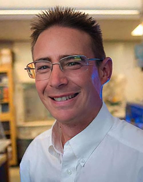
“There are a lot of approaches right now that are happening in parallel,” said Dwayne Roach, PhD (above), Assistant Professor of Bacteriophages, Infectious Disease, and Immunology at San Diego State University (SDSU), in a CNN interview. “Do we engineer phages? Do we make a phage cocktail, and then how big is the cocktail? Is it two phages or 12 phages? Should phages be inhaled, applied topically, or injected intravenously? There’s a lot of work underway on exactly how to best do this.” Clinical laboratories that test for bacterial infections may play a key role in diagnosis and treatment involving bacteriophages. (Photo copyright: San Diego State University.)
Building Libraries of Phages
When certain a bacterial species or its genotypes needs to be annihilated, a collection of phages can be created to attack it via methods that enter and weaken the bacterial cell. The bacteria will attempt to counter the intrusion by employing evasive actions, such as shedding outer skins to eliminate the docking ports utilized by the phages. These maneuvers can cause the bacteria to lose their antibiotic resistance, making them vulnerable to destruction.
Some research labs are developing libraries of phages, accumulating strains found in nature in prime breeding grounds for bacteria to locate the correct phage for a particular infection. Other labs, however, are speeding up the process by producing phages in the lab.
“Rather than just sourcing new phages from the environment, we have a bioreactor that in real time creates billions upon billions of phages,” Anthony Maresso, PhD, Associate Professor at Baylor College of Medicine in Houston told CNN. “Most of those phages won’t be active against the drug-resistant bacteria, but at some point, there will be a rare variant that has been trained, so to speak, to attack the resistant bacteria, and we’ll add that to our arsenal. It’s a next-generation approach on phage libraries.”
For the Baylor study, 12 patients were treated with phages customized to each individual’s unique bacterial profile. The antibiotic-resistant bacteria were exterminated in five of the patients, while several others showed improvement.
Clinical trials are currently being executed to test the effectiveness of phages against a variety of chronic health conditions, including:
Using a phage cocktail could be used to treat a superbug outbreak in real time, while preventing a patient from a future infection of the same superbug.
“The issue is that when patients have infections with these drug-resistant bacteria, they can still carry that organism in or on their bodies even after treatment,” Maroya Walters, PhD, epidemiologist at the federal Centers for Disease Control and Prevention (CDC) told CNN.
“They don’t show any signs or symptoms of illness, but they can get infections again, and they can also transmit the bacteria to other people,” she added.
The colorized transmission electron micrograph above shows numerous phages attached to a bacterial cell wall. Phages are known for their unique structures, which resemble a cross between NASA’s Apollo lunar lander and an arthropod. (Caption and photo copyright: Berkeley Lab.)
More Studies are Needed
According to CDC data, more than 2.8 million antimicrobial-resistant (AMR) infections occur annually in the United States. More than 35,000 people in the country will die as a result of these infections.
In addition, AMR infections are a huge global threat, associated with nearly five million deaths worldwide in 2019. Resistant infections can be extremely difficult and sometimes impossible to treat.
More research is needed before phages can be used clinically to treat superbugs. But if phages prove to be useful in fighting antibiotic-resistant bacteria, microbiologists and their clinical laboratories may soon have new tools to help protect patients from these deadly pathogens.
Studies could lead to new prognostic biomarkers and clinical laboratory diagnostics for cancer
Might fungi be involved in human cancers? Two separately published studies have found fungal DNA in various cancers in the human body. However, the researchers are unclear on how the fungi got into the cancer cells and if it is affecting the cancers’ pathology. Nevertheless, these discoveries could lead to utilizing tumor-associated fungal DNA as clinical laboratory diagnostics or prognostic biomarkers in the fight against cancer.
“The finding that fungi are commonly present in human tumors should drive us to better explore their potential effects and re-examine almost everything we know about cancer through a ‘microbiome lens,’” said Ravid Straussman, MD, PhD (above), a principal investigator at Weizmann Institute of Science and one of the authors of the study in a UCSD press release. These findings could lead to new clinical laboratory diagnostics and prognostic biomarkers. (Photo copyright: Weizmann Institute of Science.)
.
Microbiome Key to Cancer Biology and Detection
To perform their research, the team examined 17,401 samples of patient tissues, blood, and plasma across 35 different types of cancers in four independent cohorts. They discovered fungal DNA and cells in low abundances in many human cancers.
“The existence of fungi in most human cancers is both a surprise and to be expected,” said biologist Rob Knight, PhD, founding Director of the Center for Microbiome Innovation and Professor of Pediatrics and Computer Science and Engineering at UC San Diego in a UCSD press release. “It is surprising because we don’t know how fungi could get into tumors throughout the body. But it is also expected because it fits the pattern of healthy microbiomes throughout the body, including the gut, mouth and skin, where bacteria and fungi interact as part of a complex community.”
The main highlights of this study include:
Fungi detected in the different cancer types were often intracellular.
Multiple fungal-bacterial-immune ecologies were detected across tumors.
Intratumoral fungi stratified clinical outcomes, including immunotherapy response.
Cell-free fungal DNA found in both healthy and cancer patients in early-stage disease.
Fungi found on the human body appear as either environmental fungi, such as yeasts and molds, and commensal fungi, which live either on or inside the body. Both are typically harmless to most healthy people and can provide some benefits, such as improving gut health, but they may also be a contributing factor in some disease.
The researchers found that there were notable parallels between specific fungi and certain factors, such as age, tumor subtypes, smoking status, immunotherapy responses, and survival measures.
“These findings validate the view that the microbiome in its entirety is a key piece of cancer biology and may present significant translational opportunities, not only in cancer detection, but also in other biotech applications related to drug development, cancer evolution, minimal residual disease, relapse, and companion diagnostics,” said Gregory Sepich-Poore, MD, PhD, one of the study’s authors and co-founder and chief analytics officer at biotechnology company Micronoma, in the UCSD press release.
New Clinical Laboratory Tests to Identify Fungal Species in Cancer
Researchers from Duke University and Cornell University uncovered compelling evidence of fungi in multiple cancer types and focused on a detected link between Candida and gastrointestinal cancers.
They found that “several Candida species were enriched in tumor samples and tumor-associated Candida DNA was predictive of decreased survival,” according to their paper.
Their analysis of multiple body sites revealed tumor-associated mycobiomes in fungal cells. The researchers found that fungal spores known as blastomyces were associated with tumor tissues in lung cancers, and that high rates of Candida were present in stomach and colon cancers.
The Duke/Cornell researchers hope their work can provide a framework to develop new tests that can distinguish fungal species in tumors and predict cancer progression and help medical professionals and patients chose the best treatment therapies.
“These findings open up a lot of exciting research directions, from the development of diagnostics and treatments to studies of the detailed biological mechanisms of fungal relationships to cancers,” said Iliyan Iliev, PhD, Associate Professor of Microbiology and Immunology in Medicine, Weill Cornell Medicine, and one of the authors of the study, in a Weill news release.
More research is needed to determine if fungal DNA plays a role in disease pathology or if its presence does not have any causal link.
“It’s plausible that some of these fungi are promoting tumor progression and metastasis, but even if they aren’t, they could be very valuable as prognostic indicators,” Iliev said.
The insights gleaned from these two studies will be of particular interest to microbiologists, clinical laboratory professionals, and anatomic pathologists. Additional research could answer questions about how and if fungi infect tumors and if such fungi is a factor that increases cancer risk and outcomes.
Researchers surprised that process designed to detect SARS-CoV-2 also identifies monkeypox in wastewater
Early information about an outbreak in a geographical region can inform local clinical laboratories as to which infectious agents and variants they are likely to see when testing patients who have symptoms. To that end, wastewater testing has become a rich source of early clues as to where COVID-19 outbreaks are spreading and how new variants of the coronavirus are emerging.
Ongoing advances in genetic sequencing and digital technologies are making it feasible to test wastewater for infectious agents in ways that were once too time-consuming, too expensive, or simply impossible.
“Before wastewater sequencing, the only way to do this was through clinical testing, which is not feasible at large scale, especially in areas with limited resources, public participation, or the capacity to do sufficient testing and sequencing,” said Knight in a UCSD press release. “We’ve shown that wastewater sequencing can successfully track regional infection dynamics with fewer limitations and biases than clinical testing to the benefit of almost any community.” (Photo copyright: UC San Diego News.)
Same Process, Different Virus
Following August’s declaration of a state of emergency by California, San Diego County, and the federal government, UCSD researchers added monkeypox surveillance to UCSD’s existing wastewater surveillance program.
“It’s the same process as SARS-CoV-2 qPCR monitoring, except that we have been testing for a different virus. Monkeypox is a DNA virus, so it is a bit of a surprise that our process optimized for SARS-CoV-2, which is an RNA virus, works so well,” said Rob Knight, PhD, Professor of Pediatrics and Computer Science and Engineering at UCSD and one of the lead authors of the study in the press release.
According to the press release, RNA sequencing from wastewater has two specific benefits:
It avoids the potential of clinical testing biases, and
It can track changes in the prevalence of SARS-CoV-2 variants over time.
In 2020, at the height of the COVID-19 pandemic, scientists from the University of California San Diego and Scripps Research looked into genetic sequencing of wastewater. They wanted to see if it would provide insights into levels and variants of the SARS-CoV-2 within a specific community.
Individuals who have COVID-19 shed the virus in their stool.
The UCSD/Scripps researchers deployed commercial auto-sampling robots to collect wastewater samples at the main UCSD campus. They analyzed the samples for levels of SARS-CoV-2 RNA at the Expedited COVID-19 Identification Environment (EXCITE) lab at UCSD. After the success of the program on the campus, they extended their research to include other facilities and communities in the San Diego area.
“The coronavirus will continue to spread and evolve, which makes it imperative for public health that we detect new variants early enough to mitigate consequences,” said Knight in a July press release announcing the publication of their study in the journal Nature, titled, “Wastewater Sequencing Reveals Early Cryptic SARS-CoV-2 Variant Transmission.”
Detecting Pathogens Weeks Earlier than Traditional Clinical Laboratory Testing
In July, the scientists successfully determined the genetic mixture of SARS-CoV-2 variants present in wastewater samples by examining just two teaspoons of raw sewage. They found they could accurately identify new variants 14 days before traditional clinical laboratory testing. They detected the presence of the Omicron variant 11 days before it was first reported clinically in the community.
During the study, the team collected and analyzed 21,383 sewage samples, with most of those samples (19,944) being taken from the UCSD campus. They performed genomic sequencing on 600 of the samples and compared them to genomes obtained from clinical swabs. They also compared 31,149 genomes from clinical genomic surveillance to 837 wastewater samples taken from the community.
The scientists distinguished specific viral lineages present in the samples by sequencing the viruses’ complete set of genetic instructions. Mutational differences between the various SARS-CoV-2 variants can be minute and subtle, but also have notable biological deviations.
“Nothing like this had been done before. Sampling and detection efforts began modestly but grew steadily with increased research capacity and experience. Currently, we’re monitoring almost 350 buildings on campus,” said UCSD’s Chancellor Pradeep Khosla, PhD, in the July press release.
“The wastewater program was an essential element of UC San Diego Health’s response to the COVID pandemic,” said Robert Schooley, MD, Infectious Disease Specialist at UC San Diego Health, in the press release. Schooley is also a professor at UCSD School of Medicine, and one of the authors of the study.
“It provided us with real-time intelligence about locations on campus where virus activity was ongoing,” he added. “Wastewater sampling essentially allowed us to ‘swab the noses’ of every person upstream from the collector every day and to use that information to concentrate viral detection efforts at the individual level.”
Monkeypox Added to UCSD Wastewater Surveillance
In August, UCSD officially added the surveillance of the monkeypox virus to their ongoing wastewater surveillance program. A month earlier, the researchers had discerned 10,565.54 viral copies per liter of wastewater. They observed the levels fluctuating and increasing.
On August 2, the scientists detected 189,309.81 viral copies per liter of wastewater. However, it is not yet clear if the monitoring of monkeypox viral loads in wastewater will enable the researchers to accurately predict future infections or case rates.
“We don’t yet know if the data will anticipate case surges like with COVID,” Knight said in the August UCSD press release announcing the addition of monkeypox to the surveillance program. “It depends on when the virus is shed from the body relative to how bad the symptoms are that cause people to seek care. This is, in principle, different for each virus, although in practice wastewater seems to be predictive for multiple viruses.”
Utilization of genetic sequencing of wastewater sampling will continue to develop and improve. “It’s fairly easy to add new pathogens to the process,” said Smruthi Karthikeyan, PhD, an environmental engineer and postdoctoral researcher in Knight’s lab who has overseen wastewater monitoring at UC San Diego. “It’s doable on short notice. We can get more information in the same turnaround time.”
Thus, clinical laboratories engaged in testing programs for COVID-19 may soon see the addition of monkeypox to those processes.
Research could lead to clinical laboratory tests in service of precision medicine therapies to reduce a person’s susceptibility to being targeted by blood-sucking insects
Ever wonder why some people attract mosquitoes while others do not? Could biting insects pick their victims by smell? Scientists in California believe the answers to these questions could lead to new precision medicine therapies and clinical laboratory tests.
The research revealed evidence that some blood-sucking insects may identify their prey by homing in on the “scent” of chemicals produced by bacteria located in the skin microbiome of animals and humans.
This is yet another example of research into one area of the human microbiome that might someday lead to a new clinical laboratory test, in this case to determine if a person is more likely to attracts biting insects. If there were such a test, precision medicine therapies could be developed that change an individual’s microbiome to discourage insects from biting that individual.
Then, the clinical laboratory test would have value because it helped diagnose a health condition that is treatable.
Curiosity regarding why mosquitoes seem to gravitate towards some humans over others was the original catalyst for the UCSD Medical School research. “You know when you go to a barbeque and your friend is getting bombarded by mosquitos, but you’re fine? There is some research to support the idea that the difference in mosquito attraction is linked to your skin microbiome—the unique community of bacteria living on your skin,” said Holly Lutz, PhD (above), first author of the UCSD study. “Keeping in mind that some people are more attractive to mosquitoes than others, I wondered what makes insects attracted to some bats but not others.” Lutz’s research could lead to clinical laboratory tests that drive precision medicine therapies to alter human skin microbiomes and make people less attractive to biting insects. (Photo copyright: The Field Museum of Natural History.)
Biting Flies Prefer Specific Bats
“In these caves, I’d see all these different bat species or even taxonomic families roosting side by side. Some of them were loaded with bat flies, while others had none or only a few,” Lutz said in Phys.org. “And these flies are typically very specific to different kinds of bats—you won’t find a fly that normally feeds on horseshoe bats crawling around on a fruit bat. I started wondering why the flies are so particular. Clearly, they can crawl over from one kind of bat to another, but they don’t really seem to be doing that.”
The researchers suspected that the bacteria contained in the skin microbiomes of individual bats could be influencing which bats the flies selected to bite. The bacteria produce a distinctive odor which may make certain bats more attractive to the flies.
The type of fly assessed for the study are related to mosquitoes and most of them are incapable of flight.
“They have incredibly reduced wings in many cases and can’t actually fly,” Lutz explained. “And they have reduced eyesight, so they probably aren’t really operating by vision. So, some other sensory mechanisms must be at play, maybe a sense of smell or an ability to detect chemical cues.”
To test their hypothesis, the research team collected skin and fur samples from the bodies and wings of a variety of bat species located in various caves around Kenya and Uganda. They collected their samples at 14 field sites from August to October in 2016. They then examined the DNA of the bats as well as the microbes residing on the animals’ skin and searched for the presence of flies.
“The flies are exquisitely evolved to stay on their bat,” said Carl Dick, PhD, a professor of biology at Western Kentucky University and one of the study’s authors. “They have special combs, spines, and claws that hold them in place in the fur, and they can run quickly in any direction to evade the biting and scratching of the bats, or the efforts by researchers to capture them,” he told Phys.org.
“You brush the bats’ fur with your forceps, and it’s like you’re chasing the fastest little spider,” Lutz said. “The flies can disappear in a split second. They are fascinatingly creepy.”
Genetic Sequencing DNA of Bat Skin Bacteria
After collecting their specimens, the researchers extracted DNA from the collected bacteria and performed genetic sequencing on the samples. They created libraries of the bacteria contained in each skin sample and used bioinformatics methods to identify the bacteria and compare the samples from bats that had flies versus those that did not.
“How the flies actually locate and find their bats has previously been something of a mystery,” Dick noted. “But because most bat flies live and feed on only one bat species, it’s clear that they somehow find the right host.”
The scientists discovered that different bat families did have their own distinctive skin microbiome, even among samples collected from different locations. They found that differences in the skin microbiomes of certain bats does contribute to whether those bats have parasites. But not all their questions were answered.
“We weren’t able to collect the actual chemicals producing cue—secondary metabolites or volatile organic compounds—during this initial work. Without that information, we can’t definitively say that the bacteria are leading the flies to their hosts,” Lutz said.
Next Steps
“So, next steps will be to sample bats in a way that we can actually tie these compounds to the bacteria. In science, there is always a next step,” she added.
This research illustrates that there may be a reason why certain animals and humans tend to be more attractive to insects than others. It is also possible that an individual’s skin microbiome may explain why some people are more prone to mosquito and other types of insect bites.
More research and clinical studies on this topic are needed, but it could possibly lead to a clinical laboratory test to determine if an individual’s skin microbiome could contribute to his or her potential to being bitten by insects. Such a test would be quite beneficial, as insects can carry a variety of diseases that are harmful to humans.
Perhaps a precision medicine therapy could be developed to alter a person’s microbiome to make them invisible to blood-sucking insects. That would be a boon to regions of the world were diseases like malaria are spread by insect bites.
Skin patch technologies could enable clinical laboratories to monitor patients’ vitals and report to medical professionals in real time
Pathologists and clinical laboratory leaders have read many Dark Daily ebriefings on the development of skin patches over the years that do everything from monitoring fatigue in the military to being a complete lab-on-skin technology. Now, researchers at the University of California San Diego (UCSD) have developed a wearable patch that can monitor cardiovascular signals and other various biochemical levels in the body simultaneously.
The researchers believe there is enormous potential for such a patch in helping patients monitor conditions such as hypertension or diabetes. They also foresee a scenario where the patch could be used in settings where vitals must be constantly monitored. They hope to develop future versions of the patch that can detect more biomarkers within the body.
“This type of wearable would be very helpful for people with underlying medical conditions to monitor their own health on a regular basis,” Lu Yin, a PhD student and co-first author of the study, told New Atlas. “It would also serve as a great tool for remote patient monitoring, especially during the COVID-19 pandemic when people are minimizing in-person visits to the clinic,” she added.
Combining Precision Medicine with Telehealth and the Internet of Things
About the size of a postage stamp and consisting of stretchy polymers that conform to the skin, the UCSD patch monitors blood pressure and contains sensors that measure different biochemical levels in the body, such as:
The sensors are carefully arranged on the patch to eliminate interference between the signals, noted a UCSD press release.
In their published research, the UCSD researchers wrote of their new skin patch monitoring device, “Intertwined with concepts of telehealth, the internet of medical things, and precision medicine, wearable sensors offer features to actively and remotely monitor physiological parameters. Wearable sensors can generate data continuously without causing any discomfort or interruptions to daily activity, thus enhancing the self-monitoring compliance of the wearer, and improving the quality of patient care.” (Photo copyright: University of California San Diego.)
“Each sensor provides a separate picture of a physical or chemical change. Integrating them all in one wearable patch allows us to stitch those different pictures together to get a more comprehensive overview of what’s going on in our bodies,” said Sheng Xu, PhD, Principle Investigator, Xu Research Group at UCSD, Assistant Professor in the Department of NanoEngineering Department, and a co-first author of the study, in the press release.
The UCSD researchers developed their skin patch to monitor specific biomarkers that can affect blood pressure.
“Let’s say you are monitoring your blood pressure and you see spikes during the day and think that something is wrong,” co-first author Juliane Sempionatto, PhD, a postdoctoral researcher at California Institute of Technology (Caltech) and co-first author of the study, told New Atlas. “But a biomarker reading could tell you if those spikes were due to an intake of alcohol or caffeine. This combination of sensors can give you that type of information,” she added.
The blood pressure sensor sits near the center of the patch and consists of a set of small transducers welded to the patch via a conductive link. Voltage applied to the transducers send ultrasound waves through the body which bounce off arteries and create echoes that are detected by the sensor and converted into an accurate blood pressure reading.
The chemical sensor releases the drug pilocarpine into the skin to induce sweat and then measures the chemicals contained in the sweat to provide readings of certain biochemical levels.
The glucose sensor located in the patch emits a mild electrical current to the body that stimulates the release of interstitial fluid and then reads the glucose level in that fluid.
“The novelty here is that we take completely different sensors and merge them together on a single small platform as small as a stamp,” Joseph Wang, D.Sc, SAIC Endowed Chair, Distinguished Professor of NanoEngineering, Director of the Center for Wearable Sensors at UCSD, and co-author of the study told New Atlas. “We can collect so much information with this one wearable and do so in a non-invasive way, without causing discomfort or interruptions to daily activity.” (Photo copyright: University of Southern California San Diego.)
Skin Patch Measurements Closely Match Those of Traditional Devices
Test subjects wore the patch on their neck while performing various combinations of the following tasks:
exercising on a stationary bicycle,
eating a high-sugar meal,
drinking an alcoholic beverage, and
drinking a caffeinated beverage.
The results of the measurements taken from the patch closely matched measurements collected by traditional monitoring devices such as a:
For now, the patch must be connected to an external power source which transmits the reading to a counter-top machine, but the researchers hope to create a wireless version in the future.
“There are opportunities to monitor other biomarkers associated with various diseases,” Sempionatto said in the UCSD press release. “We are looking to add more clinical value to this device.”
Other Similar Skin Patch Monitoring Technologies
Though an important breakthrough, the UCSD’s device is not the first skin patch monitor to be developed.
Multiple research and clinical studies are underway that hope to prove the accuracy and safety of wearable devices at detecting and monitoring certain health conditions. It’s a worthy goal.
Skin patches, such as the one created at UCSD, could enable clinical laboratories to provide value-added service to medical professionals and patients alike. Medical labs could potentially monitor skin patch readings in real-time and notify physicians and patients of changes in biomarkers that require attention.
Further, as this technology is developed, it will likely find a ready market with the latest generation of consumers who are more willing than previous generations to buy their own diagnostic tests for home use. These “next-generation” healthcare consumers have demonstrated their willingness to use Apple watches, Fitbits, and similar wearable devices to monitor their condition during exercise and other health metrics.
Pathologists and clinical laboratory managers should not overlook the potential for robust consumer demand to accelerate development and market adoption of such skin patches.