Researchers used CRISPR-based assays to develop new clinical laboratory point-of-care blood test which boasts accuracy, affordability, and accessibility
According to UPI, the test can “distinguish between influenza A and influenza B—the two main types of seasonal flu—as well as identifying more virulent strains like H1N1 and H3N2.”
Many research teams are working to develop paper-based diagnostic screening tests because of their lower cost to produce and usefulness in remote locations. Should this near-patient point-of-care test become clinically viable, it could mean shorter times to answer, enabling speedier diagnoses and earlier start of treatment.
It also means patient specimens do not have to be transported to a clinical laboratory for testing. And reduced cost per test makes it possible to test more people. This serves the public health aspect of monitoring outbreaks of influenza and other diseases and gives hope for improved treatment outcomes.
“Being able to tease apart what strain or subtype of influenza is infecting a patient has repercussions both for treating them and public health interventions, said Jon Arizti Sanz, PhD, co-lead study author and postdoctoral researcher at the Broad Institute of Harvard and MIT, in a Broad Institute news release.
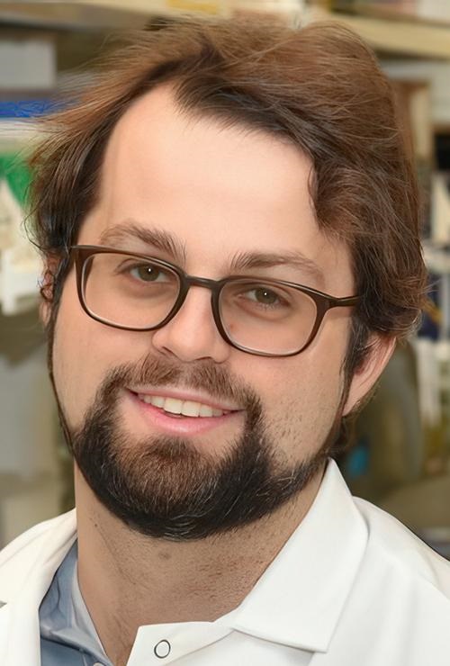
“Ultimately, we hope these tests will be as simple as rapid antigen tests, and they’ll still have the specificity and performance of a nucleic acid test that would normally be done in a laboratory setting,” Cameron A. Myhrvold, PhD (above), Assistant Professor of Molecular Biology at Princeton University in New Jersey, told CIDRAP. Influenza tests that can be performed at the point of care and in remote locations may reduce the number of screening tests performed by clinical laboratories. (Photo copyright: Michael James Butts/Hertz Foundation.)
Her team developed their tests using Streamlined Highlighting of Infections to Navigate Epidemics (SHINE), “a clustered regularly interspaced short palindromic repeats (CRISPR)-based RNA detection platform,” the researchers wrote in their Journal of Molecular Diagnostics paper.
“SHINE has a runtime of 90 minutes, can be used at room temperature and only requires an inexpensive heat block to heat the reaction. The SHINE technology has previously been used to identify SARS-CoV-2 and later to distinguish between the Delta and Omicron variants,” Bioanalysis Zone reported.
“The test uses genetically engineered enzymes to identify specific sequences of viral RNA in samples,” the researchers told UPI. Originally designed to detect COVID-19, the team adapted the technology to detect influenza in 2022 “with the aim of creating a screening tool that could be used in the field or in clinics rather than hospitals or high-tech diagnostic labs,” they said.
Influenza A and B as well as H1N1 and H3N2 subtypes were the targets of the four SHINE assays. “When tested on clinical samples, these optimized assays achieved 100% concordance with quantitative RT-PCR. Duplex Cas12a/Cas13a SHINE assays were also developed to detect two targets simultaneously,” the researchers wrote in their paper.
The team used “20 nasal swabs from people with flu-like symptoms during the 2020-2021 flu season, nasal fluid from healthy people as the control, and 2016-2021 influenza sequences downloaded from the National Center for Biotechnology Information Influenza (NICB) database. They compared the results with those from quantitative reverse transcription-polymerase chain reaction (RT-PCR) tests,” CIDRAP reported.
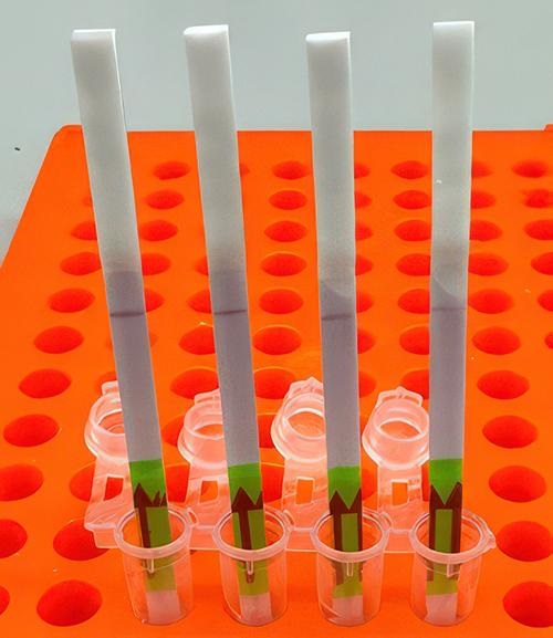
The original 2020 test (shown above) takes 90 minutes to develop at room temperature. The test developers aim to drop this down to 15 minutes. In comparison, typical polymerase chain reaction (PCR) testing requires medical laboratories to have specialized equipment, trained staff, and prolonged processing times, the Broad Institute news release notes. (Photo copyright: Broad Institute.)
Implications of the New Tests
The ease of the new tests is an important development since approximately only 1% of individuals who come down with the flu see doctors for testing, according to the news release. And researchers had this in mind, looking at speed, accuracy, and affordability as a means to “improve outbreak response and infection care around the world,” UPI reported.
There are great benefits to strain differentiation that be achieved with the new test. Doctors are hopeful the test will help dial in the best treatment plans for patients since some strains are resistant to the antiviral medication oseltamivir (Tamiflu), UPI noted. This is significant since Tamiflu “is a common antiviral,” said Sanz in the Broad Institute news release.
“These assays have the potential to expand influenza detection outside of clinical laboratories for enhanced influenza diagnosis and surveillance,” the Journal of Molecular Diagnostics paper noted. This allows for more strategic treatment planning.
“Using a paper strip readout instead of expensive fluorescence machinery is a big advancement, not only in terms of clinical care but also for epidemiological surveillance purposes,” said Ben Zhang, an MD candidate in the Health Sciences and Technology at Harvard and co-first author of the study, in the Broad Institute news release.
Future Plans for Tests
“With further development, the test strip could be reprogrammed to distinguish between SARS-CoV-2 and flu and recognize swine flu and avian flu, including the H5N1 subtype currently causing an outbreak in US dairy cattle,” the study authors told CIDRAP.
The team is also looking at ways to help prevent H5N1 from crossing into human contamination, Sanz told UPI.
The new Princeton/MIT/Harvard tests echo the trend to bring in affordability and ease-of-use with accurate results as an end goal. Faster results mean the best treatments for each person can start sooner and may render the transport of specimens to a clinical laboratory as a second step unnecessary.
As research teams work to develop paper-based viral tests for their plethora of benefits, clinical laboratories will want to pay close attention to this development as it can have a big implication on assisting with future outbreaks.
Additional research is needed before these tests are going to be commonplace in homes worldwide, but this first step brings inspiration and hope of what’s to come.
Use of artificial intelligence in clinical laboratory testing could improve the diagnosis of cancer worldwide
In a proof of concept study, scientists at Shanghai Jiao Tong University in China have developed a clinical laboratory test that utilizes artificial intelligence (AI) to diagnose three types of cancer from a single drop of dried blood. The paper-based test was able to identify patients with colorectal, gastric, and pancreatic cancers and distinguish between patients with and without cancer.
The team’s goal was to develop a way to diagnose cancer while the disease is still in the earlier stages, especially in rural areas.
“Over a billion people across the world experience a high rate of missed disease diagnosis, an issue that highlights the need for diagnostic tools showing increased accuracy and affordability. In addition, such tools could be used in ecologically fragile and energy-limited regions, pointing to the need for developing solutions that can maximize health gains under limited resources for enhanced sustainability,” the researchers wrote in an article published in the journal Nature Sustainability titled, “A Sustainable Approach to Universal Metabolic Cancer Diagnosis.”
The researchers determined that by using less than 0.05 millimeters of dried blood, their test could accurately and quickly identify if a patient had cancer between 82% to 100% of the time.
According to Chaoyuan Kuang, MD, PhD (above), an oncologist at Montefiore Health System and assistant professor at the Albert Einstein College of Medicine, unlike liquid blood, dried serum can be “collected, stored, and transported at much lower cost and with much simpler equipment,” Live Science reported. “This could help democratize the availability of cancer early detection testing across the world,” he added. A paper-based clinical laboratory test that can detect and distinguish one cancer type from another would be a boon to cancer diagnosis worldwide. (Photo copyright: Albert Einstein College of Medicine.)
Improving Cancer Screening in Rural Areas
An earlier study conducted in China in 2022 examined results from 1,570 cancer survivors from both urban and rural areas of China. That study showed that 84.1% of the patients were diagnosed with cancer only after developing symptoms and that urban patients were more likely to be diagnosed in the early stages of cancer. In addition, rural patients also had less screening and treatment options available to them.
The researchers in this latest Chinese study tested their AI model on blood donors with and without cancer and compared the results to traditional liquid-blood biopsy tests.
“Based on modeling they performed, they reported the new tool could reduce the estimated proportion of undiagnosed cases of pancreatic, gastric, and colorectal cancers by about 20% to 50% if it was used for population-level cancer screening in rural China,” Live Science reported.
The scientists used dried serum spots (DSS) and machine learning to perform the research. According to their Nature Sustainability paper, DSS can be challenging in cancer research because sensitive biomarkers in the samples are often degraded or have inadequate amount of blood for proper analysis. To circumvent these issues, the researchers used nanoparticle-enhanced laser desorption/ionization mass spectrometry (NPELDI MS) to increase reliability and sensitivity. Inorganic nanoparticles were applied to the samples to strengthen selectivity and refine metabolic compounds from the samples.
However, the study authors noted that “the adaptation of NPELDI MS to dried spot analysis has not been validated,” Interesting Engineering reported.
A ‘Great Start’
The machine learning algorithm the Chinese scientists created demonstrates that DSS samples can be used to preserve important biological markers and could be beneficial in the diagnosis of cancer.
Their research indicated an overall reduction rate of undiagnosed cancers in the range of 20.35% to 55.10%. The researchers estimated the implementation of their AI tool could reduce the proportion of specific undiagnosed cancer cases in rural China by:
84.30% to 29.20% for colorectal cancer,
77.57% to 57.22% for gastric cancer, and
34.56% to 9.30% for pancreatic cancer.
It’s a “great start,” Chaoyuan Kuang, MD, PhD, an oncologist at Montefiore Health System and assistant professor at the Albert Einstein College of Medicine told Live Science. “This cancer test won’t enter use for a long time,” he said. Nevertheless, the potential of the tool is “immense,” he added, but that “we are still years away from being able to offer this test to patients.
“With further development, it could theoretically be used for the early detection of other types of cancer or for other diseases, or to monitor the progression of disease in patients who have already been diagnosed,” he noted.
Further research and clinical trials are needed before this AI tool can be used in a clinical diagnostic setting. This study is another example of researchers looking for cancer biomarkers in specimen types that are not tissue and further supports the hope that machine learning may one day detect cancer in earlier stages, increase survival rates, and save healthcare costs.
One factor motivating this type of research in China is the fact that the nation has more than 36,000 hospitals and approximately 20,000 anatomic pathologists. Of this total, only a minority of these pathologists have been trained to the standards of North America and Northern Europe.
Like other nations, China’s demand for subspecialist pathology services outstrips its supply of such pathologists. This is the reason why researchers in that country want to develop diagnostic assays for cancer and other diseases that are faster, cheaper, and comparable to a human pathologist in accuracy.
Study results from Switzerland come as clinical laboratory scientists seek new ways to tackle the problem of antimicrobial resistance in hospitals
Microbiologists and clinical laboratory scientists engaged in the fight against antibiotic-resistant (aka, antimicrobial resistant) bacteria will be interested in a recent study conducted at the University of Basel and University Hospital Basel in Switzerland. The epidemiologists involved in the study discovered that some of these so-called “superbugs” can remain in the body for as long as nine years continuing to infect the host and others.
The researchers wanted to see how two species of drug-resistant bacteria—K. pneumoniae and E. coli—changed over time in the body, according to a press release from the university. They analyzed samples of the bacteria collected from patients who were admitted to the hospital over a 10-year period, focusing on older individuals with pre-existing conditions. They found that K. pneumoniae persisted for up to 4.5 years (1,704 days) and E. coli persisted for up to nine years (3,376 days).
“These patients not only repeatedly become ill themselves, but they also act as a source of infection for other people—a reservoir for these pathogens,” said Lisandra Aguilar-Bultet, PhD, the study’s lead author, in the press release.
“This is crucial information for choosing a treatment,” explained Sarah Tschudin Sutter, MD, Head of the Division of Infectious Diseases and Hospital Epidemiology, and of the Division of Hospital Epidemiology, who specializes in hospital-acquired infections and drug-resistant pathogens. Sutter led the Basel University study.
“The issue is that when patients have infections with these drug-resistant bacteria, they can still carry that organism in or on their bodies even after treatment,” said epidemiologist Maroya Spalding Walters, MD (above), who leads the Antimicrobial Resistance Team in the Division of Healthcare Quality Promotion at the federal Centers for Disease Control and Prevention (CDC). “They don’t show any signs or symptoms of illness, but they can get infections again, and they can also transmit the bacteria to other people.” Clinical laboratories working with microbiologists on antibiotic resistance will want to follow the research conducted into these deadly pathogens. (Photo copyright: Centers for Disease Control and Prevention.)
COVID-19 Pandemic Increased Antibiotic Resistance
The Basel researchers looked at 76 K. pneumoniae isolates recovered from 19 patients and 284 E. coli isolates taken from 61 patients, all between 2008 and 2018. The study was limited to patients in which the bacterial strains were detected from at least two consecutive screenings on admission to the hospital.
“DNA analysis indicates that the bacteria initially adapt quite quickly to the conditions in the colonized parts of the body, but undergo few genetic changes thereafter,” the Basel University press release states.
The researchers also discovered that some of the samples, including those from different species, had identical mechanisms of drug resistance, suggesting that the bacteria transmitted mobile genetic elements such as plasmids to each other.
One limitation of the study, the authors acknowledged, was that they could not assess the patients’ exposure to antibiotics.
Meanwhile, recent data from the World Health Organization (WHO) suggests that the COVID-19 pandemic might have exacerbated the challenges of antibiotic resistance. Even though COVID-19 is a viral infection, WHO scientists found that high percentages of patients hospitalized with the disease between 2020 and 2023 received antibiotics.
“While only 8% of hospitalized patients with COVID-19 had bacterial co-infections requiring antibiotics, three out of four or some 75% of patients have been treated with antibiotics ‘just in case’ they help,” the WHO stated in a press release.
WHO uses an antibiotic categorization system known as AWaRe (Access, Watch, Reserve) to classify antibiotics based on risk of resistance. The most frequently prescribed antibiotics were in the “Watch” group, indicating that they are “more prone to be a target of antibiotic resistance and thus prioritized as targets of stewardship programs and monitoring.”
“When a patient requires antibiotics, the benefits often outweigh the risks associated with side effects or antibiotic resistance,” said Silvia Bertagnolio, MD, Unit Head in the Antimicrobial resistance (AMR) Division at the WHO in the press release. “However, when they are unnecessary, they offer no benefit while posing risks, and their use contributes to the emergence and spread of antimicrobial resistance.”
Citing research from the National Institutes of Health (NIH), NPR reported that in the US, hospital-acquired antibiotic-resistant infections increased 32% during the pandemic compared with data from just before the outbreak.
“While that number has dropped, it still hasn’t returned to pre-pandemic levels,” NPR noted.
The UPenn researchers have already developed an antimicrobial treatment derived from guava plants that has proved effective in mice, Vox reported. They’ve also trained an AI model to scan the proteomes of extinct organisms.
“The AI identified peptides from the woolly mammoth and the ancient sea cow, among other ancient animals, as promising candidates,” Vox noted. These, too, showed antimicrobial properties in tests on mice.
These findings can be used by clinical laboratories and microbiologists in their work with hospital infection control teams to better identify patients with antibiotic resistant strains of bacteria who, after discharge, may show up at the hospital months or years later.
This AI platform has the potential to also reduce workload of radiologists, but also of anatomic pathologists and oncologists allowing them to be more productive
When the UK’s National Health Service (NHS) recently tested an artificial intelligence (AI) platform’s ability to analyze mammograms, the AI found early signs of breast cancer that “human doctors” had previously missed, the BBC reported. This level of ability by AI might soon be adapted to aid overworked anatomic pathologists and cancer doctors in the United Kingdom.
Out of 10,000 mammograms MIA analyzed, the AI platform found “tiny signs of breast cancer in 11 women” which had not been spotted during earlier examinations, the BBC noted, adding that the cancers “were practically invisible to the human eye.”
This is a significant development in AI’s role in healthcare. Anatomic pathologists and clinical laboratory leaders will note that ongoing advancements in AI are enabling technology developers to apply their solutions to assessing radiology images, as well as in whole slide imaging used in digital pathology. In the UK, use of AI, the BBC noted, may also help ease doctor’s workloads.
“This is just the beginning of our work with Kheiron,” said Ben Glocker, PhD (above), Professor in Machine Learning for Imaging at Imperial College London and Head of ML Research at Kheiron Medical, in a news release. “We are actively working on new methodologies for the safe deployment and continuous monitoring of MIA to support a US and UK rollout. We are working hard to make sure that as many women as possible will benefit from the use of this new technology within the next year.” AI tools such as MIA may soon take much of the load from anatomic pathologists and radiologists. (Photo copyright: Imperial College London.)
MIA Cloud-based AI Platform
Kheiron was founded in 2016 and MIA was named one of the seven biggest medical breakthroughs in 2023 by ABC News. A study conducted by Imperial College London in 2023 found that MIA “could significantly increase the early detection of breast cancers in a European healthcare setting by up to 13%,” according to an Imperial news release.
“The study was conducted over three phases (two pilot phases and a live roll-out). Overall across the three phases, the AI reader found 24 more cancers than the standard human reading—a 7% relative increase—and resulted in 70 more women recalled (0.28% relative increase),” the news release reported. “Of the additional recalls, six (initial pilot), 13 (extended pilot), and 11 (live use) additional cancers were found, increasing relative cancer detection rate by 13%, 10%, and 5% respectively. [The researchers] found that 83% of the additional cancers detected using MIA in real clinical practice were invasive, showing that MIA can detect cancers where early detection is particularly vital.”
Supported by Microsoft’s Azure Cloud, MIA came together over six years based on training encompassing millions of mammograms worldwide, Healthcare Digital reported.
“AI tools are generally pretty good at spotting symptoms of a specific disease if they are trained on enough data to enable them to be identified. This means feeding the program with as many different anonymized images of those symptoms as possible, from as diverse a range of people as possible,” Sarah Kerruish, Chief Strategy Officer, Kheiron, told Healthcare Digital.
MIA has been trained to “recognize subtle patterns and anomalies” that can point to “cancerous cells even in their earliest stages of development,” Dataconomy reported.
MIA Finds Early Cancer Signs
In the pilot study, MIA examined mammograms from 10,889 women. Each image had previously been reviewed by two radiologists, the BBC reported.
Findings include the following according to Healthcare Digital:
MIA “flagged” all people the physicians previously identified with symptoms.
The AI platform discovered 11 people with cancer the doctors did not identify.
The cancer MIA discovered—and the doctors did not—suggested cancer in early stages.
So, how did the doctors miss the cancer that MIA spotted? Gerald Lip, MD, Clinical Director for Breast Screening in North East Scotland who led the pilot study for the NHS, told Healthcare Digital, “part of the power of AI is it’s not prone to exhaustion or distraction.
“There is an element of fatigue,” he said. “You get disruptions, someone’s coming in, someone’s chatting in the background. There are lots of things that can probably throw you off your regular routine as well. And in those days when you have been distracted, you go, ‘how on earth did I miss that?’ It does happen.”
Lip is also the Chief Investigator in the Mammography Artificial Intelligence Project in the Industrial Center for Artificial Intelligence and Digital Diagnostics in Scotland.
“I see MIA as a friend and an augmentation to my practice,” he told Healthcare Digital. “MIA isn’t perfect. It had no access to patient history so [it] would flag cysts that had already been identified by previous scans and designated harmless.”
AI as a Safety Net
In the 2023 study, researchers from Imperial College London deployed MIA as an extra reader for mammograms of 25,065 women who visited screening sites in Hungary between April 2021 and January 2023, according to a news release.
“Our prospective real-world usage data in Hungary provides evidence for a significant, measurable increase of early breast cancer detection when MIA is used in clinical practice,” said Peter Kecskemethy, PhD, CEO and co-founder of Kheiron Medical, in the news release.
“Our study shows that AI can act as an effective safety net—a tool to prevent subtler signs of cancer from falling through the cracks,” said Ben Glocker, PhD, Professor in Machine Learning for Imaging at Imperial College London and Head of ML Research at Kheiron Medical, in the news release.
More studies are needed before MIA can be used in clinical settings. Nevertheless, use of AI in radiology—specifically mammograms—where the AI tool can identify very small cancers typically undetectable by radiologists, would be a boon to cancer doctors and the patients they treat.
So far, the research suggests that the AI-powered MIA has benefits to deployment in breast cancer screening. Eventually, it may also make impressive contributions to medical diagnosis and patient care, particularly if MIA eventually proves to be effective at analyzing the whole slide images used by anatomic pathologists.
With further study, this research may provide clinical laboratories with a new proteomic biomarker for dementia screenings that identifies risk more than 10 years before symptoms appear
Researchers at the University of Warwick in the UK and Fudan University in Shanghai, China, identified four protein biomarkers in blood that they say can predict dementia up to 15 years before diagnosis. They say these biomarkers may lead to clinical laboratory blood tests that offer alternatives to costly brain scans and lumbar punctures for diagnosis of dementia.
The scientists “used the largest cohort of blood proteomics and dementia to date,” according to a University of Warwick news release. This included taking blood from 52,645 “healthy” people without dementia who participated in the UK Biobank—a population-based study cohort, the new release noted.
“The proteomic biomarkers are [easy] to access and non-invasive, and they can substantially facilitate the application of large-scale population screening,” said neurovegetative disease specialist Jin-tai Yu, MD, PhD, a professor at Fudan University and co-author of the study, in the news release.
“The advent of proteomics offers an unprecedented opportunity to predict dementia onset,” the researchers wrote.
“This is a well-conducted study that adds to what we know about changes in blood that occur very early in diseases that cause dementia, which will be important for early diagnosis in the future,” said Tara Spires-Jones, PhD, in a post from the Science Media Center in the UK. “However,” she added, “it is important to note that these are still scientific research studies and that there are currently no blood tests available for routine use that can diagnose dementia with certainty.
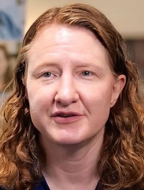
“Based on this study, it does seem likely that blood tests will be developed that can predict risk for developing dementia over the next 10 years, although individuals at higher risk often have difficulty knowing how to respond,” Suzanne Schindler, MD, PhD (above), told Reuters. Schindler, an Associate Professor of Neurology at Washington University in St. Louis, was not involved in the research. Clinical laboratories may soon have a new blood test for dementia. (Photo copyright: VJDementia.)
Predicting Onset of Dementia with 90% Accuracy
The researchers analyzed 52,645 blood samples from the UK Biobank (UKBB). The samples were collected between 2006 and 2010 from healthy individuals who at that time were without dementia.
By March 2023, 1,417 of the study participants had developed Alzheimer’s disease or some other form of dementia. The researchers looked at 1,463 proteins and identified four that were present in high levels among those people:
“Individuals with higher GFAP levels were 2.32 times more likely to develop dementia,” the researchers wrote in Nature Aging. “Notably, GFAP and LTBP2 were highly specific for dementia prediction. GFAP and NEFL began to change at least 10 years before dementia diagnosis.”
When adding known risk factors such as age, sex, and genetics, the researchers said they could predict onset of dementia with 90% accuracy, according to the University of Warwick news release.
“Our findings strongly highlight GFAP as an optimal biomarker for dementia prediction, even more than 10 years before the diagnosis, with implications for screening people at high risk for dementia and for early intervention,” the researchers wrote.
The news release also noted that smaller studies had already identified some of the proteins as potential biomarkers, “but this new research was much larger and conducted over several years.”
Further Validation Needed
Amanda Heslegrave, PhD, of the UK Dementia Research Institute, University College London described the UKBB as “an excellent resource” in the Science Media Center (SMC) post. However, she noted, it’s “a highly curated biobank and may not capture all populations that we need to know the risk for. The new biomarkers identified will need further validation before being used as screening tools.”
Another expert raised additional questions about the University of Warwick/Fudan University study in the SMC post.
“These results may help researchers understand the biological systems involved in the development of dementia,” said David Curtis, MD, PhD, of the UCL Genetics Institute at University College London. “However in my view the strengths of the reported associations are not really strong enough to say that these would form a useful test for predicting who will get dementia in the future.”
Conversely, Curtis pointed to other studies suggesting that phosphorylated tau (p-tau) proteins are better candidates for developing a simple blood test.
P-tau “provides a very good indicator of whether the pathological processes leading to Alzheimer’s disease are present in the brain,” he said. “When effective treatments for Alzheimer’s disease are developed it will be very helpful indeed to have simple blood tests—such as measuring phosphorylated tau—available in order to identify who could benefit.”
At least two blood tests based on the p-tau217 variant—from ALZpath and C2N—are currently available to US clinicians as laboratory developed tests (LDT).
The UK Biobank continues to be used by researchers both in the UK and abroad because of the full sets of data on large numbers of patients over many years. There are few other sources of such data elsewhere in the world. The UK Biobank is a large-scale biomedical database and research resource. It contains de-identified genetic, lifestyle and health information, and biological samples from 500,000 UK participants.
On its website, the UK Biobank states, “It is the most comprehensive and widely-used dataset of its kind and is globally accessible to approved researchers who are undertaking health-related research that is in the public interest, whether they are from academic, commercial, government or charitable settings.”
Thus, clinical laboratory managers and pathologists can expect a continuing stream of published studies that identify biomarkers associated with different health conditions and to see where the data used in these analyses came from the UK’s biobank.
The ASBMB story notes that nanopore technology depends on differences in charges on either side of the membrane to force DNA or RNA through the hole. This is one reason why proteins pose such a challenge.
“Think of a cell as a miniature city, with proteins as its inhabitants. Each protein-resident has a unique identity, its own characteristics, and function. If there was a database cataloging the fingerprints, job profiles, and talents of the city’s inhabitants, such a database would undoubtedly be invaluable!” said Behzad Mehrafrooz, PhD (above), Graduate Research Assistant at University of Illinois at Urbana-Champaign in an article he penned for the university website. This research should be of interest to the many clinical laboratories that do protein testing. (Photo copyright: University of Illinois.)
How the Maglia Process Works
In a Groningen University news story, Maglia said protein is “like cooked spaghetti. These long strands want to be disorganized. They do not want to be pushed through this tiny hole.”
His technique, developed in collaboration with researchers at the University of Rome Tor Vergata, uses electrically charged ions to drag the protein through the hole.
“We didn’t know whether the flow would be strong enough,” Maglia stated in the news story. “Furthermore, these ions want to move both ways, but by attaching a lot of charge on the nanopore itself, we were able to make it directional.”
The researchers tested the technology on what Maglia described as a “difficult protein” with many negative charges that would tend to make it resistant to flow.
“Previously, only easy-to-thread proteins were analyzed,” he said in the news story. “But we gave ourselves one of the most difficult proteins as a test. And it worked!”
Maglia now says that he intends to commercialize the technology through a new startup called Portal Biotech.
Detecting Post-Translational Modifications in the UK
In another recent study, researchers at the University of Oxford reported that they have adapted nanopore technology to detect post-translational modifications (PTMs) in protein chains. The term refers to changes made to proteins after they have been transcribed from DNA, explained an Oxford news story.
“The ability to pinpoint and identify post-translational modifications and other protein variations at the single-molecule level holds immense promise for advancing our understanding of cellular functions and molecular interactions,” said contributing author Hagan Bayley, PhD, Professor of Chemical Biology at University of Oxford, in the news story. “It may also open new avenues for personalized medicine, diagnostics, and therapeutic interventions.”
Bayley is the founder of Oxford Nanopore Technologies, a genetic sequencing company in the UK that develops and markets nanopore sequencing products.
The news story notes that the new technique could be integrated into existing nanopore sequencing devices. “This could facilitate point-of-care diagnostics, enabling the personalized detection of specific protein variants associated with diseases including cancer and neurodegenerative disorders,” the story states.
In another recent study, researchers at the University of Washington reported that they have developed their own method for protein sequencing with nanopore technology.
“This opens up the possibility for barcode sequencing at the protein level for highly multiplexed assays, PTM monitoring, and protein identification!” Motone wrote.
Single-cell proteomics, enabled by nanopore protein sequencing technology, “could provide higher sensitivity and wider throughput, digital quantification, and novel data modalities compared to the current gold standard of protein MS [mass spectrometry],” they wrote. “The accessibility of these tools to a broader range of researchers and clinicians is also expected to increase with simpler instrumentation, less expertise needed, and lower costs.”
There are approximately 20,000 human genes. However, there are many more proteins. Thus, there is strong interest in understanding the human proteome and the role it plays in health and disease.
Technology that makes protein testing faster, more accurate, and less costly—especially with a handheld analyzer—would be a boon to the study of proteomics. And it would give clinical laboratories new diagnostic tools and bring some of that testing to point-of-care settings like doctor’s offices.