Scientists reported positive Phase 1 trial results of their “intratumoral microdevice” in patients with glioma tumors
Here is an example of new microtechnology which has the potential to greatly shorten the time and improve the ability of physicians to determine which anti-cancer drug is most effective for an individual patient’s glioblastoma. As it is further developed, this technology could give anatomic pathologists and clinical laboratories an increased role in assessing the data produced by microdevices and helping physicians determine the most appropriate anti-cancer drug for specific patients.
In a news release, researchers at Brigham and Women’s Hospital (BWH) in Boston said they have developed an implantable “intratumoral microdevice” (IMD) that functions as a “lab in a patient,” capable of gauging the effectiveness of multiple drugs that target brain tumors. In a Phase 1 clinical trial, they tested the IMD on six patients with glioma tumors.
“In order to make the greatest impact on how we treat these tumors, we need to be able to understand, early on, which drug works best for any given patient,” study co-author Pier Paolo Peruzzi, MD, PhD, told the Harvard Gazette. “The problem is that the tools that are currently available to answer this question are just not good enough. So, we came up with the idea of making each patient their own lab, by using a device which can directly interrogate the living tumor and give us the information that we need.”
Peruzzi is Principal Investigator at the Harvey Cushing Neuro-Oncology Laboratories and Assistant Professor of Neurosurgery at Harvard Medical School.
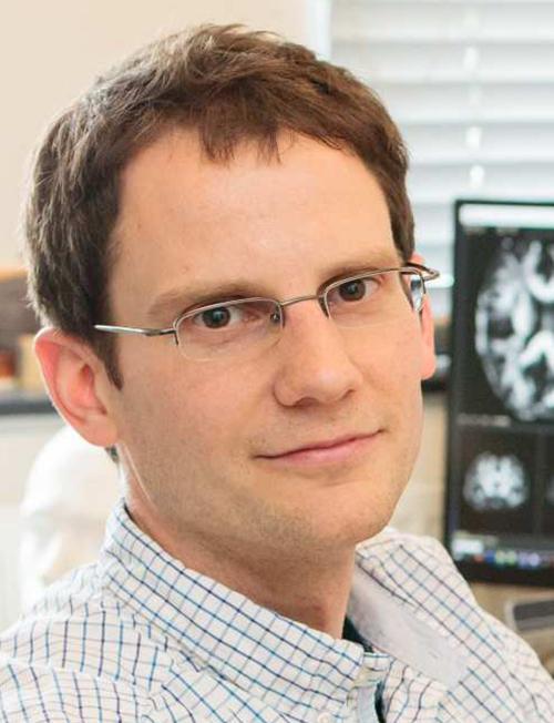
“Our goal is for the placement of these devices to become an integral part of tumor surgery,” said Pier Paolo Peruzzi, MD PhD (above) of Brigham and Women’s Hospital and Harvard Medical School in an article he co-wrote for Healio. “Then, with the data that we have from these microdevices, we can choose the best systemic chemotherapy to give to that patient.” Pathologists and clinical laboratories may soon play a role in helping doctors interpret data gathered by implantable microdevices and choose the best therapies for their patients. (Photo copyright: Dana-Farber Cancer Institute.)
New Perspective on Tumor Treatments
In a news story he co-wrote for Healio, Peruzzi explained that the microdevice—about the size and shape of a grain of rice—contains up to 30 tiny reservoirs that the researchers fill with the drugs they want to test. Surgeons implant the device during a procedure to remove the tumors.
The surgery takes two to three hours to perform, and during that time, the device releases “nanodoses” of the drugs into confined areas of the tumor. Near the end of the procedure, the device is removed along with tissue specimens. The researchers can then analyze the tissue to determine the effectiveness of each drug.
“This is not in the lab, and not in a petri dish,” Peruzzi told Harvard Gazette. “It’s actually in real patients in real time, which gives us a whole new perspective on how these tumors respond to treatment.”
The Healio story notes that gliomas are “among the deadliest brain cancers and are notoriously difficult to treat.” With current approaches, testing different therapies has posed a challenge, Peruzzi wrote.
“Right now, the only way these drugs are tested in patients is through what are called window-of-opportunity studies, where we give one drug to the patient before we resect the tumor and analyze the effect of the drug,” he said. “We can only do this with one drug at a time.”
Determining Safety of Procedure
The primary goal of the Phase 1 trial was to determine the safety of the procedure, Peruzzi noted. “To be in compliance with standard clinical practice and minimize risks to the patients, we needed to integrate the placement and retrieval of the device during an otherwise standard operation.”
The trial consisted of three men and three women ranging from 27 to 86 years old, with a median age of 76. Five were diagnosed with glioblastoma and one with grade 4 astrocytoma.
“None of the six enrolled patients experienced adverse events related to the IMD, and the exposed tissue was usable for downstream analysis for 11 out of 12 retrieved specimens,” the researchers wrote in Science Translational Medicine. They noted that application of the IMD added about 32 minutes to the time required for the surgery, equating to a cost increase of $7,800.
One drug they tested was temozolomide (TMZ), “the most widely used agent in this patient population,” they wrote. “Several patients in our trial received it systemically, either before or after IMD insertion, as part of the standard of care. Thus, although our trial was not designed to choose chemotherapy agents based on IMD data, we still could compare the observed clinical-radiological response to systemic TMZ with the patient-specific response to TMZ in the IMD-exposed tissue.”
One patient, the researchers noted, had not benefited from the drug “in concordance with the poor tissue response observed in the IMD analysis.” But in another patient, a 72-year-old woman, “IMD analysis showed a marked response to TMZ,” and she survived for 20 months after receiving the treatment “with radiological evidence of tumor response. This was despite having a subtotal tumor resection, in itself an unfavorable prognostic factor. The patient expired because of an unrelated cardiovascular event, although she had remained neurologically stable.”
Drug Duration Limitation
One limitation of the study was that testing the device during the tumor removal procedure limited the duration of the drug treatments, Peruzzi said. The Harvard Gazette noted that following their initial study, the researchers were testing a variation of the procedure in which the device is implanted three days before the main surgery in a minimally invasive technique. This gives the drugs more time to work.
Cancer researchers have theorized that common treatments for tumors can impair the immune system, Peruzzi wrote in Healio. “One thing we want to look at is which drugs can kill the tumor without killing the immune system as well,” he noted.
Future studies will determine the effectiveness of implanting microdevices into tumors to test therapies in vivo. Should they become viable, clinical laboratories and anatomic pathologists will likely be involved in receiving, interpreting, storing, and transmitting the data gathered by these devices to the patient’s doctors.
Discovery highlights how ongoing microbiome research points to new opportunities that can lead to development of more effective cancer screening clinical laboratory tests
New research from the Fred Hutchinson Cancer Center in Seattle once again demonstrates that the human microbiome plays a sophisticated role in many biological processes. Microbiologists and anatomic pathologists who diagnose tissue/biopsies will find this study’s findings intriguing.
This breakthrough in colon cancer research came from the discovery that a “subspecies” of a common type of a bacteria that resides in the mouth and causes dental plaque also “shields tumor cells from cancer treatment,” according to NBC News.
The scientists inspected colorectal cancer (CRC) tumors and found that 50% of those examined featured a subspecies of Fusobacterium nucleatum (F. nucleatum or Fn) and that this anaerobic bacterium was “shielding tumor cells from cancer-fighting drugs,” NBC News noted. Many of these tumors were considered aggressive cases of cancer.
“The discovery, experts say, could pave the way for new treatments and possibly new methods of screening,” NBC News reported.
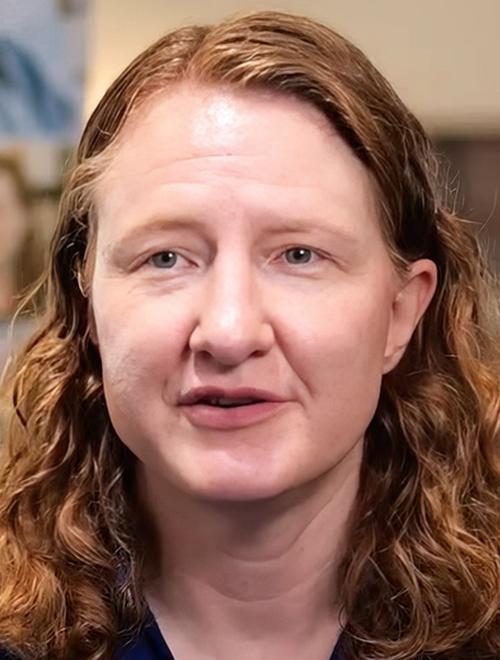
“Patients who have high levels of this bacteria in their colorectal tumors have a far worse prognosis,” Susan Bullman, PhD (above), who jointly supervised the Fred Hutch research team and who is now Associate Professor of Immunology at MD Anderson Cancer Center, told NBC News. “They don’t respond as well to chemotherapy, and they have an increased risk of recurrence,” she added. Microbiologists and clinical laboratories working with oncologists on cancer treatments will want to follow this research as it may lead to new methods for screening cancer patients. (Photo copyright: Fred Hutchinson Cancer Center.)
Developing Effective Treatments
Susan Bullman, PhD, Associate Professor of Immunology at MD Anderson Cancer Center, who along with her husband and fellow researcher Christopher D. Johnston, PhD, Assistant Professor at Fred Hutchinson Cancer Center, jointly supervised an international team of scientists that examined the genomes of 80 F. nucleatum strains from the mouths of cancer-free patients and 55 strains from tumors in patients with colorectal cancer, according to the National Institutes of Health (NIH). The NIH funded the research.
The researchers targeted a subspecies of F. nucleatum called F. nucleatum animalis (Fna) that “was more likely to be present in colorectal tumors. Further analyses revealed that there were two distinct types of Fna. Both were present in mouths, but only one type, called Fna C2, was associated with colorectal cancer” the NIH wrote in an article on its website titled, “Gum Disease-related Bacteria Tied to Colorectal Cancer.”
“Tumor-isolated strains predominantly belong to Fn subspecies animalis (Fna). However, genomic analyses reveal that Fna, considered a single subspecies, is instead composed of two distinct clades (Fna C1 and Fna C2). Of these, only Fna C2 dominates the CRC tumor niche,” the Fred Hutch researchers wrote in their Nature paper.
“We have pinpointed the exact bacterial lineage that is associated with colorectal cancer, and that knowledge is critical for developing effective preventive and treatment methods,” Johnston told the NIH.
How Bacteria Got from Mouth to Colon Not Fully Understood
Traditionally, F. nucleatum makes its home in the mouth in minute quantities. Thus, it is not fully understood how these bacteria travel from the mouth to the colon. However, the Fred Hutch researchers showed that Fna C2 could survive in acidic conditions, like those found in the gut, longer than the other types of Fna. This suggests that the bacteria may travel along a direct route through the digestive tract.
The study, which focused on participants over 50, comes at a time when colorectal cancer rates are trending upward. Rates are doubling for those under 55, jumping from 11% in 1995 to 20% in 2019. CRC is the second-leading cancer death and over 53,000 will succumb to the disease in 2024, according to NBC News.
Many of the newer diagnoses are in later stages with no clear reason why, and the Fred Hutch scientists are trying to understand how their findings tie into the increase of younger cases of colon cancer.
Bullman says it will be important to look at “whether there are elevated levels of this bacterium in young onset colorectal cancer, which is on the rise globally for unknown reasons,” she told NBC News.
Possibility of More Effective Cancer Screening
There is hope that scientists equipped with this knowledge can develop new and more effective screening and treatment options for colon cancer, as well as studying the microbiome’s impact on other diseases.
On the prevention side, researchers have seen that in mice the addition of Fna “appeared to cause precancerous polyps to form, one of the first warning signs of colorectal cancer, though Bullman added that this causation hasn’t yet been proven in humans.” NBC reported.
Future research may find that screening for Fna could determine if colorectal tumors will be aggressive, NIH reported.
“It’s possible that scientists could identify the subspecies while it’s still in the mouth and give a person antibiotics at that point, wiping it out before it could travel to the colon,” Bullman told NBC News. “Even if antibiotics can’t successfully eliminate the bacteria from the mouth, its presence there could serve as an indication that someone is at higher risk for aggressive colon cancer.”
There is also the thought of developing antibiotics to target a specific subtype of bacteria. Doing so would eliminate the need to be “wiping out both forms of the bacteria or all of the bacteria in the mouth. Further, it’s relevant to consider the possibility of harnessing the bacteria to do the cancer-fighting work,” NBC noted.
“The subtype has already proven that it can enter cancer cells quite easily, so it might be possible to genetically modify the bacteria to carry cancer-fighting drugs directly into the tumors,” Bullman told NBC News.
Further studies and research are needed. However, the Fred Hutch researchers’ findings highlight the sophistication of the human microbiome and hint at the potential role it can play in the diagnosis of cancer by clinical laboratories and pathology groups, along with better cancer treatments in the future.
Scientists turned to metabolomics to find cause of biological aging and release index of 25 metabolites that predict healthy and rapid agers
Researchers at the University of Pittsburg Medical Center and the University of Pittsburgh School of Medicine have identified biomarkers in human blood which appear to affect biological aging (aka, senescence). Since biological aging is connected to a person’s overall condition, further research and studies confirming UPMC’s findings will likely lead to a new panel of tests clinical laboratories can run to support physicians’ assessment of their patients’ health.
UPMC’s research “points to pathways and compounds that may underlie biological age, shedding light on why people age differently and suggesting novel targets for interventions that could slow aging and promote health span, the length of time a person is healthy,” according to a UPMC news release.
“We decided to look at metabolites because they’re very dynamic,” Aditi Gurkar, PhD, the study’s senior author, told the Pittsburgh Post-Gazette. Gurkar is Assistant Professor of Medicine, Division of Geriatric Medicine, Aging Institute at the University of Pittsburg. “They can change because of the diet, they can change because of exercise, they can change because of lifestyle changes like smoking,” she added.
The scientists identified 25 metabolites that “showed clear differences” in the metabolomes of both healthy and rapid agers. Based on those findings, the researchers developed the Healthy Aging Metabolic (HAM) Index, a panel of metabolites that predicted healthy agers regardless of gender or race.
“Age is more than just a number,” said Aditi Gurkar, PhD (above), Assistant Professor of Geriatric Medicine at University of Pittsburg School of Medicine and the study’s senior author in a news release. “Imagine two people aged 65: One rides a bike to work and goes skiing on the weekends and the other can’t climb a flight of stairs. They have the same chronological age, but very different biological ages. Why do these two people age differently? This question drives my research.” Gurkar’s research may one day lead to new clinical laboratory tests physicians will order when evaluating their patients’ health. (Photo copyright: University of Pittsburg.)
Clear Differences in Metabolites
According to the National Cancer Institute, a metabolite is a “substance made or used when the body breaks down food, drugs, or chemicals, or its own tissue (for example, fat or muscle tissue). This process, called metabolism, makes energy and the materials needed for growth, reproduction, and maintaining health. It also helps get rid of toxic substances.”
The UPMC researchers used metabolomics—the study of chemical process in the body that involves metabolites, other processes, and biproducts of cell metabolism—to create a “molecular fingerprint” of blood drawn from individuals in two separate study groups.
They included:
People over age 75 able to walk a flight of stairs or walk for 15 minutes without a break, and
People, age 65 to 75, who needed to rest during stair climbing and walk challenges.
The researchers found “clear differences” in the metabolomes of healthy agers as compared to rapid agers, suggesting that “metabolites in the blood could reflect biological age,” according to the UPMC news release.
“Other studies have looked at genetics to measure biological aging, but genes are very static. The genes you’re born with are the genes you die with,” said Gurkar in the news release.
Past studies on aging have explored other markers of biological age such as low grade-inflammation, muscle mass, and physical strength. But those markers fell short in “representing complexity of biological aging,” the UPMC study authors wrote in Aging Cell.
“One potential advantage of metabolomics over other ‘omic’ approaches is that metabolites are the final downstream products, and changes are closely related to the immediate (path) physiologic state of an individual,” they added.
The researchers used an artificial intelligence (AI) model that could identify “potential drivers of biological traits” and found three metabolites “that were most likely to promote healthy aging or drive rapid aging. In future research, they plan to delve into how these metabolites, and the molecular pathways that produce them, contribute to biological aging and explore interventions that could slow this process,” the new release noted.
“While it’s great that we can predict biological aging in older adults, what would be even more exciting is a blood test that, for example, can tell someone who’s 35 that they have a biological age more like a 45-year-old,” Gurkar said. “That person could then think about changing aspects of their lifestyle early—whether that’s improving their sleep, diet or exercise regime—to hopefully reverse their biological age.”
Looking Ahead
The UPMC scientists plan more studies to explore metabolites that promote healthy aging and rapid aging, and interventions to slow disease progression.
It’s possible that the blood-based HAM Index may one day become a diagnostic tool physicians and clinical laboratories use to aid monitoring of chronic diseases. As a commonly ordered blood test, it could help people find out biological age and make necessary lifestyle changes to improve their health and longevity.
With the incidence of chronic disease a major problem in the US and other developed countries, a useful diagnostic and monitoring tool like HAM could become a commonly ordered diagnostic procedure. In turn, that would allow clinical laboratories to track the same patient over many years, with the ability to use multi-year lab test data to flag patients whose biomarkers are changing in the wrong direction—thus enabling physicians to be proactive in treating their patients.
Ten year collaboration between Google and Harvard may lead to a deeper understanding of the brain and new clinical laboratory diagnostics
With all our anatomic pathology and clinical laboratory science, we still do not know that much about the structure of the brain. But now, scientists at Harvard University and Google Research studying the emerging field of connectomics have published a highly detailed 3D reconstruction of human brain tissue that allows visualization of neurons and their connections at unprecedented nanoscale resolutions.
Further investigation of the nano-connections within the human brain could lead to novel insights about the role specific proteins and molecules play in the function of the brain. Though it will likely be years down the road, data derived from this study could be used to develop new clinical laboratory diagnostic tests.
The data to generate the model came from Google’s use of artificial intelligence (AI) algorithms to color-code Harvard’s electron microscope imaging of a cubic millimeter of neural tissue—equivalent to a half-grain of rice—that was surgically removed from an epilepsy patient.
“That tiny square contains 57,000 cells, 230 millimeters of blood vessels, and 150 million synapses, all amounting to 1,400 terabytes of data,” according to the Harvard Gazette, which described the project as “the largest-ever dataset of human neural connections.”
“A terabyte is, for most people, gigantic, yet a fragment of a human brain—just a minuscule, teeny-weeny little bit of human brain—is still thousands of terabytes,” said neuroscientist Jeff W. Lichtman, MD, PhD, Jeremy R. Knowles Professor of Molecular and Cellular Biology, whose Lichtman Lab at Harvard University collaborated on the project with researchers from Google. The two labs have been working together for nearly 10 years on this project, the Harvard Gazette reported.
Lichtman’s lab focuses on the emerging field of connectomics, defined “as understanding how individual neurons are connected to one another to form functional networks,” said neurobiologist Wei-Chung Allen Lee, PhD, Assistant Professor of Neurology, Harvard Medical School, in an interview with Harvard Medical News. “The goal is to create connectomes—or detailed structural maps of connectivity—where we can see every neuron and every connection.” Lee was not involved with the Harvard/Google Research study.
“The human brain uses no more power than a dim incandescent light bulb, yet it can accomplish feats still not possible with the largest artificial computing systems,” wrote Google Research scientist Viren Jain, PhD (above), in a blog post. “To understand how requires a level of understanding more profound than knowing what part of the brain is responsible for what function. The field of connectomics aims to achieve this by precisely mapping how each cell is connected to others.” Google’s 10-year collaboration with Harvard University may lead to new clinical laboratory diagnostics. (Photo copyright: Google Research.)
Study Data and Tools Freely Available
Along with the Science paper, the researchers publicly released the data along with analytic and visualization tools. The study noted that the dataset “is large and incompletely scrutinized,” so the scientists are inviting other researchers to assist in improving the model.
“The ability for other researchers to proofread and refine this human brain connectome is one of many ways that we see the release of this paper and the associated tools as not only the culmination of 10 years of work, but the beginning of something new,” wrote Google Research scientist Viren Jain, PhD, in a blog post that included links to the online resources.
One of those tools—Neuroglancer—allows any user with a web browser to view 3D models of neurons, axons, synapses, dendrites, blood vessels, and other objects. Users can rotate the models in xyz dimensions.
Users with the requisite knowledge and skills can proofread and correct the models by signing up for a CAVE (Connectome Annotation Versioning Engine) account.
Researchers Found Several Surprises
To perform their study, Lichtman’s team cut the neural tissue into 5,000 slices, each approximately 30 nanometers thick, Jain explained in the blog post. They then used a multibeam scanning electron microscope to capture high-resolution images, a process that took 326 days.
Jain’s team at Google used AI tools to build the model. They “stitched and aligned the image data, reconstructed the three dimensional structure of each cell, including its axons and dendrites, identified synaptic connections, and classified cell types,” he explained.
Jain pointed to “several surprises” that the reconstruction revealed. For example, he noted that “96.5% of contacts between axons and their target cells have just one synapse.” However, he added, “we found a class of rare but extremely powerful synaptic connections in which a pair of neurons may be connected by more than 50 individual synapses.”
In their Science paper, the researchers suggest that “these powerful connections are not the result of chance, but rather that these pairs had a reason to be more strongly connected than is typical,” Jain wrote in the blog post. “Further study of these connections could reveal their functional role in the brain.”
Mysterious Structures
Another anomaly was the presence of “axon whorls,” as Jain described them, “beautiful but mysterious structures in which an axon wraps itself into complicated knots.”
Because the sample came from an epilepsy patient, Jain noted that the whorls could be connected to the disease or therapies or could be found in all brains.
“Given the scale and complexity of the dataset, we expect that there are many other novel structures and characteristics yet to be discovered,” he wrote. “These findings are the tip of the iceberg of what we expect connectomics will tell us about human brains.”
The researchers have a larger goal to create a comprehensive high-resolution map of a mouse’s brain, Harvard Medical News noted. This would contain approximately 1,000 times the data found in the 1-cubic-millimeter human sample.
Dark Daily has been tracking the different fields of “omics” for years, as research teams announce new findings and coin new areas of science and medicine to which “omics” is appended. Connectomics fits that description.
Though the Harvard/Google research is not likely to lead to diagnostic assays or clinical laboratory tests any time soon, it is an example of how advances in technologies are enabling researchers to investigate smaller and smaller elements within the human body.
This AI platform has the potential to also reduce workload of radiologists, but also of anatomic pathologists and oncologists allowing them to be more productive
When the UK’s National Health Service (NHS) recently tested an artificial intelligence (AI) platform’s ability to analyze mammograms, the AI found early signs of breast cancer that “human doctors” had previously missed, the BBC reported. This level of ability by AI might soon be adapted to aid overworked anatomic pathologists and cancer doctors in the United Kingdom.
Out of 10,000 mammograms MIA analyzed, the AI platform found “tiny signs of breast cancer in 11 women” which had not been spotted during earlier examinations, the BBC noted, adding that the cancers “were practically invisible to the human eye.”
This is a significant development in AI’s role in healthcare. Anatomic pathologists and clinical laboratory leaders will note that ongoing advancements in AI are enabling technology developers to apply their solutions to assessing radiology images, as well as in whole slide imaging used in digital pathology. In the UK, use of AI, the BBC noted, may also help ease doctor’s workloads.
“This is just the beginning of our work with Kheiron,” said Ben Glocker, PhD (above), Professor in Machine Learning for Imaging at Imperial College London and Head of ML Research at Kheiron Medical, in a news release. “We are actively working on new methodologies for the safe deployment and continuous monitoring of MIA to support a US and UK rollout. We are working hard to make sure that as many women as possible will benefit from the use of this new technology within the next year.” AI tools such as MIA may soon take much of the load from anatomic pathologists and radiologists. (Photo copyright: Imperial College London.)
MIA Cloud-based AI Platform
Kheiron was founded in 2016 and MIA was named one of the seven biggest medical breakthroughs in 2023 by ABC News. A study conducted by Imperial College London in 2023 found that MIA “could significantly increase the early detection of breast cancers in a European healthcare setting by up to 13%,” according to an Imperial news release.
“The study was conducted over three phases (two pilot phases and a live roll-out). Overall across the three phases, the AI reader found 24 more cancers than the standard human reading—a 7% relative increase—and resulted in 70 more women recalled (0.28% relative increase),” the news release reported. “Of the additional recalls, six (initial pilot), 13 (extended pilot), and 11 (live use) additional cancers were found, increasing relative cancer detection rate by 13%, 10%, and 5% respectively. [The researchers] found that 83% of the additional cancers detected using MIA in real clinical practice were invasive, showing that MIA can detect cancers where early detection is particularly vital.”
Supported by Microsoft’s Azure Cloud, MIA came together over six years based on training encompassing millions of mammograms worldwide, Healthcare Digital reported.
“AI tools are generally pretty good at spotting symptoms of a specific disease if they are trained on enough data to enable them to be identified. This means feeding the program with as many different anonymized images of those symptoms as possible, from as diverse a range of people as possible,” Sarah Kerruish, Chief Strategy Officer, Kheiron, told Healthcare Digital.
MIA has been trained to “recognize subtle patterns and anomalies” that can point to “cancerous cells even in their earliest stages of development,” Dataconomy reported.
MIA Finds Early Cancer Signs
In the pilot study, MIA examined mammograms from 10,889 women. Each image had previously been reviewed by two radiologists, the BBC reported.
Findings include the following according to Healthcare Digital:
MIA “flagged” all people the physicians previously identified with symptoms.
The AI platform discovered 11 people with cancer the doctors did not identify.
The cancer MIA discovered—and the doctors did not—suggested cancer in early stages.
So, how did the doctors miss the cancer that MIA spotted? Gerald Lip, MD, Clinical Director for Breast Screening in North East Scotland who led the pilot study for the NHS, told Healthcare Digital, “part of the power of AI is it’s not prone to exhaustion or distraction.
“There is an element of fatigue,” he said. “You get disruptions, someone’s coming in, someone’s chatting in the background. There are lots of things that can probably throw you off your regular routine as well. And in those days when you have been distracted, you go, ‘how on earth did I miss that?’ It does happen.”
Lip is also the Chief Investigator in the Mammography Artificial Intelligence Project in the Industrial Center for Artificial Intelligence and Digital Diagnostics in Scotland.
“I see MIA as a friend and an augmentation to my practice,” he told Healthcare Digital. “MIA isn’t perfect. It had no access to patient history so [it] would flag cysts that had already been identified by previous scans and designated harmless.”
AI as a Safety Net
In the 2023 study, researchers from Imperial College London deployed MIA as an extra reader for mammograms of 25,065 women who visited screening sites in Hungary between April 2021 and January 2023, according to a news release.
“Our prospective real-world usage data in Hungary provides evidence for a significant, measurable increase of early breast cancer detection when MIA is used in clinical practice,” said Peter Kecskemethy, PhD, CEO and co-founder of Kheiron Medical, in the news release.
“Our study shows that AI can act as an effective safety net—a tool to prevent subtler signs of cancer from falling through the cracks,” said Ben Glocker, PhD, Professor in Machine Learning for Imaging at Imperial College London and Head of ML Research at Kheiron Medical, in the news release.
More studies are needed before MIA can be used in clinical settings. Nevertheless, use of AI in radiology—specifically mammograms—where the AI tool can identify very small cancers typically undetectable by radiologists, would be a boon to cancer doctors and the patients they treat.
So far, the research suggests that the AI-powered MIA has benefits to deployment in breast cancer screening. Eventually, it may also make impressive contributions to medical diagnosis and patient care, particularly if MIA eventually proves to be effective at analyzing the whole slide images used by anatomic pathologists.
With further study, this research may provide clinical laboratories with a new proteomic biomarker for dementia screenings that identifies risk more than 10 years before symptoms appear
Researchers at the University of Warwick in the UK and Fudan University in Shanghai, China, identified four protein biomarkers in blood that they say can predict dementia up to 15 years before diagnosis. They say these biomarkers may lead to clinical laboratory blood tests that offer alternatives to costly brain scans and lumbar punctures for diagnosis of dementia.
The scientists “used the largest cohort of blood proteomics and dementia to date,” according to a University of Warwick news release. This included taking blood from 52,645 “healthy” people without dementia who participated in the UK Biobank—a population-based study cohort, the new release noted.
“The proteomic biomarkers are [easy] to access and non-invasive, and they can substantially facilitate the application of large-scale population screening,” said neurovegetative disease specialist Jin-tai Yu, MD, PhD, a professor at Fudan University and co-author of the study, in the news release.
“The advent of proteomics offers an unprecedented opportunity to predict dementia onset,” the researchers wrote.
“This is a well-conducted study that adds to what we know about changes in blood that occur very early in diseases that cause dementia, which will be important for early diagnosis in the future,” said Tara Spires-Jones, PhD, in a post from the Science Media Center in the UK. “However,” she added, “it is important to note that these are still scientific research studies and that there are currently no blood tests available for routine use that can diagnose dementia with certainty.
“Based on this study, it does seem likely that blood tests will be developed that can predict risk for developing dementia over the next 10 years, although individuals at higher risk often have difficulty knowing how to respond,” Suzanne Schindler, MD, PhD (above), told Reuters. Schindler, an Associate Professor of Neurology at Washington University in St. Louis, was not involved in the research. Clinical laboratories may soon have a new blood test for dementia. (Photo copyright: VJDementia.)
Predicting Onset of Dementia with 90% Accuracy
The researchers analyzed 52,645 blood samples from the UK Biobank (UKBB). The samples were collected between 2006 and 2010 from healthy individuals who at that time were without dementia.
By March 2023, 1,417 of the study participants had developed Alzheimer’s disease or some other form of dementia. The researchers looked at 1,463 proteins and identified four that were present in high levels among those people:
“Individuals with higher GFAP levels were 2.32 times more likely to develop dementia,” the researchers wrote in Nature Aging. “Notably, GFAP and LTBP2 were highly specific for dementia prediction. GFAP and NEFL began to change at least 10 years before dementia diagnosis.”
When adding known risk factors such as age, sex, and genetics, the researchers said they could predict onset of dementia with 90% accuracy, according to the University of Warwick news release.
“Our findings strongly highlight GFAP as an optimal biomarker for dementia prediction, even more than 10 years before the diagnosis, with implications for screening people at high risk for dementia and for early intervention,” the researchers wrote.
The news release also noted that smaller studies had already identified some of the proteins as potential biomarkers, “but this new research was much larger and conducted over several years.”
Further Validation Needed
Amanda Heslegrave, PhD, of the UK Dementia Research Institute, University College London described the UKBB as “an excellent resource” in the Science Media Center (SMC) post. However, she noted, it’s “a highly curated biobank and may not capture all populations that we need to know the risk for. The new biomarkers identified will need further validation before being used as screening tools.”
Another expert raised additional questions about the University of Warwick/Fudan University study in the SMC post.
“These results may help researchers understand the biological systems involved in the development of dementia,” said David Curtis, MD, PhD, of the UCL Genetics Institute at University College London. “However in my view the strengths of the reported associations are not really strong enough to say that these would form a useful test for predicting who will get dementia in the future.”
Conversely, Curtis pointed to other studies suggesting that phosphorylated tau (p-tau) proteins are better candidates for developing a simple blood test.
P-tau “provides a very good indicator of whether the pathological processes leading to Alzheimer’s disease are present in the brain,” he said. “When effective treatments for Alzheimer’s disease are developed it will be very helpful indeed to have simple blood tests—such as measuring phosphorylated tau—available in order to identify who could benefit.”
At least two blood tests based on the p-tau217 variant—from ALZpath and C2N—are currently available to US clinicians as laboratory developed tests (LDT).
The UK Biobank continues to be used by researchers both in the UK and abroad because of the full sets of data on large numbers of patients over many years. There are few other sources of such data elsewhere in the world. The UK Biobank is a large-scale biomedical database and research resource. It contains de-identified genetic, lifestyle and health information, and biological samples from 500,000 UK participants.
On its website, the UK Biobank states, “It is the most comprehensive and widely-used dataset of its kind and is globally accessible to approved researchers who are undertaking health-related research that is in the public interest, whether they are from academic, commercial, government or charitable settings.”
Thus, clinical laboratory managers and pathologists can expect a continuing stream of published studies that identify biomarkers associated with different health conditions and to see where the data used in these analyses came from the UK’s biobank.