New research shows a single toxic exposure during pregnancy may drive disease risk across generations, highlighting emerging opportunities for clinical labs to leverage epigenetic biomarkers for earlier, preventative diagnostics.
A new study from Washington State University suggests that a single exposure to a toxic fungicide during pregnancy may influence disease risk for up to 20 generations, with implications for how clinical laboratories understand chronic disease and prevention strategies.
Epigenetic Inheritance Expands the Diagnostic Timeline
The study found that exposure to vinclozolin—a fungicide commonly used in agriculture—triggered disease patterns in rats that persisted for 20 generations. Notably, disease incidence not only continued but worsened in later generations, with severe reproductive complications emerging.
“This study really does say that this is not going to go away,” Skinner said. “We need to do something about it. We can use epigenetics to move us away from reactionary medicine and toward preventative medicine.” (Photo credit: Washington State University)
For clinical laboratories, these findings show a growing shift toward understanding disease not just as an immediate or genetic condition, but as one influenced by ancestral environmental exposures.
Germline Changes Drive Long-Term Risk
Unlike traditional toxicology models, the study highlights how disease risk is transmitted through epigenetic changes in germline cells—sperm and eggs—rather than direct exposure alone.
“Essentially, when a gestating female is exposed, the fetus is exposed,” Skinner explained. “And then the germline inside the fetus is also exposed… Once it’s programmed in the germline, it’s as stable as a genetic mutation.”
This mechanism suggests that clinical labs may need to consider multi-generational risk factors when interpreting biomarkers or assessing patient risk profiles.
Disease Burden Intensifies Over Generations
While disease prevalence remained relatively stable across early generations, researchers observed a sharp increase in severity beginning around the 15th generation.
“By the 16th, 17th, 18th generations, disease became very prominent and we started to see abnormalities during the birth process,” Skinner noted. “Either the mother would die, or all the pups would die, so it was a really lethal sort of pathology.”
These findings suggest that long-term population health trends—such as rising chronic disease rates—may have roots in historical environmental exposures.
Implications for Clinical Laboratories
The research aligns with broader epidemiological trends showing increased rates of chronic diseases, including cancer and cardiovascular conditions. According to the CDC, more than three-quarters of Americans now live with at least one chronic disease.
For laboratories, the study underscores the potential value of epigenetic biomarkers in predicting disease susceptibility well before clinical symptoms appear.
Moving Toward Preventative Diagnostics
As clinical laboratories continue to expand their role in precision medicine, epigenetic testing may offer a pathway to earlier intervention and improved patient outcomes.
By identifying individuals at elevated risk decades in advance, labs could support a shift toward preventative care models—helping clinicians intervene before disease onset rather than reacting after diagnosis.
For lab leaders and pathologists, the study highlights that diagnostics may soon extend beyond the individual patient to include inherited environmental risk factors spanning generations.
This article was created with the assistance of Generative AI and has undergone editorial review before publishing.
Walk-in lab testing in West Virginia is giving patients faster, more affordable access to diagnostics—while pushing clinical labs to adapt to a more consumer-driven care model.
Across the clinical laboratory industry, the shift toward consumer-directed healthcare continues to gain momentum. A recent report by the Charleston Gazette-Mail highlights a growing trend in West Virginia of walk-in laboratory services that allow patients to bypass traditional physician referrals for routine diagnostic testing.
For pathologists and clinical lab professionals, this shift represents a significant evolution in the traditional diagnostic workflow. Facilities like Any Lab Test Now and local hospital-affiliated outreach centers are increasingly offering direct-to-consumer (DTC) options, allowing individuals to purchase tests such as lipid panels, glucose levels, DNA, and toxicology screens.
Key Drivers of Walk-In Testing
The article identifies several factors pushing patients toward walk-in labs:
Cost transparency: Many patients with high-deductible health plans are choosing walk-in labs that offer transparent, upfront pricing, often avoiding the “sticker shock” of traditional hospital billing.
Convenience and speed: The ability to walk in without an appointment and receive results via secure online portals—often within 24 to 48 hours—appeals to the modern healthcare consumer.
Proactive health management: There is a growing demographic of proactive patients who wish to monitor chronic conditions or wellness markers more frequently than their annual, insurance-covered physical allows.
“This gives [patients] an opportunity to manage their own health,” said Matt Brooks, director of clinical laboratory services at Marshall Health Network based in Huntington, W.V. “And it gives patients the opportunity to pay for the test without having to go through their insurance.” (Photo credit: Marshall Health Network).
The Changing Role of the Provider
While the convenience is clear, the trend raises questions regarding the interpretation of results. Patients have access to more data, yet they still require professional guidance to put that data into clinical context.
Most walk-in models encourage patients to share their results with their primary care physicians, but the “patient-as-the-customer” model places the initial responsibility for action squarely on the individual.
Implications for Clinical Labs
For traditional clinical laboratories, the growth of walk-in testing in regions like West Virginia serves as a signal to adapt. As patients become more accustomed to retail-style healthcare experiences, laboratories may need to invest more heavily in user-friendly digital interfaces and transparent pricing structures to remain competitive.
This trend also underscores a broader national movement. As more states relax regulations regarding DTC testing, the laboratory’s role is shifting from a behind-the-scenes diagnostic provider to a front-facing participant in the patient’s healthcare.
WHO introduces faster, more accessible TB testing strategies while CDC maintains a targeted, risk-based approach in the United States.
The World Health Organization (WHO) has issued new recommendations aimed at improving access to faster, more efficient tuberculosis (TB) diagnostics by introducing near point-of-care molecular testing, alternative sample collection methods, and pooled testing strategies, according to a news release.
For the first time, WHO is recommending a new class of near point-of-care nucleic acid amplification tests (NPOC-NAATs) that can be deployed in decentralized settings such as primary care clinics and community health centers. These systems are designed to deliver faster results at lower cost compared to traditional laboratory-based molecular platforms, potentially shifting more TB testing closer to the patient.
Clinical laboratory scientists should note that the WHO’s guidelines diverge noticeably from those of the Centers for Disease Control and Prevention (CDC).
The updated guidance also endorses tongue swabs as an alternative specimen type for TB detection, particularly for patients unable to produce sputum. In parallel, WHO recommends sputum pooling as a strategy to improve efficiency and reduce costs, allowing laboratories to increase throughput while conserving reagents in resource-constrained environments.
“These new WHO recommendations mark a major step forward in making TB testing faster and more accessible,” said Tereza Kasaeva, director of WHO’s Department for HIV, TB, Hepatitis & STIs. “WHO urges countries and partners to work together to roll out these guidelines to close persistent diagnostic gaps and ensure that everyone with TB can be diagnosed early and start life-saving treatment without delay.” (Photo credit: WHO)
The recommendations arrive as global diagnostic gaps persist despite international commitments to expand access to rapid molecular testing. Many patients still experience delays due to reliance on sputum samples, centralized laboratory infrastructure, and the high cost of testing platforms.
New WHO Recommendations Emphasize Access and Efficiency
WHO’s updated Module 3: Diagnosis guidelines, expected later this year, reflect a broader shift toward decentralization and scalability in TB diagnostics.
By enabling testing at peripheral healthcare levels, the new NPOC-NAAT systems could reduce turnaround times and expand access in underserved regions. Tongue swabs further simplify sample collection, while pooling strategies offer laboratories a practical way to stretch limited resources without sacrificing diagnostic reach.
Supporting materials, including an operational handbook and implementation toolkit, will guide laboratories and national TB programs through adoption, training, and workflow integration.
CDC Maintains Targeted Testing Approach in the US
In contrast to WHO’s global push for expanded access, the CDC continues to emphasize a targeted testing strategy for tuberculosis in the United States, focusing on high-risk individuals rather than universal screening.
TB case counts and rates have been increasing since 2021, the CDC noted in late 2025. The US saw a 7.9% increase in case count and a 6.9% increase in rate in 2024 as compared to a year earlier. In 2024, there were 10,388 TB cases in the US with a corresponding incidence rate of 3.1 per 100,000 population.
Two Types of TB Infection Tests
The CDC recognizes two primary methods to detect TB infection, though neither distinguishes between latent infection and active disease:
TB blood tests: Preferred for most individuals, particularly those vaccinated with Bacille Calmette-Guérin (BCG). (BCG is primarily used to prevent severe childhood TB particularly in high-prevalence countries. BCG is generally not recommended in the US.)
TB skin test: Still used in certain cases, especially for children under age five, and for baseline testing scenarios requiring a two-step approach.
Five Components of a Full Diagnostic Evaluation
If a patient tests positive or presents symptoms such as chronic cough, night sweats, or weight loss, the CDC recommends a comprehensive evaluation that includes:
Medical history and risk assessment
Physical examination
Chest X-ray
Bacteriologic testing using sputum samples (typically three), including:
NAAT for rapid detection
Sputum smear microscopy
Culture
Drug susceptibility testing to guide treatment decisions
Updated Guidance for Healthcare Personnel
Recent CDC guidance, developed with the National Tuberculosis Controllers Association, reflects a shift in screening practices for healthcare workers:
Baseline TB testing is required upon hire
Routine annual testing is no longer recommended for most healthcare workers
Post-exposure testing is advised immediately and again eight to 10 weeks later if initial results are negative
For 2026, the CDC emphasizes several important nuances for clinicians and laboratories interpreting TB test results. Blood-based interferon-gamma release assays (IGRAs) are strongly preferred for individuals who have received the BCG vaccine, as they are less likely to produce false-positive results compared to skin tests. In addition, for individuals considered low risk for TB, a positive result should be confirmed with a second test—ideally using a different method—before treatment is initiated, helping to avoid unnecessary therapy and ensure diagnostic accuracy.
Implications for Clinical Laboratories
Together, WHO and CDC guidance illustrate a divergence in strategy shaped by global versus domestic needs. WHO’s recommendations prioritize expanded access, decentralization, and cost efficiency—particularly in high-burden or resource-limited settings—while CDC guidance reflects a more targeted, risk-based approach within a lower-incidence environment.
For clinical laboratories, the evolving landscape signals both opportunity and complexity: Adoption of decentralized molecular platforms, validation of alternative specimen types, and optimization of high-throughput workflows such as pooling may become increasingly important as TB diagnostic strategies continue to evolve.
This article was created with the assistance of Generative AI and has undergone editorial review before publishing.
The research was published in Cell Metabolism and reported by The Scientist, a sibling brand of Dark Daily.
For clinical laboratory professionals, the approach highlights a possible shift away from time-intensive stool-based sequencing toward faster, more scalable testing workflows. “One of the key barriers to integrating our knowledge of the microbiome into clinical care is the time it takes to analyze the data on the microbiome,” said Ariel Hernandez-Leyva, an MD/PhD student working for gut microbiome researcher Andrew Kau’s group at Washington University School of Medicine. (Photo credit: Kau Lab)
Breath-Based Testing Could Streamline Microbiome Workflows
The research team found that volatile organic compounds (VOCs) in breath samples closely correlate with gut microbiome activity, suggesting a streamlined alternative that could reduce turnaround times and expand access to microbiome-informed diagnostics. In both human and mouse studies, breath “volatilome” profiles mirrored microbial metabolites in the gut.
In a proof-of-concept analysis, VOC patterns distinguished children with asthma from healthy controls and predicted levels of a gut bacterial species associated with the condition. Such capabilities could support earlier clinical decision-making and reduce reliance on complex sequencing workflows.
If validated in larger studies, breath-based diagnostics could offer clinical labs a practical pathway to integrate microbiome insights into routine testing, with potential applications in pediatric care, infectious disease risk assessment, and chronic disease management.
This article was created with the assistance of Generative AI and has undergone editorial review before publishing.
Set for April 28–29 in New Orleans, the 31st Annual Executive War College will bring lab leaders together for practical, execution-focused strategies across reimbursement, staffing, compliance, and emerging technologies, with new emphasis on digital pathology and AI-driven operations.
The 31st Annual Executive War College on Diagnostics, Clinical Laboratory, and Pathology Management, April 28–29 in New Orleans, will bring together clinical laboratory leaders to address the most pressing challenges shaping the industry in 2026. This year’s event emphasizes practical, execution-focused strategies across financial performance, workforce development, compliance, and emerging technologies.
A key addition to the 2026 program is the inaugural Executive Forum on Digital Pathology Management, a dedicated session exploring digital workflows, artificial intelligence (AI), and data integration. Designed as an interactive and collaborative experience, the forum will highlight real-world implementation strategies and provide attendees with actionable insights into adopting new technologies.
Recently, the Dark Report highlighted what’s to come at the event. Further, Dark Daily reported on key sessions that attendees won’t want to miss.
Six Major Themes Shaping the Industry
The conference agenda is structured around six strategic themes reflecting the evolving laboratory landscape.
Financial strategy sessions will focus on improving reimbursement, strengthening payer relationships, and using analytics to drive revenue growth.
Workforce discussions will address staffing shortages, automation, and leadership development.
Compliance sessions will offer frameworks for managing regulatory risk and embedding compliance into daily operations.
Innovation and technology will play a central role, with case studies demonstrating how laboratories can leverage molecular diagnostics, automation, and informatics to enhance clinical value and operational efficiency.
AI will receive particular attention, with sessions examining both its opportunities and challenges, including governance, validation, and return on investment.
Additionally, experts will explore trends in mergers and acquisitions and strategic partnerships, providing guidance on growth, valuation, and long-term positioning.
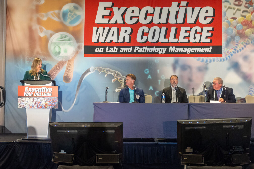
Healthcare attorney Elizabeth Sullivan of McDonald Hopkins leads a panel discussion at last year’s Executive War College. Sullivan will return for two sessions at the upcoming 2026 conference. (Photo credit: EWC)
2025 Executive War College Highlights
Workforce challenges persist in 2026 and will again be a key theme at the event. The 2025 Executive War College highlighted several innovative approaches to staffing.
For example, the Dark Report reported on a 2025 Executive War College presentation by Jennifer Fralick, vice president anatomic pathology and clinical laboratories at Stanford Health Care. Fralick noted that clinical labs are addressing severe staffing shortages by focusing on internal talent development through career ladders, training programs, and smarter staffing models that shift routine tasks away from licensed professionals. These strategies improve efficiency, reduce burnout, and help labs build sustainable, long-term workforce pipelines instead of relying solely on external hiring. (Fralick is returning to this year’s event to discuss an AI playbook for labs.)
Operational solutions will also be highlighted in the 2026 agenda. Last year, as the Dark Report noted in an article, Shashirekha Shetty, PhD, professor in the Department of Pathology at Case Western University, presented on how up to 70% of laboratory errors occur in the pre-analytical phase, often due to incorrect test orders, improper sample handling, and poor communication, making it a major risk to patient care and lab efficiency. Shetty emphasized that labs must take full ownership of this phase by implementing standardized workflows, strengthening training and collaboration with clinicians, and embedding pre-analytic quality into their overall quality management systems.
Attendees can expect updated solutions for these challenges and more presented by experts at this year’s Executive War College, which is just a short month away. With nearly 80 sessions and around 150 speakers, the program is designed to equip attendees with practical tools, real-world case studies, and operational playbooks. Laboratory executives will leave with clear, actionable roadmaps to navigate financial pressures, regulatory scrutiny, and rapid technological change.