New artificial intelligence model agrees with interpretations of human medical technologists and microbiologists with extraordinary accuracy
Microbiology laboratories will be interested in news from Brescia University in Italy, where researchers reportedly have developed a deep learning model that can visually identify and analyze bacterial species in culture plates with a high level of agreement with interpretations made by medical technologists.
They initially trained and tested the system to digitally identify pathogens associated with urinary tract infections (UTIs). UTIs are the source for a large volume of clinical laboratory microbiological testing.
The system, known as DeepColony, uses hierarchical artificial intelligence technology. The researchers say hierarchical AI is better suited to complex decision-making than other approaches, such as generative AI.
In their Nature paper, the researchers explained that microbiologists use conventional methods to visually examine culture plates that contain bacterial colonies. The scientists hypothesize which species of bacteria are present, after which they test their hypothesis “by regrowing samples from each colony separately and then employing mass spectroscopy techniques,” to confirm their hypotheses.
However, DeepColony—which was designed for use with clinical laboratory automation systems—looks at high-resolution digital scans of cultured plates and attempts to identify the bacterial strains and analyze them in much the same way a microbiologist would. For example, it can identify species based on their appearance and determine which colonies are suitable for analysis, the researchers explained.
“Working on a large stream of clinical data, and a complete set of 32 pathogens, the proposed system is capable of effectively assisting plate interpretation with a surprising degree of accuracy in the widespread and demanding framework of urinary tract infections,” the study authors wrote. “Moreover, thanks to the rich species-related generated information, DeepColony can be used for developing trustworthy clinical decision support services in laboratory automation ecosystems from local to global scale.”
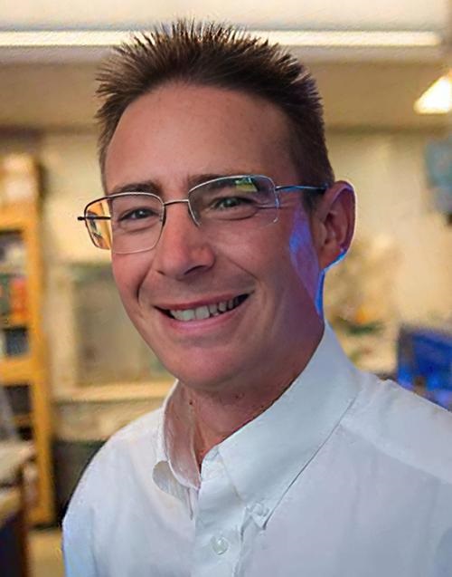
“Compared to the most common solutions based on single convolutional neural networks (CNN), multi-network architectures are attractive in our case because of their ability to fit into contexts where decision-making processes are stratified into a complex structure,” wrote the study’s lead author Alberto Signoroni, PhD (above), Associate Professor of Computer Science, University of Brescia, and his researcher team in their Nature paper. “The system must be designed to generate useful and easily interpretable information and to support expert decisions according to safety-by-design and human-in-the-loop policies, aiming at achieving cost-effectiveness and skill-empowerment respectively.” Microbiologists and clinical laboratory managers will want to follow the further development of this technology. (Photo copyright: University of Brescia.)
How Hierarchical AI Works
Writing in LinkedIn, patent attorney and self-described technology expert David Cain, JD, of Hauptman Ham, LLP, explained that hierarchical AI systems “are structured in layers, each with its own distinct role yet interconnected in a way that forms a cohesive whole. These systems are significant because they mirror the complexity of human decision-making processes, incorporating multiple levels of analysis and action. This multi-tiered approach allows for nuanced problem-solving and decision-making, akin to a seasoned explorer deftly navigating through a multifaceted terrain.”
DeepColony, the researchers wrote, consists of multiple convolutional neural networks (CNNs) that exchange information and cooperate with one another. The system is structured into five levels—labeled 0 through 4—each handling a different part of the analysis:
At level 0, the system determines the number of bacterial colonies and their locations on the plate.
At level 1, the system identifies “good colonies,” meaning those suitable for further identification and analysis.
At level 2, the system assigns each good colony to a bacterial species “based on visual appearance and growth characteristics,” the researchers wrote, referring to the determination as being “pathogen aware, similarity agnostic.”
The CNN used at this stage was trained by using images of 26,213 isolated colonies comprising 32 bacterial species, the researchers wrote in their paper. Most came from clinical laboratories, but some were obtained from the American Type Culture Collection (ATCC), a repository of biological materials and information resources available to researchers.
At level 3, the system attempts to improve accuracy by looking at the larger context of the plate. The goal here is to “determine if observed colonies are similar (pure culture) or different (mixed cultures),” the researchers wrote, describing this step as “similarity aware, pathogen agnostic.” This enables the system to recognize variants of the same strain, the researchers noted, and has the effect of reducing the number of strains identified by the system.
At this level, the system uses two “Siamese CNNs,” which were trained with a dataset of 200,000 image pairs.
Then, at level 4, the system “assesses the clinical significance of the entire plate,” the researchers added. Each plate is labeled as:
“Positive” (significant bacterial growth),
“No significant growth” (negative), or
“Contaminated,” meaning it has three or more “different colony morphologies without a particular pathogen that is prevalent over the others,” the researchers wrote.
If a plate is labeled as “positive,” it can be “further evaluated for possible downstream steps,” using MALDI-TOF mass spectrometry or tests to determine susceptibility to antimicrobial measures, the researchers stated.
“This decision-making process takes into account not only the identification results but also adheres to the specific laboratory guidelines to ensure a proper supportive interpretation in the context of use,” the researchers wrote.
Nearly 100% Agreement with Medical Technologists
To gauge DeepColony’s accuracy, the researchers tested it on a dataset of more than 5,000 urine cultures from a US laboratory. They then compared its analyses with those of human medical technologists who had analyzed the same samples.
Agreement was 99.2% for no-growth cultures, 95.6% for positive cultures, and 77.1% for contaminated or mixed growth cultures, the researchers wrote.
The lower agreement for contaminated cultures was due to “a deliberately precautionary behavior, which is related to ‘safety by design’ criteria,” the researchers noted.
Lead study author Alberto Signoroni, PhD, Associate Professor of Computer Science, University of Brescia, wrote in Nature that many of the plates identified by medical technologists as “contaminated” were labeled as “positive” by DeepColony. “We maximized true negatives while allowing for some false positives, so that DeepColony [can] focus on the most relevant or critical cases,” he said.
Will DeepColony replace medical technologists in clinical laboratories any time soon? Not likely. But the Brescia University study indicates the direction AI in healthcare is headed, with high accuracy and increasing speed. The day may not be far off when pathologists and microbiologists regularly employ AI algorithms to diagnose disease.
Dogs’ acute sense of smell can even surpass effectiveness of some clinical laboratory testing in detecting certain diseases in humans
When it comes to COVID-19 testing, a recent Italian study demonstrates that trained dogs can detect SARS-CoV-2 with accuracy comparable to rapid molecular tests used in clinical laboratories. The researchers wanted to determine if dogs could be more effective at screening people for COVID-19 at airports, schools, and other high-traffic environments as a way to detect the coronavirus and reduce the spread of this infectious disease.
Scientists at the State University of Milan in Italy conducted a study that shows dogs can be trained to accurately identify the presence of the COVID-19 infection from both biological samples and by simply smelling an individual.
For their validation study, the Italian team trained three dogs named Nala, Otto, and Helix, “to detect the presence of SARS-CoV-2 in sweat samples from infected people. At the end of the training, the dogs achieved an average sensitivity of 93% and a specificity of 99%, showing a level of accuracy highly consistent with that of the RT-PCR [reverse transcription polymerase chain reaction] used in molecular tests and a moderate to strong reproducibility over time,” Nature reported.
RT-PCR tests are the current gold-standard for SARS-CoV-2 detection. This is yet another example of scientists training dogs to smell a disease with “acceptable” accuracy. This time for COVID-19.
“We only recruited dogs that showed themselves predisposed and positively motivated to carry out this type of activity. One of the fundamental aspects was not to cause stress or anxiety in the subjects used,” Federica Pirrone, PhD (above), Associate Professor, Department of Veterinary Medicine and Animal Sciences, University of Milan, and one of the authors of the study told Lifegate. “Training always takes place using positive reinforcement of a food nature: whether it’s a particularly appetizing morsel, a biscuit, or something that associates the dog’s search with a rewarding prize.” In some instances, dogs have been shown to be as good or more effective at detecting certain diseases than clinical laboratory testing. (Photo copyright: Facebook.)
Dogs More Accurate than Rapid Antigen Testing
Nala and four other dogs (Nim, Hope, Iris and Chaos) were later trained by canine technicians from Medical Detection Dogs Italy (MDDI) to identify the existence of the SARS-CoV-2 virus by directly smelling people waiting in line in pharmacies to get a nasal swab to test for the coronavirus.
Working with their handlers, the five dogs accurately signaled the presence or absence of the virus with 89% sensitivity and 95% specificity. That rate is “well above the minimum required by the WHO [World Health Organization] for rapid swabs for SARS-CoV-2,” according to Nature.
“The results of studies published so far on the accuracy of canine smell in detecting the presence of SARS-CoV-2 in biological samples (e.g., saliva, sweat, urine, trachea-bronchial secretions) from infected people suggest that sniffer dogs might reach percentages of sensitivity and specificity comparable to, or perhaps even higher, than those of RT-PCR,” the scientists wrote in Scientific Reports.
“However, although most of these studies are of good quality, none of them provided scientific validation of canine scent detection, despite this being an important requirement in the chemical analysis practice. Therefore, further applied research in this field is absolutely justified to provide definitive validation of this biodetection method,” the researchers concluded.
Other Studies into Using Dogs for Detecting Disease
Scientists from the Division of Biological and Health Sciences, Department of Agriculture and Livestock at the University of Sonora; and the Canine Training Center Obi-K19, both in Hermosillo, Mexico, conducted the study “as part of a Frontiers of Science Project of the National Council of Science and Technology (CONACYT), in which in addition to analyzing sweat compounds, trained dogs are put to sniff the samples and make detections in people who show symptoms or could be positive for coronavirus,” Mexico Daily Post reported.
The researchers trained four dogs with sweat samples and three dogs with saliva samples of COVID-19 positive patients. The samples were obtained from a health center located in Hermosillo, Sonora, in Mexico. The dogs were restricted to spend five minutes per patient and the researchers calculated the performance of the dogs by measuring sensitivity, specificity, and their 95% confidence intervals (CI).
The researchers concluded that all four of the dogs could detect COVID-19 from either sweat or saliva samples “with sensitivity and specificity rates significantly different from random [sampling] in the field.” According to the Frontiers in Medicine study, the researchers found their results promising because, they said, it is reasonable to expect the detection rate would improve with longer exposure to the samples.
The objective of the Mexican researchers is for the dogs to ultimately reach the sensitivity range requested by WHO for the performance of an antigen test, which is at least 80% sensitivity and 97% specificity. If that goal is achieved, dogs could become important partners in the control of the COVID-19 pandemic, the scientists wrote.
Data obtained so far from these studies indicate that biosensing dogs may represent an effective method of screening for COVID-19 as well as other diseases. More studies and clinical trials are needed before the widespread use of dogs might become feasible. Nevertheless, scientists all over the world are finding that Man’s best friend can be a powerful ally in the fight against the spread of deadly diseases.
In the meantime, the gold standard in COVID-19 testing will continue to be the FDA-cleared assays used by clinical laboratories throughout the United States.
Experts cite high vaccination rates and behavioral changes among at-risk groups, but warn about complacency; clinical laboratories should remain vigilant
In July, Scott Gottlieb, MD, Commissioner of the US Food and Drug Administration (FDA) from May 2017 to April 2019, wrote an op-ed in The New York Times titled, “Monkeypox Is About to Become the Next Public Health Failure.” In it, he wrote, “Our country’s response to monkeypox has been plagued by the same shortcomings we had with COVID-19.” But has it improved? Clinical laboratory leaders and pathology group managers will find it informative to find out what has taken place since Gottlieb made his stark prediction.
The global monkeypox outbreak that emerged last spring appears to have subsided in the US and Europe, though it remains to be seen if the disease can be completely eradicated, according to multiple media reports. As of Oct. 26, 2022, the Centers for Disease Control and Prevention (CDC) reported a 7-day rolling average of 30 cases per day in the US, down from a peak of nearly 440/day in early August.
Cases are also down in cities that earlier reported heavy outbreaks. For example, the New York City Health Department reported a 7-day average of just two cases per day on Oct. 25, compared with 73/day on July 30.
And the San Francisco Department of Public Health announced on Oct. 20 that it would end the city’s public health emergency on monkeypox (MPX) effective on Oct. 31. “MPX cases have slowed to less than one case per day and more than 27,000 San Franciscans are now vaccinated against the virus,” the agency stated in a press release.
“Once again, we caution that a declining outbreak can be the most dangerous outbreak, because it can tempt us to think that the crisis is over and to let down our guard,” said World Health Organization (WHO) Director-General Tedros Adhanom Ghebreyesus, PhD, in an Oct. 12 global press briefing. “That’s not what WHO is doing. We are continuing to work with countries around the world to increase their testing capacity, and to monitor trends in the outbreak.” Clinical laboratories should not assume the outbreak has passed but continue to be vigilant and prepared for increased demand in monkeypox testing. (Photo copyright: ITU Pictures.)
Changing Behavior Lowers Infection Rates
In addition to high vaccination rates, public health experts have attributed the decline to behavioral changes among at-risk groups. “There were really substantial changes among men who have sex [with] men,” infectious disease physician Shira Doron, MD, of Tufts Medical Center in Boston, told ABC News.
On September 2, the CDC published the results of a survey indicating that about half of men who have sex with men “reported reducing their number of sex partners, one-time sexual encounters, and use of dating apps because of the monkeypox outbreak.”
Another likely factor is the disease’s limited transmissibility. “Initially, there was a lot of concern that monkeypox could spread widely at daycares or in schools, but, overall, there has been very little spread among children,” NPR reported.
But citing multiple studies, the NPR story noted “that often there isn’t very much virus in the upper respiratory tract,” where it might spread through talking or coughing. “Instead, the highest levels of virus occur on sores found on the skin and inside the anus.”
These studies, along with earlier research, “explain why monkeypox is spreading almost exclusively through contact during sex, especially anal and oral sex, during the current outbreak,” NPR reported.
Monkeypox Could Mutate, experts say
Despite the promising numbers, public health experts are warning that monkeypox could remain as a long-term threat to public health. According to an article in Nature, “At best, the outbreak might fizzle out over the next few months or years. At worst, the virus could become endemic outside Africa by reaching new animal reservoirs, making it nearly impossible to eradicate.”
In addition to the limited transmissibility of the virus, Nature noted that the outbreak stems from a relatively mild form of the pathogen and is rarely fatal. As of Oct. 28, the CDC reported a total of just six confirmed deaths in the US out of a total of 28,302 confirmed cases since the first infections were reported in May.
It is possible that the virus could mutate into a more contagious form, but Nature noted that monkeypox is a DNA virus, and that they tend to mutate more slowly than RNA viruses such as SARS-CoV-2 and HIV. Nevertheless, University of Alabama at Birmingham School of Medicine bioinformatician Elliot Lefkowitz, PhD, warned that a “worrisome mutation” could arise if the outbreak continues for much longer.
“I have no confidence that all the people who need to be tested are being tested,” she told Nature. She expressed concerns that people could resume risky behavior if they think the danger has passed.
Another question is whether currently available vaccines offer long-lasting protection. And though reported case numbers are down in the US and Europe, they are rising in parts of Africa and South America, Nature noted.
Gottlieb’s Dire Prediction
The decline in new infections followed dire warnings last summer about the possible consequences of the outbreak. In his New York Times op-ed, former Gottlieb criticized the CDC for being slow to test for the virus. He wrote, “[I]f monkeypox gains a permanent foothold in the United States and becomes an endemic virus that joins our circulating repertoire of pathogens, it will be one of the worst public health failures in modern times not only because of the pain and peril of the disease but also because it was so avoidable.”
At the time of his writing, Gottlieb was right to be concerned. On July 29, the CDC reported a seven-day moving average of 390 reported cases per day. According to the federal agency, a reported case “Includes either the positive laboratory test report date, CDC call center reporting date, or case data entry date into CDC’s emergency response common operating platform, DCIPHER.”
Quashing the outbreak, Gottlieb estimated, would have required about 15,000 tests per week among people presenting symptoms resembling monkeypox. But between mid-May and the end of June, he noted, the CDC had tested only about 2,000 samples, according to the federal agency’s July 15 Morbidity and Mortality Weekly Report (MMWR).
As a remedy, Gottlieb called on the Biden administration to re-focus the CDC’s efforts more on disease control “by transferring some of its disease prevention work to other agencies,” including the FDA.
Researchers surprised that process designed to detect SARS-CoV-2 also identifies monkeypox in wastewater
Early information about an outbreak in a geographical region can inform local clinical laboratories as to which infectious agents and variants they are likely to see when testing patients who have symptoms. To that end, wastewater testing has become a rich source of early clues as to where COVID-19 outbreaks are spreading and how new variants of the coronavirus are emerging.
Ongoing advances in genetic sequencing and digital technologies are making it feasible to test wastewater for infectious agents in ways that were once too time-consuming, too expensive, or simply impossible.
“Before wastewater sequencing, the only way to do this was through clinical testing, which is not feasible at large scale, especially in areas with limited resources, public participation, or the capacity to do sufficient testing and sequencing,” said Knight in a UCSD press release. “We’ve shown that wastewater sequencing can successfully track regional infection dynamics with fewer limitations and biases than clinical testing to the benefit of almost any community.” (Photo copyright: UC San Diego News.)
Same Process, Different Virus
Following August’s declaration of a state of emergency by California, San Diego County, and the federal government, UCSD researchers added monkeypox surveillance to UCSD’s existing wastewater surveillance program.
“It’s the same process as SARS-CoV-2 qPCR monitoring, except that we have been testing for a different virus. Monkeypox is a DNA virus, so it is a bit of a surprise that our process optimized for SARS-CoV-2, which is an RNA virus, works so well,” said Rob Knight, PhD, Professor of Pediatrics and Computer Science and Engineering at UCSD and one of the lead authors of the study in the press release.
According to the press release, RNA sequencing from wastewater has two specific benefits:
It avoids the potential of clinical testing biases, and
It can track changes in the prevalence of SARS-CoV-2 variants over time.
In 2020, at the height of the COVID-19 pandemic, scientists from the University of California San Diego and Scripps Research looked into genetic sequencing of wastewater. They wanted to see if it would provide insights into levels and variants of the SARS-CoV-2 within a specific community.
Individuals who have COVID-19 shed the virus in their stool.
The UCSD/Scripps researchers deployed commercial auto-sampling robots to collect wastewater samples at the main UCSD campus. They analyzed the samples for levels of SARS-CoV-2 RNA at the Expedited COVID-19 Identification Environment (EXCITE) lab at UCSD. After the success of the program on the campus, they extended their research to include other facilities and communities in the San Diego area.
“The coronavirus will continue to spread and evolve, which makes it imperative for public health that we detect new variants early enough to mitigate consequences,” said Knight in a July press release announcing the publication of their study in the journal Nature, titled, “Wastewater Sequencing Reveals Early Cryptic SARS-CoV-2 Variant Transmission.”
Detecting Pathogens Weeks Earlier than Traditional Clinical Laboratory Testing
In July, the scientists successfully determined the genetic mixture of SARS-CoV-2 variants present in wastewater samples by examining just two teaspoons of raw sewage. They found they could accurately identify new variants 14 days before traditional clinical laboratory testing. They detected the presence of the Omicron variant 11 days before it was first reported clinically in the community.
During the study, the team collected and analyzed 21,383 sewage samples, with most of those samples (19,944) being taken from the UCSD campus. They performed genomic sequencing on 600 of the samples and compared them to genomes obtained from clinical swabs. They also compared 31,149 genomes from clinical genomic surveillance to 837 wastewater samples taken from the community.
The scientists distinguished specific viral lineages present in the samples by sequencing the viruses’ complete set of genetic instructions. Mutational differences between the various SARS-CoV-2 variants can be minute and subtle, but also have notable biological deviations.
“Nothing like this had been done before. Sampling and detection efforts began modestly but grew steadily with increased research capacity and experience. Currently, we’re monitoring almost 350 buildings on campus,” said UCSD’s Chancellor Pradeep Khosla, PhD, in the July press release.
“The wastewater program was an essential element of UC San Diego Health’s response to the COVID pandemic,” said Robert Schooley, MD, Infectious Disease Specialist at UC San Diego Health, in the press release. Schooley is also a professor at UCSD School of Medicine, and one of the authors of the study.
“It provided us with real-time intelligence about locations on campus where virus activity was ongoing,” he added. “Wastewater sampling essentially allowed us to ‘swab the noses’ of every person upstream from the collector every day and to use that information to concentrate viral detection efforts at the individual level.”
Monkeypox Added to UCSD Wastewater Surveillance
In August, UCSD officially added the surveillance of the monkeypox virus to their ongoing wastewater surveillance program. A month earlier, the researchers had discerned 10,565.54 viral copies per liter of wastewater. They observed the levels fluctuating and increasing.
On August 2, the scientists detected 189,309.81 viral copies per liter of wastewater. However, it is not yet clear if the monitoring of monkeypox viral loads in wastewater will enable the researchers to accurately predict future infections or case rates.
“We don’t yet know if the data will anticipate case surges like with COVID,” Knight said in the August UCSD press release announcing the addition of monkeypox to the surveillance program. “It depends on when the virus is shed from the body relative to how bad the symptoms are that cause people to seek care. This is, in principle, different for each virus, although in practice wastewater seems to be predictive for multiple viruses.”
Utilization of genetic sequencing of wastewater sampling will continue to develop and improve. “It’s fairly easy to add new pathogens to the process,” said Smruthi Karthikeyan, PhD, an environmental engineer and postdoctoral researcher in Knight’s lab who has overseen wastewater monitoring at UC San Diego. “It’s doable on short notice. We can get more information in the same turnaround time.”
Thus, clinical laboratories engaged in testing programs for COVID-19 may soon see the addition of monkeypox to those processes.
Officials also worry about diminishing smallpox vaccinations, which offered people protection against the infectious disease
Monkeypox challenges from the current outbreak have dogged public health agencies even though the disease was first identified more than 50 years ago. That is because the virus has found new avenues of infection. These developments will be relevant for the nation’s clinical laboratories, which are often the first healthcare providers to confirm a suspected case is positive for monkeypox and notify a public health laboratory about the positive test result.
The latest monkeypox numbers from the federal Centers for Disease Control and Prevention (CDC) indicate that, as of September 6, the US has identified 19,962 cases in the 2022 outbreak, while worldwide the case number is 52,037.
In “When It Comes to Monkeypox Testing, Clinical Laboratories Should Be Aware of Five Significant Developments,” Dark Daily wrote about steps being taken to identify and control infections in America as well as trends in medical laboratory testing for monkeypox. This included reports of phlebotomists refusing to draw monkeypox blood samples and how social stigma surrounding the disease can affect who gets a medical laboratory test.
Workers at clinical laboratories and anatomic pathology groups will gain from understanding why monkeypox has spread beyond its traditional geography.
“Monkeypox symptoms include swollen lymph nodes, fever, and body aches that result in red bumps on hands, feet, mouth, and genitals,” Bodhraj Acharya, PhD (above), of the Laboratory Alliance of Central New York, told Dark Daily. “It spreads by close contact, respiratory droplets, lesions, and bodily fluids.” Clinical laboratories engaged in testing for monkeypox will want to stay alert to patients presenting with such symptoms. (Photo copyright: Laboratory Alliance of Central New York.)
African Public Health Officials Saw New Monkeypox Challenges Coming
Researchers and public health experts have been perplexed about how and why the latest monkeypox outbreak has occurred so aggressively beyond its origin in rural Central Africa.
“Monkeypox is caused by the pox virus, with a close resemblance to smallpox,” said Bodhraj Acharya, PhD, Manager of Chemistry and Referral Testing at the Laboratory Alliance of Central New York, in a conversation with Dark Daily. “Unlike COVID-19, this is an old enemy which has roots in the 1970s from Congo, when the disease was erratically endemic in Africa.”
According to the World Health Organization (WHO), most monkeypox cases since 1970 have been reported from rural rainforest regions in Central and Western Africa.
Thus, a monkeypox outbreak occurring in Europe and the United States in 2022 has puzzled virologists and microbiologists because it does not follow the historical pattern of the virus’ spread. For example, the first monkeypox case in the US arrived in May from a Massachusetts patient who had traveled to Canada, a state press release noted.
Before the Nigerian outbreak, the virus rose from rural areas where hunters came in close contact with animals. The illness resulted in lesions on the face, hands, and feet, Nature wrote of Yinka-Ogunleye’s recollections.
However, after 2017, she and other epidemiologists warned peers that the virus was spreading in new ways and in urban settings. For example, infected people sometimes had genital lesions, suggesting that the virus might spread through human sexual contact.
Now, in 2022, “the world is paying the price for not having responded adequately” in 2017, Yinka-Ogunleye told Nature.
Lack of Smallpox Vaccination Increases Monkeypox Challenges
The waning effects of smallpox vaccinations, which ended in 1980 after smallpox was basically eradicated from the world, may have opened the door for monkeypox to spread earlier this year. Smallpox vaccines provided some protection against monkeypox, but by now three generations of people have not received smallpox inoculations.
“Eyebrows were raised when multiple cases of monkeypox were reported from various non-endemic countries starting in May of 2022,” Acharya said. “Due to genetic similarity, smallpox vaccination provided some cross-protection, but the termination of smallpox vaccination could have provided ground for the recent insurgence and spread of monkeypox.”
Trying to jumpstart a new monkeypox vaccination campaign on the heels of COVID-19 shots may be met with resistance from a virus-weary public. But other options at preventing the current spread of monkeypox may present challenges as well, such as trying to curtail sexual activity among affected population, the BBC reported.
“The easiest way to prevent it is to close down all highly active sexual networks for a couple of months until it goes away, but I don’t think that will ever happen. Do you?” Paul Hunter, PhD, Professor of Medicine at the University of East Anglia in Norwich, England, told the BBC.
For medical laboratory workers and others who may find themselves testing for the disease in the future, the biggest lessons from current monkeypox challenges are twofold: The virus has invaded new geography, and discontinued smallpox vaccination campaigns may have left younger people exposed to monkeypox.