Findings could lead to new biomarkers clinical laboratories would use for identifying cancer in patients and monitoring treatments
As DNA “dark matter” (the DNA sequences between genes) continues to be studied, researchers are learning that so-called “junk DNA” (non-functional DNA) may influence multiple health conditions and diseases including cancer. This will be of interest to pathologists and clinical laboratories engaged in cancer diagnosis and may lead to new non-invasive liquid biopsy methods for identifying cancer in blood draws.
This technique could enable non-invasive monitoring of cancer treatment and cancer diagnosis, Technology Networks noted.
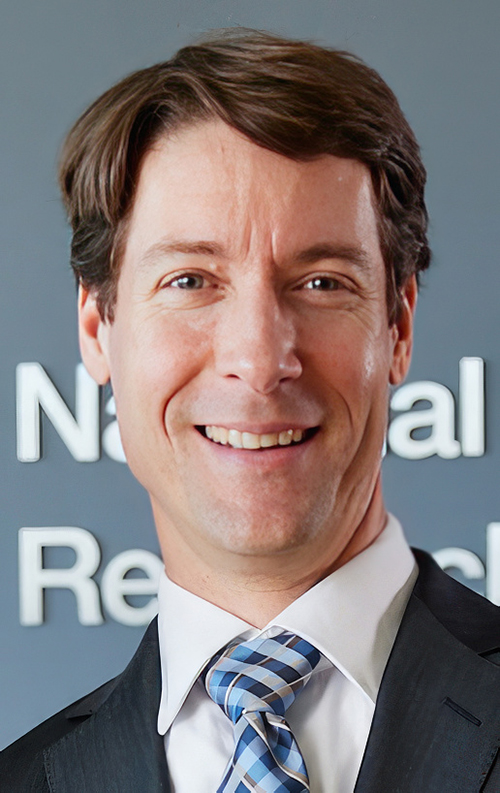
“Our study shows that ARTEMIS can reveal genomewide repeat landscapes that reflect dramatic underlying changes in human cancers,” said study co-leader Akshaya Annapragada (above), an MD/PhD student at the Johns Hopkins University School of Medicine, in a news release. “By illuminating the so-called ‘dark genome,’ the work offers unique insights into the cancer genome and provides a proof-of-concept for the utility of genomewide repeat landscapes as tissue and blood-based biomarkers for cancer detection, characterization, and monitoring.” Clinical laboratories may soon have new biomarkers for the detection of cancer. (Photo copyright: Johns Hopkins University.)
Detecting Early Lung, Liver Cancer
Artemis is a Greek word meaning “hunting goddess.” For the Johns Hopkins researchers, ARTEMIS also describes a technique “to analyze junk DNA found in tumors” and which float in the bloodstream, Financial Times explained.
“It’s like a grand unveiling of what’s behind the curtain,” said geneticist Victor Velculescu, MD, PhD, Professor of Oncology and co-director of the Cancer Genetics and Epigenetics Program at Johns Hopkins Kimmel Cancer Center, in the news release.
“Until ARTEMIS, this dark matter of the genome was essentially ignored, but now we’re seeing that these repeats are not occurring randomly,” he added. “They end up being clustered around genes that are altered in cancer in a variety of different ways, providing the first glimpse that these sequences may be key to tumor development.”
ARTEMIS could “lead to new therapies, new diagnostics, and new screening approaches for cancer,” Velculescu noted.
Repeats of DNA Sequences Tough to Study
For some time technical limitations have hindered analysis of repetitive genomic sequences by scientists.
“Genetic changes in repetitive sequences are a hallmark of cancer and other diseases, but characterizing these has been challenging using standard sequencing approaches,” the study authors wrote in their Science Translational Medicine paper.
“We developed a de novok-mer (short sequences of DNA)-finding approach called ARTEMIS to identify repeat elements from whole-genome sequencing,” the researchers wrote.
The scientists put ARTEMIS to the test in laboratory experiments.
The first analysis involved 1,280 types of repeating genetic elements “in both normal and tumor tissues from 525 cancer patients” who participated in the Pan-Cancer Analysis of Whole Genomes (PCAWG), according to Technology Networks, which noted these findings:
A median of 807 altered elements were found in each tumor.
About two-thirds (820) had not “previously been found altered in human cancer.”
Second, the researchers explored “genomewide repeat element changes that were predictive of cancer,” by using machine learning to give each sample an ARTEMIS score, according to the Johns Hopkins news release.
The scoring detected “525 PCAWG participants’ tumors from the healthy tissues with a high performance” overall Area Under the Curve (AUC) score of 0.96 (perfect score being 1.0) “across all cancer types analyzed,” the Johns Hopkins’ release states.
Liquid Biopsy Deployed
The scientists then used liquid biopsies to determine ARTEMIS’ ability to noninvasively diagnose cancer. Researchers used blood samples from:
ARTEMIS classified patients with lung cancer with an AUC of 0.82.
ARTEMIS detected people with liver cancer, as compared to others with cirrhosis or viral hepatitis, with a score of AUC 0.87.
Finally, the scientists used their “ARTEMIS blood test” to find the origin of tumors in patients with cancer. They reported their technique was 78% accurate in discovering tumor tissue sources among 12 tumor types.
“These analyses reveal widespread changes in repeat landscapes of human cancers and provide an approach for their detection and characterization that could benefit early detection and disease monitoring of patients with cancer,” the researchers wrote in Science Translational Medicine.
Large Clinical Trials Planned
Velculescu said more research is planned, including larger clinical trials.
“While still at an early stage, this research demonstrates how some cancers could be diagnosed earlier by detecting tumor-specific changes in cells collected from blood samples,” Hattie Brooks, PhD, Research Information Manager, Cancer Research UK (CRUK), told Financial Times.
Should ARTEMIS prove to be a viable, non-invasive blood test for cancer, it could provide pathologists and clinical laboratories with new biomarkers and the opportunity to work with oncologists to promptly diagnosis cancer and monitor patients’ response to treatment.
Speedy DNA sequencing and on-the-spot digital imaging may change the future of anatomic pathology procedures during surgery
Researchers at the Center for Molecular Medicine (CMM) at UMC Utrecht, a leading international university medical center in the Netherlands, have paired artificial intelligence (AI) and machine learning with DNA sequencing to develop a diagnostic tool cancer surgeons can use during surgeries to determine in minutes—while the patient is still on the operating table—whether they have fully removed all the cancerous tissue.
The method, “involves a computer scanning segments of a tumor’s DNA and alighting on certain chemical modifications that can yield a detailed diagnosis of the type and even subtype of the brain tumor,” according to The New York Times, which added, “That diagnosis, generated during the early stages of an hours-long surgery, can help surgeons decide how aggressively to operate, … In the future, the method may also help steer doctors toward treatments tailored for a specific subtype of tumor.”
This technology has the potential to reduce the need for frozen sections, should additional development and studies confirm that it accurately and reliably shows surgeons that all cancerous cells were fully removed. Many anatomic pathologists would welcome such a development because of the time pressure and stress associated with this procedure. Pathologists know that the patient is still in surgery and the surgeons are waiting for the results of the frozen section. Most pathologists would consider fewer frozen sections—with better patient outcomes—to be an improvement in patient care.
“It’s imperative that the tumor subtype is known at the time of surgery,” Jeroen de Ridder, PhD (above), associate professor in the Center for Molecular Medicine at UMC Utrecht and one of the study leaders, told The New York Times. “What we have now uniquely enabled is to allow this very fine-grained, robust, detailed diagnosis to be performed already during the surgery. It can figure out itself what it’s looking at and make a robust classification,” he added. How this discovery affects the role of anatomic pathologists and pathology laboratories during cancer surgeries remains to be seen. (Photo copyright: UMC Utrecht.)
Rapid DNA Sequencing Impacts Brain Tumor Surgeries
The UMC Utrecht scientists employed Oxford Nanopore’s “real-time DNA sequencing technology to address the challenges posed by central nervous system (CNS) tumors, one of the most lethal type of tumor, especially among children,” according to an Oxford Nanopore news release.
The researchers called their new machine learning AI application the “Sturgeon.”
According to The New York Times, “The new method uses a faster genetic sequencing technique and applies it only to a small slice of the cellular genome, allowing it to return results before a surgeon has started operating on the edges of a tumor.”
Jeroen de Ridder, PhD, an associate professor in the Center for Molecular Medicine at UMC Utrecht, told The New York Times that Sturgeon is “powerful enough to deliver a diagnosis with sparse genetic data, akin to someone recognizing an image based on only 1% of its pixels, and from an unknown portion of the image.” Ridder is also a principal investigator at the Oncode Institute, an independent research center in the Netherlands.
The researchers tested Sturgeon during 25 live brain surgeries and compared the results to an anatomic pathologist’s standard method of microscope tissue examination. “The new approach delivered 18 correct diagnoses and failed to reach the needed confidence threshold in the other seven cases. It turned around its diagnoses in less than 90 minutes, the study reported—short enough for it to inform decisions during an operation,” The New York Times reported.
But there were issues. Where the minute samples contain healthy brain tissue, identifying an adequate number of tumor markers could become problematic. Under those conditions, surgeons can ask an anatomic pathologist to “flag the [tissue samples] with the most tumor for sequencing, said PhD candidate Marc Pagès-Gallego, a bioinformatician at UMC Utrecht and a co-author of the study,” The New York Times noted.
“Implementation itself is less straightforward than often suggested,” Sebastian Brandner, MD, a professor of neuropathology at University College London, told The Times. “Sequencing and classifying tumor cells often still required significant expertise in bioinformatics as well as workers who are able to run, troubleshoot, and repair the technology,” he added.
“Brain tumors are also the most well-suited to being classified by the chemical modifications that the new method analyzes; not all cancers can be diagnosed that way,” The Times pointed out.
Thus, the research continues. The new method is being applied to other surgical samples as well. The study authors said other facilities are utilizing the method on their own surgical tissue samples, “suggesting that it can work in other people’s hands.” But more work is needed, The Times reported.
UMC Utrecht Researchers Receive Hanarth Grant
To expand their research into the Sturgeon’s capabilities, the UMC Utrecht research team recently received funds from the Hanarth Fonds, which was founded in 2018 to “promote and enhance the use of artificial intelligence and machine learning to improve the diagnosis, treatment, and outcome of patients with cancer,” according to the organization’s website.
The researchers will investigate ways the Sturgeon AI algorithm can be used to identify tumors of the central nervous system during surgery, a UMC Utrecht news release states. These type of tumors, according to the researchers, are difficult to examine without surgery.
“This poses a challenge for neurosurgeons. They have to operate on a tumor without knowing what type of tumor it is. As a result, there is a chance that the patient will need another operation,” said de Ridder in the news release.
The Sturgeon application solves this problem. It identifies the “exact type of tumor during surgery. This allows the appropriate surgical strategy to be applied immediately,” the news release notes.
The Hanarth funds will enable Jeroen and his team to develop a variant of the Sturgeon that uses “cerebrospinal fluid instead of (part of) the tumor. This will allow the type of tumor to be determined already before surgery. The main challenge is that cerebrospinal fluid contains a mixture of tumor and normal DNA. AI models will be trained to take this into account.”
The UMC Utrecht scientists’ breakthrough is another example of how organizations and research groups are working to shorten time to answer, compared to standard anatomic pathology methods. They are combining developing technologies in ways that achieve these goals.
Study findings may lead to new clinical laboratory tests, as well as vaccines and immunotherapies for neurodegenerative diseases
Research into the human genome continues to produce useful new insights. This time, a study led by researchers at Stanford University identified a genetic variation that is believed to help “slow or even stall” progression of neurodegenerative diseases, including Alzheimer’s and Parkinson’s, according to a press release. Because these genetic variations are common, it is likely that diagnostic tests can be developed for use by clinical laboratories.
Researchers at Stanford Medicine led the study which discovered that approximately one in five individuals carry the gene variant, a protective allele identified as DR4 (aka, HLA-DR4). It’s one of a large number of alleles found in a gene known as DRB1.
DRB1 is part of a family of genes collectively known as the human lymphocyte antigen complex or HLA. The HLA-DRB1 gene plays a crucial role in the ability of the immune system to see a cell’s inner contents.
“In an earlier study, we’d found that carrying the DR4 allele seemed to protect against Parkinson’s disease,” said Emmanuel Mignot, MD, PhD (above), Director of the Stanford Center for Narcolepsy, in a Stanford press release. “Now, we’ve found a similar impact of DR4 on Alzheimer’s disease.” Clinical laboratories may soon have new vaccines for both neurodegenerative diseases. (Photo copyright: Stanford University.)
DR4 Found to Impact Both Parkinson’s and Alzheimer’s Diseases
To perform their research, the team examined a large collection of medical and genetic databases from 176,000 people who had either Alzheimer’s or Parkinson’s disease. The people involved in the study were from numerous countries located in East Asia, Europe, the Middle East and South America. Their genomes were then compared with people who did not have the diseases, focusing on the incidence and age of onset.
“In an earlier study we’d found that carrying the DR4 allele seemed to protect against Parkinson’s disease,” said Mignot in the Stanford press release. “Now, we’ve found a similar impact of DR4 on Alzheimer’s disease.”
The team found that about 20% to 30% of people carry DR4, and that they have around a 10% risk reduction for developing the two diseases.
“That this protective factor for Parkinson’s wound up having the same protective effect with respect to Alzheimer’s floored me,” said Emmanuel Mignot, MD, PhD, the Craig Reynolds Professor of Sleep Medicine in the Department of Psychiatry and Behavioral Sciences at Stanford University and the Director of the Stanford Center for Narcolepsy, in the Stanford Medicine press release. “The night after we found that out, I couldn’t sleep.”
The scientists also analyzed data from autopsied brains of more than 7,000 Alzheimer’s patients and discovered that individuals who carry DR4 had fewer neurofibrillary tangles and that those tangles are composed mainly of modified tau proteins, a common biomarker for Alzheimer’s.
The presence of these tangles corresponds with the severity of Alzheimer’s disease. They are not typically seen in Parkinson’s patients, but the Stanford team found that Parkinson’s patients who did carry DR4 experienced later onset of symptoms.
Mignot stated that tau, which is essential in Alzheimer’s, may also play a role in Parkinson’s, but that further research is required to prove its function.
Both diseases are characterized by the progressive loss of certain nerve cells or neurons in the brain and are linked to an accumulation of abnormal proteins. The Stanford researchers suggested that the DR4 gene variant may help protect individuals from Alzheimer’s and Parkinson’s by preventing the buildup of tau proteins.
“This is a very interesting study, providing additional evidence of the involvement of the immune system in the pathogenesis of Alzheimer’s and Parkinson’s,” neurologist Wassim Elyaman, PhD, Assistant Professor of Neurological Sciences in Neurology, the Taub Institute and the Institute for Genomic Medicine at Columbia University, told Live Science.
New Vaccines and Immunotherapies
According to the Alzheimer’s Association, more than six million Americans are currently living with Alzheimer’s disease and approximately one in three Americans die with Alzheimer’s or another dementia.
The Parkinson’s Foundation states that nearly one million Americans are currently living with Parkinson’s disease, and that number is expected to rise to 1.2 million by 2030. Parkinson’s is the second-most common neurodegenerative disease after Alzheimer’s disease.
Even though the genetic analysis of the Stanford research is strong, more immune cell and blood-based research is needed to definitively establish how tau is connected to the two diseases.
This research could have implications for clinical laboratories by giving them biomarkers for a useful new diagnostic test, particularly for diagnosing Alzheimer’s and Parkinson’s.
Further, Mignot suggested that an effective vaccine could delay the onset or slow the progression of both diseases. He hopes to test his hypothesis on genetically modified mice and eventually human subjects.
Findings could lead to deeper understanding of why we age, and to medical laboratory tests and treatments to slow or even reverse aging
Can humans control aging by keeping their genes long and balanced? Researchers at Northwestern University in Evanston, Illinois, believe it may be possible. They have unveiled a “previously unknown mechanism” behind aging that could lead to medical interventions to slow or even reverse aging, according to a Northwestern news release.
Should additional studies validate these early findings, this line of testing may become a new service clinical laboratories could offer to referring physicians and patients. It would expand the test menu with assays that deliver value in diagnosing the aging state of a patient, and which identify the parts of the transcriptome that are undergoing the most alterations that reduce lifespan.
It may also provide insights into how treatments and therapies could be implemented by physicians to address aging.
“I find it very elegant that a single, relatively concise principle seems to account for nearly all of the changes in activity of genes that happen in animals as they change,” Thomas Stoeger, PhD, postdoctoral scholar in the Amaral Lab who led the study, told GEN. Clinical laboratories involved in omics research may soon have new anti-aging diagnostic tests to perform. (Photo copyright: Amaral Lab.)
Possible ‘New Instrument’ for Biological Testing
Researchers found clues to aging in the length of genes. A gene transcript length reveals “molecular-level changes” during aging: longer genes relate to longer lifespans and shorter genes suggest shorter lives, GEN summarized.
The phenomenon the researchers uncovered—which they dubbed transcriptome imbalance—was “near universal” in the tissues they analyzed (blood, muscle, bone, and organs) from both humans and animals, Northwestern said.
According to the National Human Genome Research Institute fact sheet, a transcriptome is “a collection of all the gene readouts (aka, transcript) present in a cell” shedding light on gene activity or expression.
The Northwestern study suggests “systems-level” changes are responsible for aging—a different view than traditional biology’s approach to analyzing the effects of single genes.
“We have been primarily focusing on a small number of genes, thinking that a few genes would explain disease,” said Luis Amaral, PhD, Senior Author of the Study and Professor of Chemical and Biological Engineering at Northwestern, in the news release.
“So, maybe we were not focused on the right thing before. Now that we have this new understanding, it’s like having a new instrument. It’s like Galileo with a telescope, looking at space. Looking at gene activity through this new lens will enable us to see biological phenomena differently,” Amaral added.
In their Nature Aging paper, Amaral and his colleagues wrote, “We hypothesize that aging is associated with a phenomenon that affects the transcriptome in a subtle but global manner that goes unnoticed when focusing on the changes in expression of individual genes.
“We show that transcript length alone explains most transcriptional changes observed with aging in mice and humans,” they continued.
In tissues studied, older animals’ long transcripts were not as “abundant” as short transcripts, creating “imbalance.”
“Imbalance” likely prohibited the researchers’ discovery of a “specific set of genes” changing.
As animals aged, shorter genes “appeared to become more active” than longer genes.
In humans, the top 5% of genes with the shortest transcripts “included many linked to shorter life spans such as those involved in maintaining the length of telomeres.”
Conversely, the researchers’ review of the leading 5% of genes in humans with the longest transcripts found an association with long lives.
Antiaging drugs—rapamycin (aka, sirolimus) and resveratrol—were linked to an increase in long-gene transcripts.
“The changes in the activity of genes are very, very small, and these small changes involve thousands of genes. We found this change was consistent across different tissues and in different animals. We found it almost everywhere,” Thomas Stoeger, PhD, postdoctoral scholar in the Amaral Lab who led the study, told GEN.
In their paper, the Northwestern scientists noted implications for creation of healthcare interventions.
“We believe that understanding the direction of causality between other age-dependent cellular and transcriptomic changes and length-associated transcriptome imbalance could open novel research directions for antiaging interventions,” they wrote.
While more research is needed to validate its findings, the Northwestern study is compelling as it addresses a new area of transcriptome knowledge. This is another example of researchers cracking open human and animal genomes and gaining new insights into the processes supporting life.
For clinical laboratories and pathologists, diagnostic testing to reverse aging and guide the effectiveness of therapies may one day be possible—kind of like science’s take on the mythical Fountain of Youth.
Clinical laboratories and pathology groups may soon have new assays for diagnosis, treatment identification, patient monitoring
It’s here at last! The human Y chromosome now has a full and complete sequence. This achievement by an international team of genetic researchers is expected to open the door to significant insights in how variants and mutations in the Y chromosome are involved in various diseases and health conditions. In turn, these insights could lead to new diagnostic assays for use by clinical laboratories and pathology groups.
Pathologists and clinical laboratories involved in genetic research will understand the significance of this accomplishment. The full Y chromosome sequence “fills in gaps across more than 50% of the Y chromosome’s length, [and] uncovers important genomic features with implications for fertility, such as factors in sperm production,” SciTechDaily noted.
This breakthrough will make it possible for other research teams to gain further understanding of the functions of the Y chromosome and how specific gene variants and mutations contribute to specific health conditions and diseases. In turn, knowledge of those genetic sequences and mutations would give clinical laboratories the assays that help diagnosis, identify relevant therapies, and monitor a patient’s progress.
“When you find variation that you haven’t seen before, the hope is always that those genomic variants will be important for understanding human health,” said Adam Phillippy, PhD, a senior investigator and head of the Genome Informatics Section at the National Human Genome Research Institute, in a press release. Clinical laboratories and anatomic pathology groups may soon have new assays based on the T2T study findings. (Photo copyright: National Human Genome Research Institute.)
Study Background and Recognition
Revolutionary thinking by the Telomere-to-Telomere (T2T) scientists led to the team’s breakthrough. The researchers “applied new DNA sequencing technologies and sequence assembly methods, as well as knowledge gained from generating the first gapless sequences for the other 23 human chromosomes,” SciTechDaily reported.
In 1977, the first complete genome of an organism was sequenced. Thus began the commencement of sequencing technology research. Twenty years ago the first human genome sequence was completed. The result was thanks to years of work through the preferred “chain termination” (aka, Sanger Sequencing) method developed by Fred Sanger and a $2.7 billion contribution from the Human Genome Project, according to a study published in the African Journal of Laboratory Medicine (AJLM).
By 2005, a new era in genomic sequencing emerged. Scientists now employed a technique called pyrosequencing and the change had great benefits. “Massively parallel or next-generation sequencing (NGS) technologies eliminated the need for multiple personnel working on a genome by automating DNA cleavage, amplification, and parallel short-read sequencing on a single instrument, thereby lowering costs and increasing throughput,” the AJLM paper noted.
The new technique brought great results. “Next-generation sequencing technologies have made sequencing much easier, faster and cheaper than Sanger sequencing,” the AJLM study authors noted.
The changes allowed more sequencing to be completed. Nevertheless, more than half of the Y chromosome sequence was still unknown until the new findings from the T2T study, SciTechDaily reported.
Why the TDT Breakthrough Is So Important
“The biggest surprise was how organized the repeats are,” said Adam Phillippy, PhD, a senior investigator and head of the NHGRI. “We didn’t know what exactly made up the missing sequence. It could have been very chaotic, but instead, nearly half of the chromosome is made of alternating blocks of two specific repeating sequences known as satellite DNA. It makes a beautiful, quilt-like pattern.”
Much can be gained in knowing more about the Y chromosome. Along with the X chromosome, it is significant in sexual development. Additionally, current research is showing that genes on the Y chromosome are linked to the risk and severity of cancer.
Might What Comes Next Give Clinical Labs New Diagnostic Tools?
The variety of new regions of the Y chromosome that the T2T team discovered bring into focus several areas of new genetic research. For instance, the “azoospermia factor region, a stretch of DNA containing several genes known to be involved in sperm production” was uncovered, and “with the newly completed sequence, the researchers studied the structure of a set of inverted repeats or palindromes in the azoospermia factor region,” SciTechDaily reported.
“This structure is very important because occasionally these palindromes can create loops of DNA. Sometimes, these loops accidentally get cut off and create deletions in the genome,” said Arang Rhie, PhD, a staff scientist at NHGRI and first author of the Nature study.
Missing regions would challenge the production of sperm, impacting fertility, so being able to finally see a complete sequence will help research in this area.
Scientists are only just beginning to recognize the value of this breakthrough to future genetic research and development. As genetic sequencing costs continue to drop, the T2T research findings could mean new treatment options for pathologists and diagnostic assays for clinical laboratories are just around the corner.