Machine learning software may help pathologists make earlier and more accurate diagnoses
In Boston, two major academic centers are teaming up to apply big data and machine learning to the problem of diagnosing cancers earlier and with more accuracy. It is research that might have major implications for the anatomic pathology profession.
A collaborative effort between teams at Beth Israel Deaconess Medical Center (BIDMC) and Harvard Medical School (HMS) has resulted in an innovation that could result in more accurate diagnoses in the pathology laboratory. The teams have been working on a machine learning software program that will eventually function as an artificial intelligence (AI) to improve the accuracy of diagnostics. They hope to someday build AI-powered computer systems that can accurately and quickly interpret pathology images. (more…)
International research team that developed swarm learning believe it could ‘significantly promote and accelerate collaboration and information exchange in research, especially in the field of medicine’
“Swarm Learning” is a technology that enables cross-site analysis of population health data while maintaining patient privacy protocols to generate improvements in precision medicine. That’s the goal described by an international team of scientists who used this approach to develop artificial intelligence (AI) algorithms that seek out and identify lung disease, blood cancer, and COVID-19 data stored in disparate databases.
Since 80% of patient records feature clinical laboratory test results, there’s no doubt this protected health information (PHI) would be curated by the swarm learning algorithms.
In their study they wrote, “Fast and reliable detection of patients with severe and heterogeneous illnesses is a major goal of precision medicine. … However, there is an increasing divide between what is technically possible and what is allowed, because of privacy legislation. Here, to facilitate the integration of any medical data from any data owner worldwide without violating privacy laws, we introduce Swarm Learning—a decentralized machine-learning approach that unites edge computing, blockchain-based peer-to-peer networking, and coordination while maintaining confidentiality without the need for a central coordinator, thereby going beyond federated learning.”
What is Swarm Learning?
Swarm Learning is a way to collaborate and share medical research toward a goal of advancing precision medicine, the researchers stated.
The technology blends AI with blockchain-based peer-to-peer networking to create information exchange across a network, the DZNE news release explained. The machine learning algorithms are “trained” to detect data patterns “and recognize the learned patterns in other data as well,” the news release noted.
“Medical research data are a treasure. They can play a decisive role in developing personalized therapies that are tailored to each individual more precisely than conventional treatments,” said Joachim Schultze, MD (above), Director, Systems Medicine at DZNE and Professor, Life and Medical Sciences Institute at the University of Bonn, in the news release. “It’s critical for science to be able to use such data as comprehensively and from as many sources as possible,” he added. This, of course, would include clinical laboratory test results data. (Photo copyright: University of Bonn.)
Since, as Dark Daily has reported many times, clinical laboratory test data comprises as much as 80% of patients’ medical records, such a treasure trove of information will most likely include medical laboratory test data as well as reports on patient diagnoses, demographics, and medical history. Swarm learning incorporating laboratory test results may inform medical researchers in their population health analyses.
“The key is that all participants can learn from each other without the need of sharing confidential information,” said Eng Lim Goh, PhD, Senior Vice President and Chief Technology Officer for AI at Hewlett Packard Enterprise (HPE), which developed base technology for swarm learning, according to the news release.
An HPE blog post notes that “Using swarm learning, the hospital can combine its data with that of hospitals serving different demographics in other regions and then use a private blockchain to learn from a global average, or parameter, of results—without sharing actual patient information.
“Under this model,” the blog continues, “‘each hospital is able to predict, with accuracy and with reduced bias, as though [it has] collected all the patient data globally in one place and learned from it,’ Goh says.”
Swarm Learning Applied in Study
The researchers studied four infectious and non-infectious diseases:
They used 16,400 transcriptomes from 127 clinical studies and assessed 95,000 X-ray images.
Data for transcriptomes were distributed over three to 32 blockchain nodes and across three nodes for X-rays.
The researchers “fed their algorithms with subsets of the respective data set” (such as those coming from people with disease versus healthy individuals), the news release noted.
Findings included:
90% algorithm accuracy in reporting on healthy people versus those diagnosed with diseases for transcriptomes.
76% to 86% algorithm accuracy in reporting of X-ray data.
Methodology worked best for leukemia.
Accuracy also was “very high” for tuberculosis and COVID-19.
X-ray data accuracy rate was lower, researchers said, due to less available data or image quality.
“Our study thus proves that swarm learning can be successfully applied to very different data. In principle, this applies to any type of information for which pattern recognition by means of artificial intelligence is useful. Be it genome data, X-ray images, data from brain imaging, or other complex data,” Schultze said in the DZNE news release.
The scientists say hospitals as well as research institutions may join or form swarms. So, hospital-based medical laboratory leaders and pathology groups may have an opportunity to contribute to swarm learning. According to Schultze, sharing information can go a long way toward “making the wealth of experience in medicine more accessible worldwide.”
By training a computer to analyze blood samples, and then automating the expert assessment process, the AI processed months’ worth of blood samples in a single day
New technologies and techniques for acquiring and transporting biological samples for clinical laboratory testing receive much attention. But what of the quality of the samples themselves? Blood products are expensive, as hospital medical laboratories that manage blood banks know all too well. Thus, any improvement to how labs store blood products and confidently determine their viability for transfusion is useful.
One such improvement is coming out of Canada. Researchers at the University of Alberta (U of A) in collaboration with scientists and academic institutions in five countries are looking into ways artificial intelligence (AI) and deep learning can be used to efficiently and quickly analyze red blood cells (RBCs). The results of the study may alter the way donated blood is evaluated and selected for transfusion to patients, according to an article in Folio, a U of A publication, titled, “AI Could Lead to Faster, Better Analysis of Donated Blood, Study Shows.”
Improving Blood Diagnostics through Precision Medicine and Deep Learning
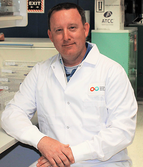
“This project is an excellent example of how we are using our world-class expertise in precision health to contribute to the interdisciplinary work required to make fundamental changes in blood diagnostics,” said Jason Acker, PhD, a senior scientist at Canadian Blood Services’ Centre for Innovation, Professor of Laboratory Medicine and Pathology at the University of Alberta, and one of the lead authors of the study, in the Folio article.
The research took more than three years to complete and involved 19 experts from 12 academic institutions and blood collection facilities located in Canada, Germany, Switzerland, the United Kingdom, and the US.
“Our study shows that artificial intelligence gives us better information about the red blood cell morphology, which is the study of how these cells are shaped, much faster than human experts,” said Jason Acker, PhD (above), Senior Research Scientist, Canadian Blood Services, and Professor of Laboratory Medicine and Pathology at the University of Alberta, in an article published on the Canadian Blood Services website. “We anticipate this technology will improve diagnostics for clinicians as well as quality assurance for blood operators such as Canadian Blood Services in the coming years,” he added. Clinical laboratories in the US may also benefit from this new blood viability process. (Photo copyright: University of Alberta.)
To perform the study, the scientists first collected and manually categorized 52,000 red blood cell images. Those images were then used to train an algorithm that mimics the way a human mind works. The computer system was next tasked with analyzing the shape of RBCs for quality purposes.
Removing Human Bias from RBC Classification
“I was happy to collaborate with a group of people with diverse backgrounds and expertise,” said Tracey Turner, a senior research assistant in Acker’s laboratory and one of the authors of the study, in a Canadian Blood Services (CBS) article. “Annotating and reviewing over 52,000 images took a long time, however, it allowed me to see firsthand how much bias there is in manual classification of cell shape by humans and the benefit machine classification could bring.”
According to the CBS article, a red blood cell lasts about 115 days in the human body and the shape of the RBC reveals its age. Newer, healthier RBCs are shaped like discs with smooth edges. As they age, those edges become jagged and the cell eventually transforms into a sphere and loses the ability to perform its duty of transporting oxygen throughout the body.
Blood donations are processed, packed, and stored for later use. Once outside the body, the RBCs begin to change their shape and deteriorate. RBCs can only be stored for a maximum of 42 days before they lose the ability to function properly when transfused into a patient.
Scientists routinely examine the shape of RBCs to assess the quality of the cell units for transfusion to patients and, in some cases, diagnose and assess individuals with certain disorders and diseases. Typically, microscope examinations of red blood cells are performed by experts in medical laboratories to determine the quality of the stored blood. The RBCs are classified by shape and then assigned a morphology index score. This can be a complex, time-consuming, and laborious process.
“One of the amazing things about machine learning is that it allows us to see relationships we wouldn’t otherwise be able to see,” Acker said. “We categorize the cells into the buckets we’ve identified, but when we categorize, we take away information.”
Human analysis, apparently, is subjective and different professionals can arrive at different results after examining the same blood samples.
“Machines are naive of bias, and AI reveals some characteristics we wouldn’t have identified and is able to place red blood cells on a more nuanced spectrum of change in shape,” Acker explained.
The researchers discovered that the AI could accurately analyze and categorize the quality of the red blood cells. This ability to perform RBC morphology assessment could have critical implications for transfusion medicine.
“The computer actually did a better job than we could, and it was able to pick up subtle differences in a way that we can’t as humans,” Acker said.
“It’s not surprising that the red cells don’t just go from one shape to another. This computer showed that there’s actually a gradual progression of shape in samples from blood products, and it’s able to better classify these changes,” he added. “It radically changes the speed at which we can make these assessments of blood product quality.”
More Precision Matching Blood Donors to Recipients
According to the World Health Organization (WHO), approximately 118.5 million blood donations are collected globally each year. There is a considerable contrast in the level of access to blood products between high- and low-income nations, which makes accurate assessment of stored blood even more critical. About 40% of all blood donations are collected in high-income countries that home to only about 16% of the world’s population.
More studies and clinical trials will be necessary to determine if U of A’s approach to using AI to assess the quality of RBCs can safely transfer to clinical use. But these early results promise much in future precision medicine treatments.
“What this research is leading us to is the fact that we have the ability to be much more precise in how we match blood donors and recipients based on specific characteristics of blood cells,” Acker stated. “Through this study we have developed machine learning tools that are going to help inform how this change in clinical practice evolves.”
The AI tools being developed at the U of A could ultimately benefit patients as well as blood collection centers, and at hospitals where clinical laboratories typically manage the blood banking services, by making the process of matching transfusion recipients to donors more precise and ultimately safer.
FDA’s LDT RULE: Steps to Prepare for Your FDA First Milestone Submission WEBINAR Thursday, November 7, 2024 at 1pm ET First compliance milestonearrives in just seven months! REGISTER NOW • Assess Your Lab’s LDTs to Keep the Winners• Master the Roadmap to Compliance• Calculate the Cost-Benefit of LDT Compliance ⇒⇒ SUMMARY: Laboratories face unprecedented challenges with this ruling and may need to make some difficult decisions regarding their LDT menus. Despite ongoing litigation against the...
Study findings could lead to new clinical laboratory diagnostics that give pathologists a more detailed understanding about certain types of cancer
New studies proving artificial intelligence (AI) can be used effectively in clinical laboratory diagnostics and personalized healthcare continue to emerge. Scientists in the UK recently trained an AI model using machine learning and deep learning to enable earlier, more accurate detection of 13 different types of cancer.
DNA stores genetic information in sequences of four nucleotide bases: A (adenine), T (thymine), G (guanine) and C (cytosine). These bases can be modified through DNA methylation. There are millions of DNA methylation markers in every single cell, and they change in the early stages of cancer development.
One common characteristic of many cancers is an epigenetic phenomenon called aberrant DNA methylation. Modifications in DNA can influence gene expression and are observable in cancer cells. A methylation profile can differentiate tumor types and subtypes and changes in the process often come before malignancy appears. This renders methylation very useful in catching cancers while in the early stages.
However, deciphering slight changes in methylation patterns can be extremely difficult. According to the scientists, “identifying the specific DNA methylation signatures indicative of different cancer types is akin to searching for a needle in a haystack.”
Nevertheless, the researchers believe identifying these changes could become a useful biomarker for early detection of cancers, which is why they built their AI models.
“Computational methods such as this model, through better training on more varied data and rigorous testing in the clinic, will eventually provide AI models that can help doctors with early detection and screening of cancers,” said Shamith Samarajiwa, PhD (above), Senior Lecturer and Group Leader, Computational Biology and Genomic Data Science, Imperial College London, in a news release. “This will provide better patient outcomes.” With additional research, clinical laboratories and pathologists may soon have new cancer diagnostics based on these AI models. (Photo copyright: University of Cambridge.)
The researchers then used a combination of machine learning and deep learning techniques to train an AI algorithm to examine DNA methylation patterns of the collected data. The algorithm identified and differentiated specific cancer types, including breast, liver, lung and prostate, from non-cancerous tissue with a 98.2% accuracy rate. The team evaluated their AI model by comparing the results to independent research.
In their Biology Methods and Protocols paper, the authors noted that their model does require further training and testing and stressed that “the important aspect of this study was the use of an explainable and interpretable core AI model.” They also claim their model could help medical professionals understand “the underlying mechanisms that contribute to the development of cancer.”
Using AI to Lower Cancer Rates Worldwide
According to the Centers for Disease Control and Prevention (CDC), cancer ranks as the second leading cause of death in the United States with 608,371 deaths reported in 2022. The leading cause of death in the US is heart disease with 702,880 deaths reported in the same year.
Globally cancer diagnoses and death rates are even more alarming. World Health Organization (WHO) data shows an estimated 20 million new cancer cases worldwide in 2022, with 9.7 million persons perishing from various cancers that year.
The UK researchers are hopeful their new AI model will help lower those numbers. They state in their paper that “most cancers are treatable and curable if detected early enough.”
More research and studies are needed to confirm the results of this study, but it appears to be a very promising line of exploration and development of using AI to detect, identify, and diagnose cancer earlier. This type of probing could provide pathologists with improved tools for determining the presence of cancer and lead to better patient outcomes.