These advances in the battle against cancer could lead to new clinical laboratory screening tests and other diagnostics for early detection of the disease
As Dark Daily reported in part one of this story, the World Economic Forum (WEF) has identified 12 new breakthroughs in the fight against cancer that will be of interest to pathologists and clinical laboratory managers.
As we noted in part one, the WEF originally announced these breakthroughs in an article first published in May 2022 and then updated in October 2024. According to the WEF, the World Health Organization (WHO) identified cancer as a “leading cause of death globally” that “kills around 10 million people a year.”
The WEF is a non-profit organization base in Switzerland that, according to its website, “engages political, business, academic, civil society and other leaders of society to shape global, regional and industry agendas.”
Monday’s ebrief focused on four advances identified by WEF that should be of particular interest to clinical laboratory leaders. Here are the others.
Personalized Cancer Vaccines in England
The National Health Service (NHS) in England, in collaboration with the German pharmaceutical company BioNTech, has launched a program to facilitate development of personalized cancer vaccines. The NHS Cancer Vaccine Launch Pad will seek to match cancer patients with clinical trials for the vaccines. The Launch Pad will be based on messenger ribonucleic acid (mRNA) technology, which is the same technology used in many COVID-19 vaccines.
The BBC reported that these cancer vaccines are treatments, not a form of prevention. BioNTech receives a sample of a patient’s tumor and then formulates a vaccine that exposes the cancer cells to the patient’s immune system. Each vaccine is tailored for the specific mutations in the patient’s tumor.
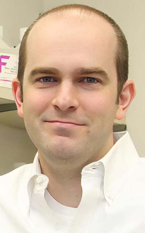
“I think this is a new era. The science behind this makes sense,” medical oncologist Victoria Kunene, MBChB, MRCP, MSc (above), trial principal investigator from Queen Elizabeth Hospital Birmingham (QEHB) involved in an NHS program to develop personalized cancer vaccines, told the BBC. “My hope is this will become the standard of care. It makes sense that we can have something that can help patients reduce their risk of cancer recurrence.” These clinical trials could lead to new clinical laboratory screening tests for cancer vaccines. (Photo copyright: Queen Elizabeth Hospital Birmingham.)
Seven-Minute Cancer Treatment Injection
NHS England has also begun treating eligible cancer patients with under-the-skin injections of atezolizumab, an immunotherapy marketed under the brand name Tecentriq, Reuters reported. The drug is usually delivered intravenously, a procedure that can take 30 to 60 minutes. Injecting the drug takes just seven minutes, Reuters noted, saving time for patients and cancer teams.
The drug is designed to stimulate the patient’s immune system to attack cancer cells, including breast, lung, liver, and bladder cancers.
AI Advances in India
One WEF component—the Center for the Fourth Industrial Revolution (C4IR)—aims to harness emerging technologies such as artificial intelligence (AI) and virtual reality. In India, the organization says the Center is seeking to accelerate use of AI-based risk profiling to “help screen for common cancers like breast cancer, leading to early diagnosis.”
Researchers are also exploring the use of AI to “analyze X-rays to identify cancers in places where imaging experts might not be available.”
Using AI to Assess Lung Cancer Risk
Early-stage lung cancer is “notoriously hard to detect,” WEF observed. To help meet this challenge, researchers at Massachusetts Institute of Technology (MIT) developed an AI model known as Sybil that analyzes low-dose computed tomography scans to predict a patient’s risk of getting the disease within the next six years. It does so without a radiologist’s intervention, according to a press release.
Using Genomics to Identify Cancer-Causing Mutations
In what has been described as the “largest study of whole genome sequencing data,” researchers at the University of Cambridge in the UK announced they have discovered a “treasure trove” of information about possible causes of cancer.
Using data from England’s 100,000 Genomes Project, the researchers analyzed the whole genome sequences of 12,000 NHS cancer patients.
This allowed them “to detect patterns in the DNA of cancer, known as ‘mutational signatures,’ that provide clues about whether a patient has had a past exposure to environmental causes of cancer such as smoking or UV light, or has internal, cellular malfunctions,” according to a press release.
The researchers also identified 58 new mutational signatures, “suggesting that there are additional causes of cancer that we don’t yet fully understand,” the press release states.
The study appeared in April 2022 in the journal Science.
Validation of CAR-T-Cell Therapy
CAR-T-cell therapy “involves removing and genetically altering immune cells, called T cells, from cancer patients,” WEF explained. “The altered cells then produce proteins called chimeric antigen receptors (CARs), which can recognize and destroy cancer cells.”
The therapy appeared to receive validation in 2022 when researchers at the University of Pennsylvania published an article in the journal Nature noting that two early recipients of the treatment were still in remission after 12 years.
However, the US Food and Drug Administration (FDA) announced in 2023 that it was investigating reports of T-cell malignancies, including lymphoma, in patients who had received the treatment.
WEF observed that “the jury is still out as to whether the therapy is to blame but, as a precaution, the drug packaging now carries a warning.”
Breast Cancer Drug Repurposed for Prevention
England’s NHS announced in 2023 that anastrozole, a breast cancer drug, will be available to post-menopausal women to help reduce their risk of developing the disease.
“Around 289,000 women at moderate or high risk of breast cancer could be eligible for the drug, and while not all will choose to take it, it is estimated that if 25% do, around 2,000 cases of breast cancer could potentially be prevented in England, while saving the NHS around £15 million in treatment costs,” the NHS stated.
The tablet, which is off patent, has been used for many years to treat breast cancer, the NHS added. Anastrozole blocks the body’s production of the enzyme aromatase, reducing levels of the hormone estrogen.
Big Advance in Treating Cervical Cancer
In October 2024, researchers announced results from a large clinical trial demonstrating that a new approach to treating cervical cancer—one that uses currently available therapies—can reduce the risk of death by 40% and the risk of relapsing by 36%.
“This is the biggest improvement in outcome in this disease in over 20 years,” said Mary McCormack, PhD, clinical oncologist at the University College London and lead investigator in the trial.
The scientists published their findings in The Lancet.
Pathologists and clinical lab managers will want to keep track of these 12 breakthrough advancements in the diagnosis and treatment of cancer highlighted by the WEF. They will likely lead to new screening tests for the disease and could save many lives.
Study results from Switzerland come as clinical laboratory scientists seek new ways to tackle the problem of antimicrobial resistance in hospitals
Microbiologists and clinical laboratory scientists engaged in the fight against antibiotic-resistant (aka, antimicrobial resistant) bacteria will be interested in a recent study conducted at the University of Basel and University Hospital Basel in Switzerland. The epidemiologists involved in the study discovered that some of these so-called “superbugs” can remain in the body for as long as nine years continuing to infect the host and others.
The researchers wanted to see how two species of drug-resistant bacteria—K. pneumoniae and E. coli—changed over time in the body, according to a press release from the university. They analyzed samples of the bacteria collected from patients who were admitted to the hospital over a 10-year period, focusing on older individuals with pre-existing conditions. They found that K. pneumoniae persisted for up to 4.5 years (1,704 days) and E. coli persisted for up to nine years (3,376 days).
“These patients not only repeatedly become ill themselves, but they also act as a source of infection for other people—a reservoir for these pathogens,” said Lisandra Aguilar-Bultet, PhD, the study’s lead author, in the press release.
“This is crucial information for choosing a treatment,” explained Sarah Tschudin Sutter, MD, Head of the Division of Infectious Diseases and Hospital Epidemiology, and of the Division of Hospital Epidemiology, who specializes in hospital-acquired infections and drug-resistant pathogens. Sutter led the Basel University study.
“The issue is that when patients have infections with these drug-resistant bacteria, they can still carry that organism in or on their bodies even after treatment,” said epidemiologist Maroya Spalding Walters, MD (above), who leads the Antimicrobial Resistance Team in the Division of Healthcare Quality Promotion at the federal Centers for Disease Control and Prevention (CDC). “They don’t show any signs or symptoms of illness, but they can get infections again, and they can also transmit the bacteria to other people.” Clinical laboratories working with microbiologists on antibiotic resistance will want to follow the research conducted into these deadly pathogens. (Photo copyright: Centers for Disease Control and Prevention.)
COVID-19 Pandemic Increased Antibiotic Resistance
The Basel researchers looked at 76 K. pneumoniae isolates recovered from 19 patients and 284 E. coli isolates taken from 61 patients, all between 2008 and 2018. The study was limited to patients in which the bacterial strains were detected from at least two consecutive screenings on admission to the hospital.
“DNA analysis indicates that the bacteria initially adapt quite quickly to the conditions in the colonized parts of the body, but undergo few genetic changes thereafter,” the Basel University press release states.
The researchers also discovered that some of the samples, including those from different species, had identical mechanisms of drug resistance, suggesting that the bacteria transmitted mobile genetic elements such as plasmids to each other.
One limitation of the study, the authors acknowledged, was that they could not assess the patients’ exposure to antibiotics.
Meanwhile, recent data from the World Health Organization (WHO) suggests that the COVID-19 pandemic might have exacerbated the challenges of antibiotic resistance. Even though COVID-19 is a viral infection, WHO scientists found that high percentages of patients hospitalized with the disease between 2020 and 2023 received antibiotics.
“While only 8% of hospitalized patients with COVID-19 had bacterial co-infections requiring antibiotics, three out of four or some 75% of patients have been treated with antibiotics ‘just in case’ they help,” the WHO stated in a press release.
WHO uses an antibiotic categorization system known as AWaRe (Access, Watch, Reserve) to classify antibiotics based on risk of resistance. The most frequently prescribed antibiotics were in the “Watch” group, indicating that they are “more prone to be a target of antibiotic resistance and thus prioritized as targets of stewardship programs and monitoring.”
“When a patient requires antibiotics, the benefits often outweigh the risks associated with side effects or antibiotic resistance,” said Silvia Bertagnolio, MD, Unit Head in the Antimicrobial resistance (AMR) Division at the WHO in the press release. “However, when they are unnecessary, they offer no benefit while posing risks, and their use contributes to the emergence and spread of antimicrobial resistance.”
Citing research from the National Institutes of Health (NIH), NPR reported that in the US, hospital-acquired antibiotic-resistant infections increased 32% during the pandemic compared with data from just before the outbreak.
“While that number has dropped, it still hasn’t returned to pre-pandemic levels,” NPR noted.
The UPenn researchers have already developed an antimicrobial treatment derived from guava plants that has proved effective in mice, Vox reported. They’ve also trained an AI model to scan the proteomes of extinct organisms.
“The AI identified peptides from the woolly mammoth and the ancient sea cow, among other ancient animals, as promising candidates,” Vox noted. These, too, showed antimicrobial properties in tests on mice.
These findings can be used by clinical laboratories and microbiologists in their work with hospital infection control teams to better identify patients with antibiotic resistant strains of bacteria who, after discharge, may show up at the hospital months or years later.
Clinical laboratories and pathology groups should be on the alert to this new digital threat; telehealth sessions and video conferencing calls particularly vulnerable to acoustic AI attacks
Banks may be the first to get hit by a new form of hacking because of all the money they hold in deposit accounts, but experts say healthcare providers—including medical laboratories—are comparably lucrative targets because of the value of patient data. The point of this hacking spear is artificial intelligence (AI) with increased capabilities to penetrate digital defenses.
AI is developing rapidly. Are healthcare organizations keeping up? The hackers sure are. An article from GoBankingRates titled, “How Hackers Are Using AI to Steal Your Bank Account Password,” reveals startling new AI capabilities that could enable bad actors to compromise information technology (IT) security and steal from customers’ accounts.
Though the article covers how the AI could conduct cyberattacks on bank information, similar techniques can be employed to gain access to patients’ protected health information (PHI) and clinical laboratory databases as well, putting all healthcare consumers at risk.
The new AI cyberattack employs an acoustic Side Channel Attack (SCA). An SCA is an attack enabled by leakage of information from a physical computer system. The “acoustic” SCA listens to keystrokes through a computer’s microphone to guess a password with 95% accuracy.
“With recent developments in deep learning, the ubiquity of microphones and the rise in online services via personal devices, acoustic side channel attacks present a greater threat to keyboards than ever,” wrote UK study authors Joshua Harrison, MEng, Durham University; Ehsan Toreini, University of Surrey; and Maryam Mehrnezhad, PhD, University of London.
Hackers could be recording keystrokes during video conferencing calls as well, where an accuracy of 93% is achievable, the authors added.
This nefarious technological advance could spell trouble for healthcare security. Using acoustic SCA attacks, busy healthcare facilities, clinical laboratories, and telehealth appointments could all be potentially compromised.
“The ubiquity of keyboard acoustic emanations makes them not only a readily available attack vector, but also prompts victims to underestimate (and therefore not try to hide) their output,” wrote Joshua Harrison, MEng (above), and his team in their IEEE Xplore paper. “For example, when typing a password, people will regularly hide their screen but will do little to obfuscate their keyboard’s sound.” Since computer keyboards and microphones in healthcare settings like hospitals and clinical laboratories are completely ubiquitous, the risk that this AI technology will be used to invade and steal patients’ protected health information is high. (Photo copyright: CNBC.)
Why Do Hackers Target Healthcare?
Ransomware attacks in healthcare are costly and dangerous. According to InstaMed, a healthcare payments and billing company owned by J.P. Morgan, healthcare data breaches increased to 29.5% in 2021 costing over $9 million. And beyond the financial implications, these attacks put sensitive patient data at risk.
Healthcare can be seen as one of the most desirable markets for hackers seeking sensitive information. As InstaMed points out, credit card hacks are usually quickly figured out and stopped. However, “medical records can contain multiple pieces of personally identifiable information. Additionally, breaches that expose this type of data typically take longer to uncover and are harder for an organization to determine in magnitude.”
With AI advancing at such a high rate, healthcare organizations may be unable to adapt older network systems quickly—leaving them vulnerable.
“Legacy devices have been an issue for a while now,” Alexandra Murdoch, medical data analyst at GlobalData PLC, told Medical Device Network, “Usually big medical devices, such as imaging equipment or MRI machines are really expensive and so hospitals do not replace them often. So as a result, we have in the network these old devices that can’t really be updated, and because they can’t be updated, they can’t be protected.”
But telehealth, according to the UK researchers, may also be one way hackers get past safeguards and into critical hospital systems.
“When trained on keystrokes recorded using the video-conferencing software Zoom, an accuracy of 93% was achieved, a new best for the medium. Our results prove the practicality of these side channel attacks via off-the-shelf equipment and algorithms,” the UK researchers wrote in IEEE Xplore.
“[AI] has worrying implications for the medical industry, as more and more appointments go virtual, the implications of deepfakes is a bit concerning if you only interact with a doctor over a Teams or a Zoom call,” David Higgins, Senior Director at information security company CyberArk, told Medical Device Network.
Higgins elaborated on why healthcare is a highly targeted industry for hackers.
“For a credit card record, you are looking at a cost of one to two dollars, but for a medical record, you are talking much more information because the gain for the purposes of social engineering becomes very lucrative. It’s so much easier to launch a ransomware attack, you don’t even need to be a coder, you can just buy ransomware off of the dark web and use it.”
Steps Healthcare Organizations Should Take to Prevent Cyberattacks
Hackers will do whatever they can to get their hands on medical records because stealing them is so lucrative. And this may only be the beginning, Higgins noted.
“I don’t think we are going to see a slowdown in attacks. What we are starting to see is that techniques to make that initial intrusion are becoming more sophisticated and more targeted,” he told Medical Device Network. “Now with things like AI coming into the mix, it’s going to become much harder for the day-to-day individual to spot a malicious email. Generative AI is going to fuel more of that ransomware and sadly it’s going to make it easier for more people to get past that first intrusion stage.”
To combat these attacks patient data needs to be encrypted, devices updated, and medical staff well-trained to spot cyberattacks before they get out of hand. These SCA attacks on bank accounts could be easily transferable to attacks on healthcare organizations’ patient records.
Clinical laboratories, anatomic pathology groups, and other healthcare facilities would be wise to invest in cybersecurity, training for workers, and updated technology. The hackers are going to stay on top of the technology, healthcare leaders need to be one step ahead of them.
If further research confirms these findings, clinical laboratory identification of cancer cells could lead to new treatments for certain childhood cancers
Can cancer cells be changed into normal healthy cells? According to molecular biologists at the Cold Spring Harbor Laboratory (CSHL) in Long Island the answer is, apparently, yes. At least for certain types of cancer. And clinical laboratories and anatomic pathologists may play a key role in identifying these specific cancer cells and then guiding physicians in selecting the most appropriate therapies.
The cancer cells in question are called rhabdomyosarcoma (RMS) and are “particularly aggressive,” according to ScienceAlert. Generally, and most sadly, the cancer primarily affects children below the age of 18. It begins in skeletal muscle, mutates throughout the body, and is often deadly.
“Treatment usually involves chemotherapy, surgery, and radiation procedures. Now, new research by scientists at Cold Spring Harbor Laboratory demonstrates differentiation therapy as a new treatment option for RMS,” Genetic Engineering and Biotechnology News (GEN) reported.
For those young cancer patients, this new research could become a lifesaving therapy as further studies validate the approach, which has been in development for six years.
“Every successful medicine has its origin story,” said Christopher Vakoc, MD, PhD (above), a molecular biologist at Cold Spring Harbor Laboratory, who led the team that develop the method for converting cancer cells into healthy cells. “And research like this is the soil from which new drugs are born.” As these findings are confirmed, it may be that clinical laboratories and anatomic pathologists will be needed to identify the specific cancer cells in patients once treatment is developed. (Photo copyright: Cold Spring Harbor Laboratory.)
Differentiation Therapy
According to an article in the Chinese Journal of Cancer on the National Library of Medicine website, “Differentiation therapy is based on the concept that a neoplasm is a differentiation disorder [aka, differentiation syndrome] or a dedifferentiation disease. In response to the induction of differentiation, tumor cells can revert to normal or nearly normal cells, thereby altering their malignant phenotype and ultimately alleviating the tumor burden or curing the malignant disease without damaging normal cells.”
Vakoc and his team first pursued differentiation therapy to treat Ewing sarcoma, a pediatric cancer that forms in soft tissues or in bone. In January 2023, GEN reported that the researchers had discovered that “Ewing sarcoma could potentially be stopped by developing a drug that blocks the protein known as ETV6.”
“This protein is present in all cells. But when you perturb the protein, most normal cells don’t care,” Vakoc told GEN. “The process by which the sarcoma forms turns this ETV6 molecule—this relatively innocuous, harmless protein that isn’t doing very much—into something that’s now controlling a life-death decision of the tumor cell.”
The researchers discovered that when ETV6 was blocked in lab-grown Ewing sarcoma cells, the cells became normal, healthy cells. “The sarcoma cell reverts back into being a normal cell again,” they told GEN. “The shape of the cell changes. The behavior of the cells changes. A lot of the cells will arrest their growth. It’s really an explosive effect.”
The scientists then turned their attention on Rhabdomyosarcoma to see if they could elicit a similar response.
“In this study, we developed a high-throughput genetic screening method to identify genes that cause rhabdomyosarcoma cells to differentiate into normal muscle. We used this platform to discover the protein NF-Y as an important molecule that contributes to rhabdomyosarcoma biology. CRISPR-based genetic targeting of NF-Y converts rhabdomyosarcoma cells into differentiated muscle, and we reveal the mechanism by which this occurs,” they wrote in PNAS.
“Scientists have successfully induced rhabdomyosarcoma cells to transform into normal, healthy muscle cells. It’s a breakthrough that could see the development of new therapies for the cruel disease, and it could lead to similar breakthroughs for other types of human cancers,” ScienceAlert reported.
“The cells literally turn into muscle,” Vakoc told ScienceAlert. “The tumor loses all cancer attributes. They’re switching from a cell that just wants to make more of itself to cells devoted to contraction. Because all its energy and resources are now devoted to contraction, it can’t go back to this multiplying state,” he added.
Promising New Therapies for Multiple Cancers in Children
Differentiation therapy as a treatment option gained popularity when “scientists noticed that leukemia cells are not fully mature, similar to undifferentiated stem cells that haven’t yet fully developed into a specific cell type. Differentiation therapy forces those cells to continue their development and differentiate into specific mature cell types,” ScienceAlert noted.
Vakoc and his team had previously “effectively reversed the mutation of the cancer cells that emerge in Ewing sarcoma.” It was those promising results from differentiation therapy that inspired the team to push further and attempt success with rhabdomyosarcoma.
Their results are “a key step in the development of differentiation therapy for rhabdomyosarcoma and could accelerate the timeline for which such treatments are expected,” ScienceAlert commented.
Developing New Therapies for Deadly Cancers
Vakoc and his team are considering differentiation therapy’s potential effectiveness for other types of cancer as well. They note that “their technique, now demonstrated on two different types of sarcoma, could be applicable to other sarcomas and cancer types since it gives scientists the tools needed to find how to cause cancer cells to differentiate,” ScienceAlert reported.
“Since many forms of human sarcoma exhibit a defect in cell differentiation, the methodology described here might have broad relevance for the investigation of these tumors,” the researchers wrote in PNAS.
Clinical laboratories and anatomic pathologist play a critical role in identifying many types of cancers. And though any treatment that comes from the Cold Spring Harbor Laboratory research is years away, it illustrates how new insights into the basic dynamics of cancer cells is helping researchers develop effective therapies for attacking those cancers.
Scientists believe the biodegradable device could someday help detect multiple saliva biomarkers. If true, it might provide a new type of test for clinical laboratories
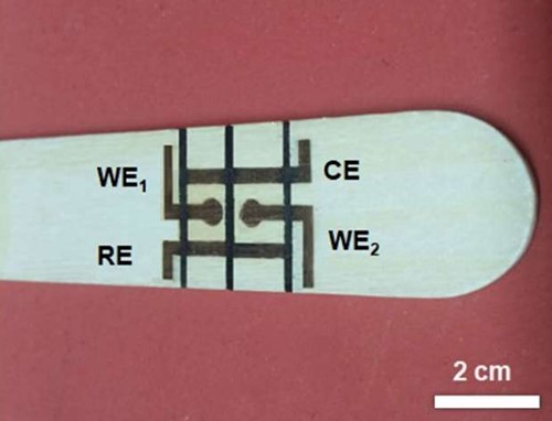
When it comes to tongue depressors, it turns out you can teach an old dog new tricks. Researchers from National and Kapodistrian University of Athens Greece (NKUA) have taken this simple wooden medical tool and developed a high-tech biosensing device that may someday be useful at the point-of-care in hospitals and as a new type of test for clinical laboratories.
Using diode laser engraving, the researchers developed an “eco-friendly disposable sensor that can measure glucose levels and other biomarkers in saliva,” according to LabMedica.
This proof-of-principle biosensing device demonstrates the feasibility of “simultaneous determination of glucose and nitrite in artificial saliva,” according to the NKUA scientists who hope it will help doctors diagnose a variety of conditions.
In their published paper, the scientists at the University of Athens wrote that their wooden electrochemical biosensing tongue depressor (above) “is an easy-to-fabricate disposable point-of-care chip with a wide scope of applicability to other bioassays,” and that “it paves the way for the low-cost and straightforward production of wooden electrochemical platforms.” Might this and other similar biosensing devices eventually find their way to clinical laboratories for use in identifying and tracking certain biomarkers for disease? (Photo copyright: University of Athens.)
How to Make a High-Tech Tongue Depressor
Though wood is affordable and accessible, it doesn’t conduct electricity very well. The researchers’ first attempt to solve this problem was to use the wood as “a passive substrate” to which they coated “metals and carbon-based inks,” LabMedica reported. After that they tried using high-powered lasers to “char specific regions on the wood, turning those spots into conductive graphite.” But that process was complicated, expensive, and a fire hazard.
The researchers eventually turned to “low-power diode lasers” which have been used successfully “to make polyimide-based sensors but have not previously been applied to wooden electronics and electrochemical sensors,” LabMedica noted.
In their Analytical Chemistry paper, the researchers wrote, “A low-cost laser engraver, equipped with a low-power (0.5 W) diode laser, programmably irradiates the surface of the WTD [wooden tongue depressor], forming two mini electrochemical cells (e-cells). The two e-cells consist of four graphite electrodes: two working electrodes, a common counter, and a common reference electrode. The two e-cells are spatially separated via programmable pen-plotting, using a commercial hydrophobic marker pen.”
In other words, the researchers “used a portable, low-cost laser engraver to create a pattern of conductive graphite electrodes on a wooden tongue depressor, without the need for special conditions. Those electrodes formed two electrochemical cells separated by lines drawn with a water-repellent permanent marker,” states a press release from the American Chemical Society.
“The biosensor was then used to quickly and simultaneously measure nitrite and glucose concentrations in artificial saliva. Nitrite can indicate oral diseases like periodontitis, while glucose can serve as a diagnostic for diabetes. The researchers suggest that these low-cost devices could be adapted to detect other saliva biomarkers and could be easily and rapidly produced on-site at medical facilities,” LabMedica reported.
Benefits of Using Wood
One of the major benefits of using wood for their biosensing device is how environmentally friendly it is. “Since wood is a renewable, biodegradable naturally occurring material, the development of conductive patterns on wood substrates is a new and innovative chapter in sustainable electronics and sensors,” the researchers wrote in Analytical Chemistry.
Additionally, the tongue depressor features “An easy-to-fabricate disposable point-of-care chip with a wide scope of applicability to other bioassays, while it paves the way for the low-cost and straightforward production of wooden electrochemical platforms,” the researchers added.
This adds to a growing trend of developing bioassay products that keep the health of our planet in mind.
“This new BC test is non-toxic, naturally biodegradable and both inexpensive and scalable to mass production, currently costing less than $4.00 per test to produce. Its cellulose fibers do not require the chemicals used to manufacture paper, and the test is almost entirely biodegradable,” a UPenn blog post noted.
New Future Tool Use in Clinical Diagnostics
Who could have predicted that the lowly wooden tongue depressor would go high tech with technology that uses lasers to convert it to an electrochemical multiplex biosensing device for oral fluid analysis? This is yet another example of technologies cleverly applied to classic devices that enable them to deliver useful diagnostic information about patients.
And while a biosensing tongue depressor is certainly a diagnostic tool that may be useful for nurses and physicians in clinic and hospital settings, with further technology advancements, it could someday be used to collect specimens that measure more than glucose and nitrites.