Newly combined digital pathology, artificial intelligence (AI), and omics technologies are providing anatomic pathologists and medical laboratory scientists with powerful diagnostic tools
Add “spatial transcriptomics” to the growing list of “omics” that have the potential to deliver biomarkers which can be used for earlier and more accurate diagnoses of diseases and health conditions. As with other types of omics, spatial transcriptomics might be a new tool for surgical pathologists once further studies support its use in clinical care.
Among this spectrum of omics is spatial transcriptomics, or ST for short.
Spatial Transcriptomics is a groundbreaking and powerful molecular profiling method used to measure all gene activity within a tissue sample. The technology is already leading to discoveries that are helping researchers gain valuable information about neurological diseases and breast cancer.
Marriage of Genetic Imaging and Sequencing
Spatial transcriptomics is a term used to describe a variety of methods designed to assign cell types that have been isolated and identified by messenger RNA (mRNA), to their locations in a histological section. The technology can determine subcellular localization of mRNA molecules and can quantify gene expression within anatomic pathology samples.
In “Spatial: The Next Omics Frontier,” Genetic Engineering and Biotechnology News (GEN) wrote, “Spatial transcriptomics gives a rich, spatial context to gene expression. By marrying imaging and sequencing, spatial transcriptomics can map where particular transcripts exist on the tissue, indicating where particular genes are expressed.”
In an interview with Technology Networks, George Emanuel, PhD, co-founder of life-science genomics company Vizgen, said, “Spatial transcriptomic profiling provides the genomic information of single cells as they are intricately spatially organized within their native tissue environment.
“With techniques such as single-cell sequencing, researchers can learn about cell type composition; however, these techniques isolate individual cells in droplets and do not preserve the tissue structure that is a fundamental component of every biological organism,” he added.
“Direct spatial profiling the cellular composition of the tissue allows you to better understand why certain cell types are observed there and how variations in cell state might be a consequence of the unique microenvironment within the tissue,” he continued. “In this way, spatial transcriptomics allows us to measure the complexity of biological systems along the axes that are most relevant to their function.”
“Although spatial genomics is a nascent field, we are already seeing broad interest among the community and excitement across a range of questions, all the way from plant biology to improving our understanding of the complex interactions of the tumor microenvironment,” George Emanuel, PhD (above), told Technology Networks. Oncologists, anatomic pathologists, and medical laboratory scientists my soon see diagnostics that take advantage of spatial genomics technologies. (Photo copyright: Vizgen.)
According to 10x Genomics, “spatial transcriptomics utilizes spotted arrays of specialized mRNA-capturing probes on the surface of glass slides. Each spot contains capture probes with a spatial barcode unique to that spot.
“When tissue is attached to the slide, the capture probes bind RNA from the adjacent point in the tissue. A reverse transcription reaction, while the tissue is still in place, generates a cDNA [complementary DNA] library that incorporates the spatial barcodes and preserves spatial information.
“Each spot contains approximately 200 million capture probes and all of the probes in an individual spot share a barcode that is specific to that spot.”
“The highly multiplexed transcriptomic readout reveals the complexity that arises from the very large number of genes in the genome, while high spatial resolution captures the exact locations where each transcript is being expressed,” Emanuel told Technology Networks.
Spatial Transcriptomics for Breast Cancer and Neurological Diagnostics
In that paper, the authors wrote “we envision that in the coming years we will see simplification, further standardization, and reduced pricing for the ST protocol leading to extensive ST sequencing of samples of various cancer types.”
Spatial transcriptomics is also being used to research neurological conditions and neurodegenerative diseases. ST has been proven as an effective tool to hunt for marker genes for these conditions as well as help medical professionals study drug therapies for the brain.
“You can actually map out where the target is in the brain, for example, and not only the approximate location inside the organ, but also in what type of cells,” Malte Kühnemund, PhD, Director of Research and Development at 10x Genomics, told Labiotech.eu. “You actually now know what type of cells you are targeting. That’s completely new information for them and it might help them to understand side effects and so on.”
The field of spatial transcriptomics is rapidly moving and changing as it branches out into more areas of healthcare. New discoveries within ST methodologies are making it possible to combine it with other technologies, such as Artificial Intelligence (AI), which could lead to powerful new ways oncologists and anatomic pathologists diagnose disease.
“I think it’s going to be tricky for pathologists to look at that data,” Kühnemund said. “I think this will go hand in hand with the digital pathology revolution where computers are doing the analysis and they spit out an answer. That’s a lot more precise than what any doctor could possibly do.”
Spatial transcriptomics certainly is a new and innovative way to look at tissue biology. However, the technology is still in its early stages and more research is needed to validate its development and results.
Nevertheless, this is an opportunity for companies developing artificial intelligence tools for analyzing digital pathology images to investigate how their AI technologies might be used with spatial transcriptomics to give anatomic pathologists a new and useful diagnostic tool.
Experts list the top challenges facing widespread adoption of proteomics in the medical laboratory industry
Year-by-year, clinical
laboratories find new ways to use mass spectrometry to
analyze clinical specimens, producing results that may be more precise than
test results produced by other methodologies. This is particularly true in the
field of proteomics.
However, though mass spectrometry is highly accurate and
fast, taking only minutes to convert a specimen into a result, it is not fully
automated and requires skilled technologists to operate the instruments.
Thus, although the science of proteomics is advancing
quickly, the average pathology laboratory isn’t likely to be using mass
spectrometry tools any time soon. Nevertheless, medical
laboratory scientists are keenly interested in adapting mass spectrometry
to medical lab test technology for a growing number of assays.
Molly Campbell, Science Writer and Editor in Genomics, Proteomics, Metabolomics, and Biopharma at Technology Networks, asked proteomics experts “what, in their opinion, are the greatest challenges currently existing in proteomics, and how can we look to overcome them?” Here’s a synopsis of their answers:
Lack of High Throughput Impacts Commercialization
Proteomics isn’t as efficient as it needs to be to be
adopted at the commercial level. It’s not as efficient as its cousin genomics. For it to become
sufficiently efficient, manufacturers must be involved.
John Yates
III, PhD, Professor, Department of Molecular Medicine at Scripps Research California
campus, told Technology
Networks, “One of the complaints from funding agencies is that you can
sequence literally thousands of genomes very quickly, but you can’t do the same
in proteomics. There’s a push to try to increase the throughput of proteomics
so that we are more compatible with genomics.”
For that to happen, Yates says manufacturers need to
continue advancing the technology. Much of the research is happening at
universities and in the academic realm. But with commercialization comes
standardization and quality control.
“It’s always exciting when you go to ASMS [the conference for the American Society
for Mass Spectrometry] to see what instruments or technologies are going to be
introduced by manufacturers,” Yates said.
There are signs that commercialization isn’t far off. SomaLogic, a privately-owned American protein
biomarker discovery and clinical diagnostics company located in Boulder, Colo.,
has reached the commercialization stage for a proteomics assay platform called SomaScan. “We’ll be
able to supplant, in some cases, expensive diagnostic modalities simply from a
blood test,” Roy
Smythe, MD, CEO of SomaLogic, told Techonomy.
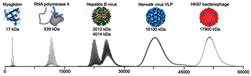
The graphic above illustrates the progression mass spectrometry took during its development, starting with small proteins (left) to supramolecular complexes of intact virus particles (center) and bacteriophages (right). Because of these developments, today’s medical laboratories have more assays that utilize mass spectrometry. (Photo copyright: Technology Networks/Heck laboratory, Utrecht University, the Netherlands.)
Achieving the Necessary Technical Skillset
One of the main reasons mass spectrometry is not more widely
used is that it requires technical skill that not many professionals possess.
“For a long time, MS-based proteomic analyses were technically demanding at
various levels, including sample processing, separation science, MS and the
analysis of the spectra with respect to sequence, abundance and
modification-states of peptides and proteins and false discovery rate
(FDR) considerations,” Ruedi
Aebersold, PhD, Professor of Systems Biology at the Institute of Molecular Systems Biology (IMSB) at
ETH Zurich, told Technology
Networks.
Aebersold goes on to say that he thinks this specific
challenge is nearing resolution. He says that, by removing the problem created
by the need for technical skill, those who study proteomics will be able to
“more strongly focus on creating interesting new biological or clinical
research questions and experimental design.”
Yates agrees. In a paper titled, “Recent Technical Advances in
Proteomics,” published in F1000 Research, a peer-reviewed open research
publishing platform for scientists, scholars, and clinicians, he wrote, “Mass
spectrometry is one of the key technologies of proteomics, and over the last
decade important technical advances in mass spectrometry have driven an
increased capability of proteomic discovery. In addition, new methods to
capture important biological information have been developed to take advantage
of improving proteomic tools.”
No High-Profile Projects to Stimulate Interest
Genomics had the Human Genome Project
(HGP), which sparked public interest and attracted significant funding. One of
the big challenges facing proteomics is that there are no similarly big,
imagination-stimulating projects. The work is important and will result in
advances that will be well-received, however, the field itself is complex and difficult
to explain.
Emanuel
Petricoin, PhD, is a professor and co-director of the Center for Applied
Proteomics and Molecular Medicine at George
Mason University. He told Technology
Networks, “the field itself hasn’t yet identified or grabbed onto a
specific ‘moon-shot’ project. For example, there will be no equivalent to the
human genome project, the proteomics field just doesn’t have that.”
He added, “The equipment needs to be in the background and
what you are doing with it needs to be in the foreground, as is what happened
in the genomics space. If it’s just about the machinery, then proteomics will
always be a ‘poor step-child’ to genomics.”
Democratizing Proteomics
Alexander
Makarov, PhD, is Director of Research in Life Sciences Mass Spectrometry
(MS) at Thermo Fisher
Scientific. He told Technology
Networks that as mass spectrometry grew into the industry we have today,
“each new development required larger and larger research and development teams
to match the increasing complexity of instruments and the skyrocketing
importance of software at all levels, from firmware to application. All this
extends the cycle time of each innovation and also forces [researchers] to
concentrate on solutions that address the most pressing needs of the scientific
community.”
Makarov describes this change as “the increasing democratization of MS,” and says that it “brings with it new requirements for instruments, such as far greater robustness and ease-of-use, which need to be balanced against some aspects of performance.”
One example of the increasing democratization of MS may be
several public proteomic datasets available to scientists. In European
Pharmaceutical Review, Juan
Antonio Viscaíno, PhD, Proteomics Team Leader at the European Bioinformatics Institute (EMBL-EBI)
wrote, “These datasets are increasingly reused for multiple applications, which
contribute to improving our understanding of cell biology through proteomics
data.”
Sparse Data and Difficulty Measuring It
Evangelia
Petsalaki, PhD, Group Leader EMBL-EBI, told Technology
Networks there are two related challenges in handling proteomic data.
First, the data is “very sparse” and second “[researchers] have trouble
measuring low abundance proteins.”
Petsalaki notes, “every time we take a measurement, we
sample different parts of the proteome or phosphoproteome and
we are usually missing low abundance players that are often the most important
ones, such as transcription
factors.” She added that in her group they take steps to mitigate those
problems.
“However, with the advances in MS technologies developed by
many companies and groups around the world … and other emerging technologies
that promise to allow ‘sequencing’ proteomes, analogous to genomes … I expect
that these will not be issues for very long.”
So, what does all this mean for clinical laboratories? At the
current pace of development, its likely assays based on proteomics could become
more common in the near future. And, if throughput and commercialization ever
match that of genomics, mass spectrometry and other proteomics tools could
become a standard technology for pathology laboratories.