If further research confirms these findings, clinical laboratory identification of cancer cells could lead to new treatments for certain childhood cancers
Can cancer cells be changed into normal healthy cells? According to molecular biologists at the Cold Spring Harbor Laboratory (CSHL) in Long Island the answer is, apparently, yes. At least for certain types of cancer. And clinical laboratories and anatomic pathologists may play a key role in identifying these specific cancer cells and then guiding physicians in selecting the most appropriate therapies.
The cancer cells in question are called rhabdomyosarcoma (RMS) and are “particularly aggressive,” according to ScienceAlert. Generally, and most sadly, the cancer primarily affects children below the age of 18. It begins in skeletal muscle, mutates throughout the body, and is often deadly.
“Treatment usually involves chemotherapy, surgery, and radiation procedures. Now, new research by scientists at Cold Spring Harbor Laboratory demonstrates differentiation therapy as a new treatment option for RMS,” Genetic Engineering and Biotechnology News (GEN) reported.
For those young cancer patients, this new research could become a lifesaving therapy as further studies validate the approach, which has been in development for six years.
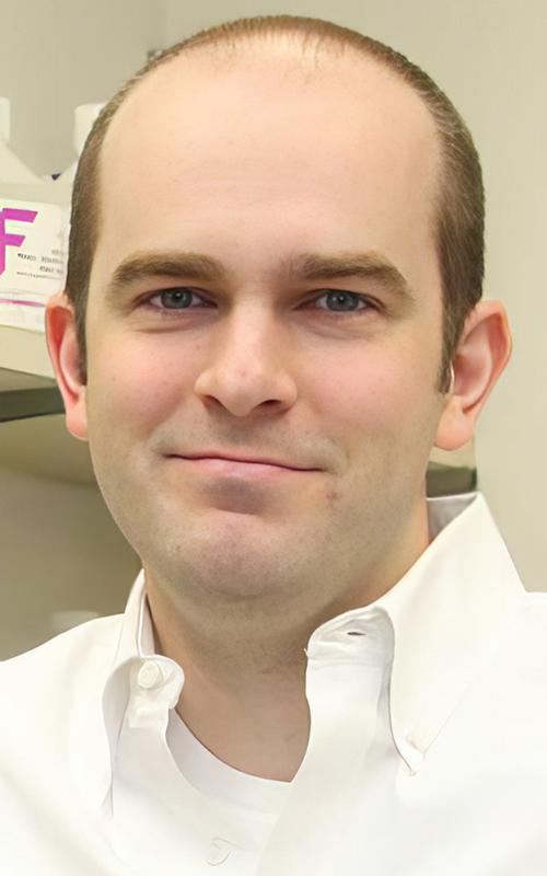
“Every successful medicine has its origin story,” said Christopher Vakoc, MD, PhD (above), a molecular biologist at Cold Spring Harbor Laboratory, who led the team that develop the method for converting cancer cells into healthy cells. “And research like this is the soil from which new drugs are born.” As these findings are confirmed, it may be that clinical laboratories and anatomic pathologists will be needed to identify the specific cancer cells in patients once treatment is developed. (Photo copyright: Cold Spring Harbor Laboratory.)
Differentiation Therapy
According to an article in the Chinese Journal of Cancer on the National Library of Medicine website, “Differentiation therapy is based on the concept that a neoplasm is a differentiation disorder [aka, differentiation syndrome] or a dedifferentiation disease. In response to the induction of differentiation, tumor cells can revert to normal or nearly normal cells, thereby altering their malignant phenotype and ultimately alleviating the tumor burden or curing the malignant disease without damaging normal cells.”
Vakoc and his team first pursued differentiation therapy to treat Ewing sarcoma, a pediatric cancer that forms in soft tissues or in bone. In January 2023, GEN reported that the researchers had discovered that “Ewing sarcoma could potentially be stopped by developing a drug that blocks the protein known as ETV6.”
“This protein is present in all cells. But when you perturb the protein, most normal cells don’t care,” Vakoc told GEN. “The process by which the sarcoma forms turns this ETV6 molecule—this relatively innocuous, harmless protein that isn’t doing very much—into something that’s now controlling a life-death decision of the tumor cell.”
The researchers discovered that when ETV6 was blocked in lab-grown Ewing sarcoma cells, the cells became normal, healthy cells. “The sarcoma cell reverts back into being a normal cell again,” they told GEN. “The shape of the cell changes. The behavior of the cells changes. A lot of the cells will arrest their growth. It’s really an explosive effect.”
The scientists then turned their attention on Rhabdomyosarcoma to see if they could elicit a similar response.
“In this study, we developed a high-throughput genetic screening method to identify genes that cause rhabdomyosarcoma cells to differentiate into normal muscle. We used this platform to discover the protein NF-Y as an important molecule that contributes to rhabdomyosarcoma biology. CRISPR-based genetic targeting of NF-Y converts rhabdomyosarcoma cells into differentiated muscle, and we reveal the mechanism by which this occurs,” they wrote in PNAS.
“Scientists have successfully induced rhabdomyosarcoma cells to transform into normal, healthy muscle cells. It’s a breakthrough that could see the development of new therapies for the cruel disease, and it could lead to similar breakthroughs for other types of human cancers,” ScienceAlert reported.
“The cells literally turn into muscle,” Vakoc told ScienceAlert. “The tumor loses all cancer attributes. They’re switching from a cell that just wants to make more of itself to cells devoted to contraction. Because all its energy and resources are now devoted to contraction, it can’t go back to this multiplying state,” he added.
Promising New Therapies for Multiple Cancers in Children
Differentiation therapy as a treatment option gained popularity when “scientists noticed that leukemia cells are not fully mature, similar to undifferentiated stem cells that haven’t yet fully developed into a specific cell type. Differentiation therapy forces those cells to continue their development and differentiate into specific mature cell types,” ScienceAlert noted.
Vakoc and his team had previously “effectively reversed the mutation of the cancer cells that emerge in Ewing sarcoma.” It was those promising results from differentiation therapy that inspired the team to push further and attempt success with rhabdomyosarcoma.
Their results are “a key step in the development of differentiation therapy for rhabdomyosarcoma and could accelerate the timeline for which such treatments are expected,” ScienceAlert commented.
Developing New Therapies for Deadly Cancers
Vakoc and his team are considering differentiation therapy’s potential effectiveness for other types of cancer as well. They note that “their technique, now demonstrated on two different types of sarcoma, could be applicable to other sarcomas and cancer types since it gives scientists the tools needed to find how to cause cancer cells to differentiate,” ScienceAlert reported.
“Since many forms of human sarcoma exhibit a defect in cell differentiation, the methodology described here might have broad relevance for the investigation of these tumors,” the researchers wrote in PNAS.
Clinical laboratories and anatomic pathologist play a critical role in identifying many types of cancers. And though any treatment that comes from the Cold Spring Harbor Laboratory research is years away, it illustrates how new insights into the basic dynamics of cancer cells is helping researchers develop effective therapies for attacking those cancers.
Study findings may lead to new clinical laboratory tests, as well as vaccines and immunotherapies for neurodegenerative diseases
Research into the human genome continues to produce useful new insights. This time, a study led by researchers at Stanford University identified a genetic variation that is believed to help “slow or even stall” progression of neurodegenerative diseases, including Alzheimer’s and Parkinson’s, according to a press release. Because these genetic variations are common, it is likely that diagnostic tests can be developed for use by clinical laboratories.
Researchers at Stanford Medicine led the study which discovered that approximately one in five individuals carry the gene variant, a protective allele identified as DR4 (aka, HLA-DR4). It’s one of a large number of alleles found in a gene known as DRB1.
DRB1 is part of a family of genes collectively known as the human lymphocyte antigen complex or HLA. The HLA-DRB1 gene plays a crucial role in the ability of the immune system to see a cell’s inner contents.
“In an earlier study, we’d found that carrying the DR4 allele seemed to protect against Parkinson’s disease,” said Emmanuel Mignot, MD, PhD (above), Director of the Stanford Center for Narcolepsy, in a Stanford press release. “Now, we’ve found a similar impact of DR4 on Alzheimer’s disease.” Clinical laboratories may soon have new vaccines for both neurodegenerative diseases. (Photo copyright: Stanford University.)
DR4 Found to Impact Both Parkinson’s and Alzheimer’s Diseases
To perform their research, the team examined a large collection of medical and genetic databases from 176,000 people who had either Alzheimer’s or Parkinson’s disease. The people involved in the study were from numerous countries located in East Asia, Europe, the Middle East and South America. Their genomes were then compared with people who did not have the diseases, focusing on the incidence and age of onset.
“In an earlier study we’d found that carrying the DR4 allele seemed to protect against Parkinson’s disease,” said Mignot in the Stanford press release. “Now, we’ve found a similar impact of DR4 on Alzheimer’s disease.”
The team found that about 20% to 30% of people carry DR4, and that they have around a 10% risk reduction for developing the two diseases.
“That this protective factor for Parkinson’s wound up having the same protective effect with respect to Alzheimer’s floored me,” said Emmanuel Mignot, MD, PhD, the Craig Reynolds Professor of Sleep Medicine in the Department of Psychiatry and Behavioral Sciences at Stanford University and the Director of the Stanford Center for Narcolepsy, in the Stanford Medicine press release. “The night after we found that out, I couldn’t sleep.”
The scientists also analyzed data from autopsied brains of more than 7,000 Alzheimer’s patients and discovered that individuals who carry DR4 had fewer neurofibrillary tangles and that those tangles are composed mainly of modified tau proteins, a common biomarker for Alzheimer’s.
The presence of these tangles corresponds with the severity of Alzheimer’s disease. They are not typically seen in Parkinson’s patients, but the Stanford team found that Parkinson’s patients who did carry DR4 experienced later onset of symptoms.
Mignot stated that tau, which is essential in Alzheimer’s, may also play a role in Parkinson’s, but that further research is required to prove its function.
Both diseases are characterized by the progressive loss of certain nerve cells or neurons in the brain and are linked to an accumulation of abnormal proteins. The Stanford researchers suggested that the DR4 gene variant may help protect individuals from Alzheimer’s and Parkinson’s by preventing the buildup of tau proteins.
“This is a very interesting study, providing additional evidence of the involvement of the immune system in the pathogenesis of Alzheimer’s and Parkinson’s,” neurologist Wassim Elyaman, PhD, Assistant Professor of Neurological Sciences in Neurology, the Taub Institute and the Institute for Genomic Medicine at Columbia University, told Live Science.
New Vaccines and Immunotherapies
According to the Alzheimer’s Association, more than six million Americans are currently living with Alzheimer’s disease and approximately one in three Americans die with Alzheimer’s or another dementia.
The Parkinson’s Foundation states that nearly one million Americans are currently living with Parkinson’s disease, and that number is expected to rise to 1.2 million by 2030. Parkinson’s is the second-most common neurodegenerative disease after Alzheimer’s disease.
Even though the genetic analysis of the Stanford research is strong, more immune cell and blood-based research is needed to definitively establish how tau is connected to the two diseases.
This research could have implications for clinical laboratories by giving them biomarkers for a useful new diagnostic test, particularly for diagnosing Alzheimer’s and Parkinson’s.
Further, Mignot suggested that an effective vaccine could delay the onset or slow the progression of both diseases. He hopes to test his hypothesis on genetically modified mice and eventually human subjects.
Device could pave the way for real-time, noninvasive breath analysis to detect and monitor diseases and be a new service medical laboratories can offer
Breathalyzer technology is not new, but until now human breath detection devices have not been comparable to clinical laboratory blood testing for disease detection and monitoring. That may soon change and there are implications for clinical laboratories, partly because breath samples are considered to be non-invasive for patients.
Scientists with JILA, a research center jointly operated by the National Institutes of Standards and Technology (NIST) and the University of Colorado Boulder, recently increased the sensitivity of their laser frequency comb breathalyzer one thousand-fold. This created a device that can detect four disease biomarkers simultaneously, with the potential to identify six more, according to an NIST news release.
Medical laboratory scientists will understand the significance of this development. JILA’s enhanced breathalyzer device could pave the way for real-time, noninvasive breath analysis to detect and monitor diseases, and potentially eliminate the need for many blood-based clinical laboratory tests.
During their research, physicist Jun Ye, PhD, and David Nesbitt, PhD, both Fellows at JILA and professors at University of Colorado Boulder, detected and monitored four biomarkers in the breath of a volunteer:
These chemicals can be indicators of various health conditions. Methane in the breath, for example, can indicate intestinal problems.
The researchers say the JILA breathalyzer also could detect six additional biomarkers of disease without any further modifications to the device. They would include:
NIST/JILA Research Fellows Jun Ye, PhD (left), and David Nesbitt, PhD (right) of the University of Colorado Boulder, “built a breathalyzer that identifies biomarkers of disease by measuring the colors and amounts of light absorbed as a laser frequency comb passes through breath samples inside a glass tube,” according to an NIST news release. Should they succeed in creating a portable version, their noninvasive device could become an option compared to conventional clinical laboratory blood testing methods used to identify and monitor diseases. (Photos copyright: University of Colorado Boulder.)
“Determining the identity and concentration of the molecules present in breath is a powerful tool to assess the overall health of a person, analogous to blood testing in clinical medicine, but in a faster and less invasive manner,” the researchers wrote in PNAS.
“The presence of a particular molecule (or combination of molecules) can indicate the presence of a certain health condition or infection, facilitating a diagnosis. Monitoring the concentration of the molecules of interest over time can help track the development (or recurrence) of a condition, as well as the effectiveness of the administered treatment,” they added.
How the JILA Breathalyzer Detects Biomarkers
According to a 2008 NIST news release, JILA researchers had developed a prototype comb breathalyzer in that year. However, the research did not continue. But then the COVID-19 pandemic brought the JILA/NIST laboratories focus back to the breathalyzer with hopes that new research could lead to a breath test for detecting the SARS-CoV-2 coronavirus and other conditions.
“We are really quite optimistic and committed to pushing this technology to real medical applications,” Ye said in the 2021 NIST news release.
Analytical Scientist explained that JILA’s new and improved breathalyzer system “fingerprints” chemicals by measuring the amount of light absorbed as a laser frequency comb passes back and forth through breath samples loaded into a mirrored glass tube.
JILA’s original 13-year-old prototype comb analyzed colors and amounts of light in the near-infrared band. However, JILA’s recent improvements include advances in optical coatings and a shift to analyzing mid-infrared band light, allowing detection sensitivity up to parts-per-trillion level, a thousand-fold improvement over the prototype.
Corresponding study author Jutta Toscano, PhD, postdoctoral researcher at the University of Basel in Switzerland and previously Lindemann fellow at JILA, told Physics World the new frequency comb can “probe the molecular fingerprint region where fundamental, and more intense, spectroscopic transitions are found.
“By matching the frequency of the comb teeth with the cavity modes—the ‘standing modes’ of the cavity—we can increase the interaction path length between molecules inside the cavity and laser light by a factor of around 4000, equivalent to an effective path length of a few kilometers,” she added. “We then probe the light that leaks out of the cavity by sending it into an FTIR [Fourier-transform infrared] spectrometer to find out which exact comb teeth have been absorbed and by how much. In turn, this tells us which molecules are present in the breath sample and their concentration.”
Even Hippocrates Studied Breath
Ye noted in the NIST statement that JILA is the only institution that has published research on comb breathalyzers.
In their PNAS paper, the researchers wrote, “Breath analysis is an exceptionally promising and rapidly developing field of research, which examines the molecular composition of exhaled breath. … Despite its distinctive advantages of being a rapid, noninvasive technique and its long history dating back to Hippocrates, breath analysis has not yet been as widely deployed for routine diagnostics and monitoring as other methods, such as blood-based analysis.
“We have shown that this technique offers unique advantages and opportunities for the detection of light biomarkers in breath,” the researchers noted, “and it is poised to facilitate real-time, noninvasive monitoring of breath for clinical studies, as well as for early detection and long-term monitoring of temporary and permanent health conditions.”
Validation of these findings and further design research to make the system portable are required before JILA’s frequency comb breathalyzer can become a competitor to clinical laboratory blood tests for disease identification and monitoring. Nevertheless, JILA’s research brings breathalyzer technology a step closer to offering real-time, non-invasive analysis of human biomarkers for disease.
The technology is similar to the concept of a liquid biopsy, which uses blood specimens to identify cancer by capturing tumor cells circulating in the blood.
According to the American Cancer Society, lung cancer is responsible for approximately 25% of cancer deaths in the US and is the leading cause of cancer deaths in both men and women. The ACS estimates there will be about 236,740 new cases of lung cancer diagnosed in the US this year, and about 130,180 deaths due to the disease.
Early-stage lung cancer is typically asymptomatic which leads to later stage diagnoses and lowers survival rates, largely due to a lack of early disease detection tools. The current method used to detect early lung cancer lesions is low-dose spiral CT imaging, which is costly and can be risky due to the radiation hazards of repeated screenings, the news release noted.
MGH’s newly developed diagnostic tool detects lung cancer from alterations in blood metabolites and may lead to clinical laboratory tests that could dramatically improve survival rates of the deadly disease, the MGH scientist noted in a news release.
“Our study demonstrates the potential for developing a sensitive screening tool for the early detection of lung cancer,” said Leo Cheng, PhD (above), in the news release. Cheng is Associate Professor of Radiology at Harvard Medical School and Associate Biophysicist in Radiology at Massachusetts General Hospital. “The predictive model we constructed can identify which people may be harboring lung cancer. Individuals with suspicious findings would then be referred for further evaluation by imaging tests, such as low-dose CT, for a definitive diagnosis,” he added. Oncologists may soon have a clinical laboratory test for screening patients with early-stage lung cancer. (Photo copyright: OCSMRM.)
Detecting Lung Cancer in Blood Metabolomic Profiles
The MGH scientists created their lung-cancer predictive model based on magnetic resonance spectroscopy which can detect the presence of lung cancer from alterations in blood metabolites.
The researchers screened tens of thousands of stored blood specimens and found 25 patients who had been diagnosed with non-small-cell lung carcinoma (NSCLC), and who had blood specimens collected both at the time of their diagnosis and at least six months prior to the diagnosis. They then matched these individuals with 25 healthy controls.
The scientists first trained their statistical model to recognize lung cancer by measuring metabolomic profiles in the blood samples obtained from the patients when they were first diagnosed with lung cancer. They then compared those samples to those of the healthy controls and validated their model by comparing the samples that had been obtained from the same patients prior to the lung cancer diagnosis.
The predictive model yielded values between the healthy controls and the patients at the time of their diagnoses.
“This was very encouraging, because screening for early disease should detect changes in blood metabolomic profiles that are intermediate between healthy and disease states,” Cheng noted.
The MGH scientists then tested their model with a different group of 54 patients who had been diagnosed with NSCLC using blood samples collected before their diagnosis. The second test confirmed the accuracy of their model.
Predicting Five-Year Survival Rates for Lung Cancer Patients
Values derived from the MGH predictive model measured from blood samples obtained prior to a lung cancer diagnosis also could enable oncologists to predict five-year survival rates for patients. This discovery could prove to be useful in determining clinical strategies and personalized treatment decisions.
The researchers plan to analyze the metabolomic profiles of the clinical characteristics of lung cancer to understand the entire metabolic spectrum of the disease. They hope to create similar models for other illnesses and have already created a model that can distinguish aggressive prostate cancer by measuring the metabolomics profiles of more than 400 patients with that disease.
In addition, they are working on a similar model to screen for Alzheimer’s disease using blood samples and cerebrospinal fluid.
More research and clinical studies are needed to validate the utilization of blood metabolomics models as early screening tools in clinical practice. However, this technology might provide pathologists and clinical laboratories with diagnostic tests for the screening of early-stage lung cancer that could save thousands of lives each year.
Scientists working to sequence all 1.66 million animal species say this is a missed opportunity to better understand our own genetics; such research would identify biomarkers useful for clinical laboratory testing
For 23 years, the world’s genomic scientists have been on a mission to sequence the genomes of all animal species. And they’ve made great progress. However, according to a recent study conducted by researchers at Washington State University (WSU) and Brigham Young University (BYU), only a fraction of the sequences are from invertebrate species. And that, according to the study’s authors, is “overlooking huge swathes of diversity and opportunity.”
The push to sequence the whole genomes of all animals began in 1998 with the sequencing of the Caenorhabditis elegans roundworm, according to a WSU news release. It was the first animal genome sequence, but it was not to be the last. Nearly 25 years later, genomic scientists have sequenced about 3,300 animal genomes. And while that’s a lot of genomic sequences, it’s a drop in bucket of the approximately 1.7 million animal species on the planet.
But here’s where the missed opportunity comes in. According to the WSU news release, “Vertebrates account for 54% of all genome sequencing assemblies, despite representing only 3.9% of animal species. In contrast, the invertebrates of the Arthropoda phylum, which includes insects and spiders, comprise only 34% of current datasets while representing 78.5% of all species.”
“We are interested in ourselves, and that’s not necessarily a bad thing,” said Paul Frandsen, PhD (above), in the news release. Frandsen is Assistant Professor of Genetics, Genomics, and Biotechnology at Brigham Young University and one of the study authors. “But to begin to understand entire ecosystems,” he continued, “we have to start sampling more of the variety of life to gain a clearer picture. Vertebrates are important components of ecosystems, but arguably insects and many other small creatures probably play an even more important role because they’re down at the base of the food web.” (Photo copyright: Brigham Young University.)
The scientists analyzed the best available genome assemblies found in GenBank, the world’s most extensive genetic database. They found that 3,278 unique animal species across 24 phyla, 64 classes, and 258 orders have been sequenced and assembled to date.
They also found that sequencing efforts have focused heavily on species that most resemble humans. The Hominidae, a taxonomic family of primates that includes humans as well as great apes, bonobos, chimpanzees, orangutans, and gorillas, has the most contiguous genome data assembled.
The team discovered that vertebrates account for 54% of the animal genome sequencing that has been performed even though they make up less than four percent of known animal species. By comparison, invertebrates of the Arthropoda phylum, which represent 78.5% of all animal species, comprise only 34% of the completed animal genome sequencing. And yet, the Arthropoda phylum is the largest phylum in the animal kingdom and includes insects, spiders, scorpions, centipedes, millipedes, crabs, crayfish, lobsters, and barnacles.
“With genome assemblies accumulating rapidly, we want to think about where we are putting our efforts. It’s not being spread evenly across the animal tree of life,” said lead author Scott Hotaling, PhD, post-doctoral researcher at WSU, in the news release. “Invertebrates are still very underrepresented, which makes sense given that people seem to care more about vertebrates, the so-called ‘charismatic megafauna.’”
The team discovered that only five arthropod groups: ants, bees, butterflies, fruit flies, and mosquitos, were well represented in genome sequencing. The longest genome sequenced so far belongs to the Australian lungfish, the only surviving member of the family Neoceratodontidae.
1,100 Years to Sequence All Eukaryotic Life
The scientists also discerned that animal genome assemblies have been produced by 52 countries on every continent with permanent inhabitants. The majority of animal genome sequencing (77%) that is being performed is mostly occurring in developed countries located in the Northern Hemisphere, often referred to as the Global North. Nearly 70% of all animal genome assemblies have been produced by just three countries: the United States, China, and Switzerland.
There are geographic differences between regions regarding the types of animals being sequenced and assembled with North America concentrating on mammals and insects, Europe focusing on fish, and birds being the main type of animals sequenced in Asia.
The scientists would like to see more animal genome sequencing happening in countries from the Global South, or Southern Hemisphere, particularly in tropical regions that contain a myriad of diversity among animal species.
“If we want to build a global discipline, we need to include a global people,” Hotaling said. “It’s just basic equity, and from a pure scientific standpoint, the people who live in areas where species are being sequenced have a lot of knowledge about those species and ecosystems. They have a lot to contribute.”
But the WSU/BYU scientists found that many species in GenBank only have low-quality assemblies available. They noted that “the quality of a genome assembly is likely the most important factor dictating its long-term value.”
Fortunately, several animal genome sequencing ventures have been announced in recent years, so the amount of available data is expected to rise exponentially. These projects include:
The Earth BioGenome Project (EBP) which aspires to sequence and catalog the genes of all the eukaryotic species on the planet within ten years.
The Vertebrate Genomes Project which seeks to generate high-quality assemblies for 70,000 extant vertebrate species.
The Bird 10K Project that seeks to generate assemblies for all extant birds.
The i5K Project which plans to produce 5,000 arthropod genome assemblies.
The authors of the PNAS paper noted that there are currently only about four genome assemblies happening each day and, at that rate, the sequencing of all eukaryotic life will not be completed until the year 3130.
So, microbiologists, clinical laboratory professionals, and genomic scientists have plenty of time to get up to speed.