Research findings could lead to new biomarkers for genetic tests and give clinical laboratories new capabilities to diagnose different health conditions
New insights continue to emerge about “junk DNA” (aka, non-coding DNA). For pathologists and clinical laboratories, these discoveries may have value and eventually lead to new biomarkers for genetic testing.
One recent example comes from researchers at Stanford Medicine in California who recently learned how non-coding DNA—which makes up 98% of the human genome—affects gene expression, the function that leads to observable characteristics in an organism (phenotypes).
The research also could lead to a better understanding of how short tandem repeats (STRs)—the number of times a gene is copied into RNA for protein use—affect gene expression as well, according to Stanford.
“We’ve known for a while that short tandem repeats or STRs, aren’t junk because their presence or absence correlates with changes in gene expression. But we haven’t known how they exert these effects,” said study lead Polly Fordyce, PhD (above), Associate Professor of Bioengineering and Genetics at Stanford University, in a news release. The research could lead to new clinical laboratory biomarkers for genetic testing. (Photo copyright: Stanford University.)
To Bind or Not to Bind
In their Science paper, the Stanford researchers described an opportunity to explore a new angle to transcription factors binding to some sequences, also known as sequence motifs.
“Researchers have spent a lot of time characterizing these transcription factors and figuring out which sequences—called motifs—they like to bind to the most,” said the study lead Polly Fordyce, PhD, Associate Professor of Bioengineering and Genetics at Stanford University, in a Stanford Medicine news release.
“But current models don’t adequately explain where and when transcription factors bind to non-coding DNA to regulate gene expression. Sometimes, no transcription factor is attached to something that looks like a perfect motif. Other times, transcription factors bind to stretches of DNA that aren’t motifs,” the news release explains.
Transcription factors are “like light switches that can turn genes on or off depending on what cells need,” notes a King’s College LondonEDIT Labblog post.
But why do transcription factors target some places in the genome and not others?
“To solve the puzzle of why transcription factors go to some places in the genome and not to others, we needed to look beyond the highly preferred motifs,” Fordyce added. “In this study, we’re showing that the STR sequence around the motif can have a really big effect on transcription factor binding, providing clues as to what these repeated sequences might be doing.”
Such information could aid in understanding certain hereditary conditions and diseases.
“Variations in STR length have been associated with changes in gene expression and implicated in several complex phenotypes such as schizophrenia, cancer, autism, and Crohn’s disease. However, the mechanism by which STRs affect transcription remains unknown,” the researchers wrote in Science.
Special Assays Explore Binding
According to their paper, the research team turned to the Fordyce Lab’s previously developed microfluidic binding assays (MITOMI, k–MITOMI, and STAMMP) to analyze the impact of different DNA sequences on transcription factor binding.
“In the experiment we asked, ‘How do these changes impact the strength of transcription factor binding?’ We saw a surprisingly large effect. Varying the STR sequence around a motif can have a 70-fold impact on the binding,” Fordyce wrote.
In an accompanying Science article titled, “Repetitive DNA Regulates Gene Expression,” Thomas Kuhlman, PhD, Assistant Professor, Physics and Astronomy, University of California, Riverside, wrote that the study “demonstrates that STRs exert their effects by directly binding transcription factor proteins, thus explaining how STRs might influence gene expression in both normal and diseased states.”
“This research unveils, for the first time, the intricate connection between how variants in the non-coding genome affect genes that are associated with blood pressure and with hypertension. What we’ve created is a kind of functional map of the regulators of blood pressure genes, “said Philipp Maass, PhD, Lead Researcher and Assistant Professor Molecular Genetics, University of Toronto, in a news release.
The research team used massively parallel reporter assay (MPRA) technology to analyze 4,608 genetic variants associated with blood pressure.
The findings could aid precision medicine for cardiovascular health and may possibly be adopted to other conditions, according to The Hospital for Sick Children.
“The variants we have characterized in the non-coding genome could be used as genomic markers for hypertension, laying the groundwork for future genetic research and potential therapeutic targets for cardiovascular disease,” Maass noted.
Why All the ‘Junk’ DNA?
Clinical laboratory scientists may wonder why genetic research has primarily focused on 20,000 genes within the genome, leaving the “junk” DNA for later investigation. So did researchers at Harvard University.
“After the Human Genome Project, scientists found that there were around 20,000 genes within the genome, a number that some researchers had already predicted. Remarkably, these genes comprise only about 1-2% of the three billion base pairs of DNA. This means that anywhere from 98-99% of our entire genome must be doing something other than coding for proteins,” they wrote in a blog post.
“Imagine being given multiple volumes of encyclopedias that contained a coherent sentence in English every 100 pages, where the rest of the space contained a smattering of uninterpretable random letters and characters. You would probably start to wonder why all those random letters and characters were there in the first place, which is the exact problem that has plagued scientists for decades,” they added.
Not only is junk DNA an interesting study subject, but ongoing research may also produce useful new biomarkers for genetic diagnostics and other clinical laboratory testing. Thus, medical lab professionals may want to keep an eye on new developments involving non-coding DNA.
The researchers unveiled a diagnostic device that uses microfluidic technology to identify cell types in blood by their size. The device also “can isolate individual cancer cells from patient blood samples,” according to a news release.
The ability to isolate circulating tumor cells could enable clinical laboratories to perform diagnostic cancer tests on liquid biopsies and blood samples. Dark Daily reported on various studies involving liquid biopsies—an alternative to invasive and costly cancer diagnostic procedures, such as surgery and tissue biopsies—in previous e-briefings.
“This new microfluidics chip lets us separate cancer cells from whole blood or minimally diluted blood. Our device is cheap and doesn’t require much specimen preparation or dilution, making it fast and easy-to-use,” said Ian Papautsky, PhD, Professor of Bioengineering at University of Illinois at Chicago, in the news release. He is shown above with members of the Papautsky Lab, which has been developing “microfluidic systems and point- of-care sensors for public health applications.” (Photo copyright: University of Illinois at Chicago.)
Searching for ‘Purity’
The UIC and QUT researchers were motivated by the
information-rich nature of circulating tumor cells. They also saw opportunity
for escalated “purity” in results, as compared to past studies.
In the paper, they acknowledged the work of other scientists
who deployed microfluidic technology affinity-based methods to differentiate
tumor cells in blood. Past studies (including previous work by the authors)
also explored tumor cells based on size and difference from white blood cells.
“While many emerging systems have been tested using patient samples, they share a common shortcoming: their purity remains to be significantly improved. High purity is in strong demand for circulating tumor cell enumeration, molecular characterization, and functional assays with less background intervention from white blood cells,” the authors wrote in their paper.
How the Device Works
The scientists say their system leverages “size-dependent
inertial migration” of cells. According to the news release:
Blood passes through “microchannels” formed in
plastic in the device;
“Inertial migration and shear-induced diffusion”
separate cancer cells from blood;
Tiny differences in size determine a cell’s
attraction to a location; and
Cells separate to column locations as the liquid
moves.
In other words, the device works as a filter sorting out, in
blood samples, the circulating tumor cells based on their unique size, New
Atlas explained.
93% of Cancer Cells Recovered by Device
When the researchers tested their new device:
Researchers placed 10 small-cell-lung cancer cells into five-milliliter samples of healthy blood;
The blood was then flowed through the device; and
93% of the cancer cells were recovered.
“A 7.5 milliliter tube of blood, which is typical volume for
a blood draw, might have 10 cancer cells and 35- to 40-billion blood cells. So,
we are really looking for a needle in a haystack,” Papautsky stated in the news
release.
The graphic above illustrates how, in the lab, the microfluidic device enabled the researchers to separate out cancer cells in six of the eight lung cancer samples they studied. (Graphic copyright: Ian Papautsky, PhD/University of Illinois at Chicago/New Atlas.)
“We report on a novel multi-flow microfluidic system for the
separation of circulating tumor cells with high purity. The microchannel takes
advantage of inertial migration of cells. The lateral migration of cells
strongly depends on cell size in our microchannel, and label-free separation of
circulating tumor cells from white blood cells is thus achieved without
sophisticated sample predation steps and external controls required by
affinity-based and active approaches,” the researchers wrote in their paper.
The researchers plan wider trials and the addition of
biomarkers to enable cancer DNA detection, New Atlas reported, which described
the UIC/QUT study as part of a “new wave of diagnostics.”
With so much focus on liquid biopsy research, it may be
possible for medical laboratories to one day not only diagnose cancer through
blood tests, but also to find the disease earlier and in a more precise way
than with traditional tissue sample analysis.
Ever shrinking “lab-on-a-…” technologies, a boon to medical laboratories and anatomic pathologists in remote resource-strapped regions, also have a place in modern labs
Researchers took another leap forward in reducing the size of clinical laboratory diagnostic tests and observational tools. This demonstration involved lab-on-a-fiber technology and showed promise in both monitoring anatomic pathology biomarkers in vivo and supplementing the abilities of existing lab-on-a-chip and microfluidic devices.
In 2013, Dark Daily reported on research into an implantable laboratory-on-a-chip (LOC) for monitoring blood chemistry during chemotherapy. It was a major breakthrough at the time, which promised new and powerful tools for cancer treatment regimens.
However, most LOC systems aren’t designed for wet environments. Also, while microfluidics and flexible membranes allow for smaller footprints and tighter placement, they are still invasive in ways that might make patients uncomfortable or make real-world use less than ideal. And, long-term use brings further complications, such as corrosion or foreign-body granulomas.
Thus, lab-on-a-fiber’s ability to function in vivo, is one of the device’s principal advantages, as ExtremeTech noted.
Lab-on-a-fiber technology addresses many concerns. It is small enough to insert directly into organs, muscle mass, or veins when used as biosensors. And the fibers can return a wealth of information by using light and reflection, while allowing for minimal discomfort and precision placement.
Schematic of the lab-on-a-fiber biosensing principle. A metallic nanostructure supporting a resonant plasmonic mode is integrated on the optical fiber tip. When a molecular binding event occurs at the sensor surface, the reflectance peak associated to the plasmonic mode shifts towards longer wavelengths. (Image and caption copyright: Analyst/The Royal Society of Chemistry.)
The Past and Future of Scaling Clinical Laboratory Testing
Developers believe lab-on-a-fiber approaches could offer further adaptability and functionality to other “lab-on-a-…” technologies. For example, as highlighted in Advanced Science News, researchers are employing lab-on-a-fiber technologies to further refine and improve LOC functions and designs.
“As the scientific world moves inexorably to smaller dimensions … The emerging concept of ‘lab‐on‐fiber’ will give the optical fiber platform additional (highly integrated) functionalities,” noted Deepak Uttamchandani, PhD, Vice Dean Research, Faculty of Engineering, and, Robert Blue, PhD, Research Fellow, both at the University of Strathclyde, Glasgow, UK, in their review paper, “Recent Advances In Optical Fiber Devices for Microfluidics Integration.” The paper, published in the Journal of Biophotonics, examined “the recent emergence of miniaturized optical fiber-based sensing and actuating devices that have been successfully integrated into fluidic microchannels that are part of microfluidic and lab‐on‐chip systems.”
In his review paper on the emerging concept of lab-on-a-fiber, Deepak Uttamchandani, PhD, notes, “The versatility of the optical fiber platform has already allowed researchers to conduct immunoassays in microchannels using both fluorescently‐labelled and label‐free formats whilst gaining advantages of reduced assay time and increased sensitivity.” (Photo copyright: University of Strathclyde.)
Lab-on-a-Fiber: Another Step Forward or a Major Change?
At each milestone in the scaling of clinical laboratory testing, experts and media outlets predicted the demise of big laboratories and the dawn of a POC-centric testing era. Yet, despite 20-plus years of progress, this has yet to happen.
While it is critical for anatomical pathology leaders and clinical laboratory managers to stay abreast of developments in testing technology, much of the innovation behind lab-on-a-fiber remains strictly in the research realm. Challenges to the commercialization of these new techniques include both physical factors, such as design and manufacture of ready-to-use tests, and regulatory concerns, including FDA clearances and payer approval of new assays and diagnostic procedures.
Until researchers and test manufacturers overcome these hurdles, threats to current standards and workflows are minimal. However, much like the gains in scale realized through incorporating lab-on-a-chip concepts into clinical laboratory testing, the research powering these innovations might prove useful in further improving and expanding medical laboratory testing options.
Research goal was to isolate circulating tumor cells in venipuncture samples with improved purity compared to standard spiral chips
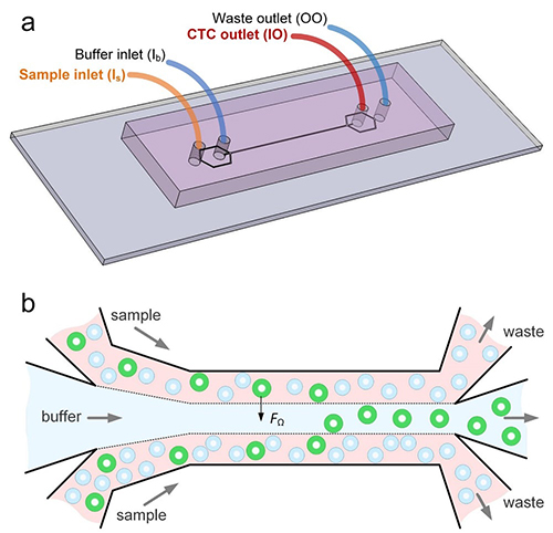
Many research teams are pursuing the goal of creating assays that detect circulating tumor cells (CTCs) that would allow earlier and more accurate diagnosis of cancer. Now comes news of a unique technology developed at the University of Michigan (U-M) Ann Arbor that showed promised in an early study.
The method of using CTCs to diagnose cancer in patients, while further analyzing specific characteristics of a given cancer case, shows promise as an innovative tool for clinical laboratories and oncologists. However, current approaches face challenges when it comes to proving accuracy and establishing thresholds that might indicate the need for further action.
Researchers at U-M believe they may have solved that problem. They created “Labyrinth,” a “label-free microfluidic device” that condenses 637mm of channels—including 11 loops and 56 corners—onto a 500μm-wide chip that uses inertia and Dean flow to separate white blood cells and CTCs from venipuncture samples at rates as high as 2.5ml per minute. These results improve upon the traditional spiral chip design.
Publishing their findings in Cell Systems, first author of the study Eric Lin, PhD, noted, “With the recent advances in tools for genomic characterization, it is more compelling than ever to look at the tumor heterogeneity to understand tumor progression and resistance to therapies. The Labyrinth device enabled high yields of CTCs without the bias induced by antibody-based selection, allowing the identification of true biological tumor heterogeneity.”
The graphic above, taken from the University of Michigan study, demonstrates the “High-throughput and label-free Labyrinth device that enables single CTC isolation and gene expression characterization.” According to the researchers, “Labyrinth offers a cell-surface marker-independent single-cell isolation platform to study heterogeneous CTC subpopulations.” The U-M study shows promise in creating tools for oncologist and clinical laboratory cancer treatment. (Image copyright: University of Michigan/Cell Systems.)
Challenges in the Isolation of CTCs
The Labyrinth chip is not the first device to assist in isolating CTCs. The U-M study notes that while immune-affinity capture is a validated approach to prognosis, therapeutic monitoring and molecular diagnostics, it does not work with all cancer cases. The researchers also note the method creates challenges in single-cell analysis later.
Existing label-free methods of isolation, such as deterministic lateral displacement, microfluidic flow fractionation, and acoustic-based separation, avoid these concerns but face issues of their own. The researchers noted, “Issues encountered with these approaches include pore clogging, high-pressure drop, pre-fixation to prevent CTC loss, low throughput, and excessive non-specific cell retention.”
The researchers further clarified that a major factor separating the Labyrinth chip from other methods is the ability to identify CTC subpopulations without the need for manual selection based on positive or negative protein expression. Thus, improving the ability to conduct further single-cell analysis from the results. Testing of the Labyrinth chip involved a variety of cancer cell lines, including:
· Human breast (MCF-7);
· Pancreatic (PANC-1);
· Prostate (PC-3); and,
· Lung (H1650).
And while standard spiral chips are already a common method for conducting size-based sorting, the purity of results is less than ideal with thousands of other cells remaining in the sample.
The researchers reported that the Labyrinth chip recovered 91.5% (plus or minus 0.9%) of cancer cells and removed 91.4% (plus or minus 3.3%) of white blood cells in a spiked buffer test.
“Bigger cells, like most cancer cells, focus pretty fast due to the curvature. But the smaller the cell is, the longer it takes to get focused,” Sunitha Nagrath, PhD, Associate Professor of Chemical Engineering and a lead developer of the Labyrinth chip, stated in a U-M news release. “The corners produce a mixing action that makes the smaller white blood cells come close to the equilibrium position much faster.”
Labyrinth also supports a series configuration of multiple chips. While testing two chips in series, researchers noted “a two-log improvement in tumor cell enrichment over the single Labyrinth.” They claim this is a higher purity than other label-free methods they studied, while adding only five minutes to processing times.
Sunitha Nagrath, PhD (above), is an Associate Professor of Chemical Engineering at the University of Michigan, and one of the lead developers of the Labyrinth chip. “You cannot put a box around these cells,” she noted in the U-M news release. “The markers for them are so complex, there is no one marker we could target for all these stages.” (Photo copyright: University of Michigan.)
Current Testing Using the Labyrinth Chip
The chip is already in use in a clinical trial for an aggressive form of breast cancer by Max Wicha, MD, Madeline and Sidney Forbes Professor of Oncology, Founding Director Emeritus, University of Michigan Comprehensive Cancer Center, and co-author of the Cell Systems study, who lead the study along with Nagrath.
The trial involves the attempted activation of adult system cells by blocking the signaling molecule interleukin-6. Wicha suspects the molecule enables cancer stem cells as well. “We think that this may be a way to monitor patients in clinical trials,” he said in the U-M news release. “Rather than just counting the cells, by capturing them, we can perform molecular analysis [to] know what we can target with treatments.”
The news release further highlights how this chip is specifically suited to such a task. As cancer stem cells transition from stem-like cells to more ordinary cell types, their gene expression shifts as well. This creates an issue when using conventional cell targeting. Nagrath notes this concern, stating, “The markers for [cancer stem cells] are so complex, there is no one marker we could target for all these stages.”
The Labyrinth chip shows potential for overcoming one of the biggest hurdles to leveraging CTCs to diagnose cancers and develop personalized therapies. Currently, the chip can output to Fluidigm, DEPArray by Silicon Biosystems, and RainDance Technologies’ RainDrop Digital PCR System.
The U-M researchers hope that future research will yield additional applications and compatible systems to further improve the ability for medical laboratories to use CTCs in the early detection and monitoring of cancer cases.
Researchers believe newly developed optomechanical technology might eventually be used by medical laboratories
Pathologists and medical laboratory scientists have long been aware of the parallel between cancer and the mechanical properties located in cells. However, a diagnostic tool to assess these properties has until now been unavailable. This may soon change.
A team at the University of Illinois at Urbana-Champaign (UIUC) recently created a technique involving “OptoMechanoFluidics” that might increase understanding of how diseases reshape the mechanical attributes of cells in the human body. The researchers’ innovative opto-mechano-fluidic approach could provide a new way to study how human cells congregate in tissue and bones by examining high-speed photonic sensing of free-flowing particles in the body at rates potentially reaching 10,000 particles per second.