Viral reservoir could be behind persistence, says study, which also suggests a blood biomarker could be found for clinical laboratory testing
Microbiologists and virologists working closely with physicians treating long COVID-19 patients will gain new insights in a study that found coronavirus spike protein in COVID-19 patients’ blood up to 12 months after diagnosis. The researchers believe their findings could be used to develop a clinical laboratory biomarker for long COVID-19.
Researchers at Brigham and Women’s Hospital and Massachusetts General Hospital said medical experts are not sure why some people have unwelcome symptoms weeks and months after a positive COVID-19 diagnosis, while others clear the infection without lingering effects.
The scientists believe if this work is validated, clinical laboratories might gain an assay to use in the diagnosis of long COVID-19.
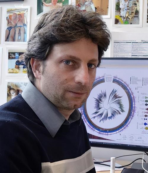
“The half-life of spike protein in the body is pretty short, so its presence indicates that there must be some kind of active viral reservoir,” said David Walt, PhD (above), Professor of Pathology, Brigham and Women’s Hospital, and lead author of the study that found coronavirus spike protein in long COVID patients. The study findings indicate a potential clinical laboratory biomarker for long COVID-19. (Photo copyright: Brigham and Women’s Hospital.)
Viral Reservoir Possibly Behind Long COVID-19
The study suggests that SARS-CoV-2 finds a home in the body, particularly the gastrointestinal tract, “through viral reservoirs, where it continues to release spike protein and trigger inflammation,” Medical News Today reported.
Lead author of the study David Walt, PhD, Professor of Pathology, Brigham and Women’s Hospital and the Hansjörg Wyss Professor Biologically Inspired Engineering at Harvard Medical School, told The Guardian he “was motivated to carry out the study after earlier research by his colleagues detected genetic material from the COVID virus (viral RNA) in stool samples from children with multisystem inflammatory syndrome (a rare but serious condition that often strikes around four weeks after catching COVID) as well as spike protein and a marker of gut leakiness in their blood.”
Long COVID—also known as long-haul COVID, post-COVID-19, or its technical name, post-acute sequelae of COVID-19 or PASC—can involve health problems continuing weeks, months, or even years after a positive diagnosis, according to the federal Centers for Disease Control and Prevention (CDC).
Symptoms of long COVID, according to the researchers, include:
fatigue,
loss of smell,
memory loss,
gastrointestinal distress, and
shortness of breath.
“If someone could somehow get to that viral load and eliminate it, it might lead to resolution of symptoms,” Walt told the Boston Globe, which noted that the researchers may explore a clinical trial involving antiviral drugs for treatment of long COVID-19.
Clues from Earlier Studies on Long COVID-19
Medical conditions that persisted following a COVID-19 infection have been studied for some time. In fact, in an earlier study, Walt and others found children who developed a multisystem inflammation syndrome weeks after being infected by SARS-CoV-2, according to their 2021 paper published in The Journal of Clinical Investigation, titled, “Multisystem Inflammatory Syndrome in Children Is Driven by Zonulin-Dependent Loss of Gut Mucosal Barrier.”
Although these earlier studies provided clues, the cause of PASC remains unclear, the researchers noted. They planned to take a more precise look at PASC biology by using appropriate sampling and patient recruitment.
“Disentangling the complex biology of PASC will rely on the identification of biomarkers that enable classification of patient phenotypes. Here, we analyze plasma samples collected from PASC and COVID-19 patients to determine the levels of SARS-CoV-2 antigens and cytokines and identify a blood biomarker that appears in the majority of PASC patients,” the researchers wrote.
Finding a Marker of a Persistent Infection
The researchers used plasma samples from 63 people with a previous SARS-CoV-2 diagnosis (37 also had PASC), Medical News Today reported. Over a 12-month period, the researchers’ findings included:
Detection in 65% of PASC samples of full-length spike, S1 spike, and nucleocapsid throughout the year of testing.
Spike detected in 60% of PASC patient samples, and not found in the COVID-19 samples.
In an interview with Scientific American, bioengineer Zoe Swank PhD, post-doctoral researcher, Brigham and Women’s Hospital, and co-author of the study, said, “Our main hypothesis is that the spike protein is not causing the symptoms, but it’s just a marker that is released because you still have infection of some cells with SARS-CoV-2.”
In that article, Swank shared the scientists’ intent to do more research involving hundreds of samples over the course of the COVID-19 pandemic from many hospitals and people.
COVID-19 Not the Only Virus That Hangs On
Having a long-haul COVID-19 marker is a “game-changer,” according to an infectious disease expert who was not involved in the study.
“There has not so far been a clear, objective marker that is measurable in the blood of people experiencing long COVID-19,” Michael Peluso, MD, Assistant Professor, Medicine, University of California San Francisco, told Scientific American. “I hope their findings will hold up. It really would make a difference for a lot of people if a marker like this could be validated,” he added.
However, COVID-19 is not the only virus that could persist. Ebola also may linger in areas that skirt the immune system, such as the eye interior and central nervous system, according to a World Health Organization fact sheet.
Thus, medical laboratory leaders may want to follow the Brigham and Women’s Hospital research to see if the scientists validate their finding, discover a biomarker for long-haul COVID-19, and pursue a clinical trial for antiviral drugs. Such discoveries could have implications for how diagnostic professionals work with physicians to care for long COVID patients.
Experts say it is time ‘to restore our confidence in vaccines’ as many medical laboratories take steps to support testing for the polio virus
Clinical laboratories and microbiologists in the state of New York will want to know that, in July, a man in New York was diagnosed with polio and subsequently the virus was detected in the wastewater of two New York counties.
The area, Rockland County, N.Y., just north of New York City, was also at the forefront of a measles outbreak that occurred in 2018 and 2019. The outbreak was attributed to low vaccination rates within the community.
The unidentified, immunocompetent young man was admitted to a New York hospital after experiencing a low-grade fever, neck stiffness, back and abdominal pain, constipation, and lower extremity weakness. He eventually developed paralysis from the disease, which is irreversible.
Poliomyelitis, commonly known as polio, is a disabling and life-threatening disease that is caused by the poliovirus. Though it rarely surfaces in the United States, there is now confirmation of the first US case since 2013.
“The polio vaccine is safe and effective, protecting against this potentially debilitating disease, and it has been part of the backbone of required, routine childhood immunizations recommended by health officials and public health agencies nationwide,” said Mary T. Bassett, MD (left), Health Commissioner at the New York Department of Health, in a press release. Clinical laboratories and microbiologists in New York may want to prepare for an increase in vaccination requests. (Photo copyright: Time.)
Is Polio Back in America? Clinical Laboratories Will Want to Be Prepared
“I think it’s concerning because it can spread,” epidemiologist Walter Orenstein, MD, Professor, Department of Medicine, Division of Infectious Diseases at Emory University School of Medicine told STAT. “If there are unvaccinated communities, it can cause a polio outbreak.”
According to the federal Centers for Disease Control and Prevention (CDC), public health experts are working diligently to discover how and where the infected individual contracted polio. The CDC website states that the risk for people who have received the polio vaccine is very low, but there is concern for those who have not received the recommended doses of the vaccine.
“Most of the US population has protection against polio because they were vaccinated during childhood, but in some communities with low vaccine coverage, there are unvaccinated people at risk,” the CDC noted. “Polio and its neurologic effects cannot be cured but can be prevented through vaccination.”
The US uses an injectable polio vaccine for the poliovirus which contains killed viruses. The vaccine “instructs” the immune system to recognize and combat the virus. This inactivated polio vaccine (IPV) is administered to children as a shot in the arm or leg and is typically given in four separate doses.
“The inactivated polio vaccine we have is very effective and very safe and could have prevented this,” Orenstein told STAT. “We need to restore our confidence in vaccines.”
“Based on what we know about this case, and polio in general, the (New York) Department of Health strongly recommends that unvaccinated individuals get vaccinated or boosted with the FDA-approved IPV polio vaccine as soon as possible,” said Mary T. Bassett, MD, Health Commissioner at the New York Department of Health in a press release.
Poliovirus Found in Wastewater via Use of Gene Sequencing
Poliovirus is very contagious and is transmitted through person-to-person contact. The virus lives in an infected person’s throat and intestines and can contaminate food and water in unsanitary conditions. According to the CDC, typical symptoms of the illness include flu-like symptoms such as:
Sore throat
Fever
Tiredness
Nausea
Headache
Stomach pain
Most of these symptoms will disappear within five days, but polio can invade the nervous system and cause more serious complications, such as meningitis, paralysis, and even death.
After confirmation of the new case of polio, wastewater surveillance detected the presence of the poliovirus in Rockland and Orange counties, New York.
Wastewater analysis can uncover pathogens within a community and has been used in the fight against other infectious diseases, including:
“In some regards, wastewater is a public health dream scenario,” said Mark Siedner, MD, an infectious disease doctor at Massachusetts General Hospital and associate professor at Harvard Medical School, in an interview with Fortune. “Everyone poops, and most people poop every day. It provides real-time data on infection rates. In that regard, it’s an extremely powerful tool, particularly good at detecting early warning signs. Before people get sick, we might get a signal.”
Wastewater analysis can provide insights regarding the types of viruses that people within a community are shedding and if the volume of those viruses are increasing. This information can provide scientists with an early marker for an outbreak of an illness that is on the verge of spreading.
Microbiologists and clinical laboratories should be aware of the specific types of infectious agents public health authorities are detecting in wastewater, even as they perform screening and diagnostic tests on their patients for similar infectious diseases.
Polio is Appearing Worldwide
The Global Polio Eradication Initiative (GPEI) has announced that new cases of polio have been reported in Israel and the United Kingdom. These are countries where polio cases are extremely rare.
This indicates that microbiologists and clinical laboratories managers will want to be on constant alert for uncommon infectious diseases that may appear suddenly, even if those illnesses are rare. Accurate and immediate diagnoses of such infectious diseases could play a major role in triggering a public health response to control potential outbreaks while they are in their earlier stages.
The technology is similar to the concept of a liquid biopsy, which uses blood specimens to identify cancer by capturing tumor cells circulating in the blood.
According to the American Cancer Society, lung cancer is responsible for approximately 25% of cancer deaths in the US and is the leading cause of cancer deaths in both men and women. The ACS estimates there will be about 236,740 new cases of lung cancer diagnosed in the US this year, and about 130,180 deaths due to the disease.
Early-stage lung cancer is typically asymptomatic which leads to later stage diagnoses and lowers survival rates, largely due to a lack of early disease detection tools. The current method used to detect early lung cancer lesions is low-dose spiral CT imaging, which is costly and can be risky due to the radiation hazards of repeated screenings, the news release noted.
MGH’s newly developed diagnostic tool detects lung cancer from alterations in blood metabolites and may lead to clinical laboratory tests that could dramatically improve survival rates of the deadly disease, the MGH scientist noted in a news release.
“Our study demonstrates the potential for developing a sensitive screening tool for the early detection of lung cancer,” said Leo Cheng, PhD (above), in the news release. Cheng is Associate Professor of Radiology at Harvard Medical School and Associate Biophysicist in Radiology at Massachusetts General Hospital. “The predictive model we constructed can identify which people may be harboring lung cancer. Individuals with suspicious findings would then be referred for further evaluation by imaging tests, such as low-dose CT, for a definitive diagnosis,” he added. Oncologists may soon have a clinical laboratory test for screening patients with early-stage lung cancer. (Photo copyright: OCSMRM.)
Detecting Lung Cancer in Blood Metabolomic Profiles
The MGH scientists created their lung-cancer predictive model based on magnetic resonance spectroscopy which can detect the presence of lung cancer from alterations in blood metabolites.
The researchers screened tens of thousands of stored blood specimens and found 25 patients who had been diagnosed with non-small-cell lung carcinoma (NSCLC), and who had blood specimens collected both at the time of their diagnosis and at least six months prior to the diagnosis. They then matched these individuals with 25 healthy controls.
The scientists first trained their statistical model to recognize lung cancer by measuring metabolomic profiles in the blood samples obtained from the patients when they were first diagnosed with lung cancer. They then compared those samples to those of the healthy controls and validated their model by comparing the samples that had been obtained from the same patients prior to the lung cancer diagnosis.
The predictive model yielded values between the healthy controls and the patients at the time of their diagnoses.
“This was very encouraging, because screening for early disease should detect changes in blood metabolomic profiles that are intermediate between healthy and disease states,” Cheng noted.
The MGH scientists then tested their model with a different group of 54 patients who had been diagnosed with NSCLC using blood samples collected before their diagnosis. The second test confirmed the accuracy of their model.
Predicting Five-Year Survival Rates for Lung Cancer Patients
Values derived from the MGH predictive model measured from blood samples obtained prior to a lung cancer diagnosis also could enable oncologists to predict five-year survival rates for patients. This discovery could prove to be useful in determining clinical strategies and personalized treatment decisions.
The researchers plan to analyze the metabolomic profiles of the clinical characteristics of lung cancer to understand the entire metabolic spectrum of the disease. They hope to create similar models for other illnesses and have already created a model that can distinguish aggressive prostate cancer by measuring the metabolomics profiles of more than 400 patients with that disease.
In addition, they are working on a similar model to screen for Alzheimer’s disease using blood samples and cerebrospinal fluid.
More research and clinical studies are needed to validate the utilization of blood metabolomics models as early screening tools in clinical practice. However, this technology might provide pathologists and clinical laboratories with diagnostic tests for the screening of early-stage lung cancer that could save thousands of lives each year.
One key finding of interest to clinical laboratory scientists is that this research study indicates that the human microbiome may more closely correlate with blood markers of metabolic disease than the genome of individuals
In the search for more sensitive diagnostic biomarkers (meaning the ability to detect disease with smaller samples and smaller quantities of the target biomarker), an international team of researchers has teased out a finding that a panel of multiple biomarkers in the human microbiome is more closely correlated with metabolic disease than genetic markers.
The team also discovered that the foods an individual ate had a more powerful impact on their microbiomes than their genes. The study participants included several sets of identical twins. The researchers found that identical twins shared only about 34% of the same gut microbes. People who were unrelated shared 30% of the same gut microbes.
This is a fascinating insight for pathologists and microbiologists involved in the study of the human microbiome for use in development of precision medicine clinical laboratory testing and drug therapies.
Microbiome Markers for Obesity, Heart Disease, and More
The study began in 2018, when an international team of researchers analyzed the gut microbiomes, diets, and blood biomarkers for cardiometabolic health obtained from 1,100 mostly healthy adults in the United Kingdom (UK) and the United States (US). They collected blood samples from the participants before and after meals to examine blood sugar levels, hormones, cholesterol, and inflammation levels. Sleep and activity levels also were monitored. Participants had to wear a continuous glucose monitor for two weeks during the research period.
The scientists discovered that the composition of a healthy gut microbiome is strongly linked to certain foods, food groups, nutrients, and diet composition. They identified markers for obesity, impaired glucose tolerance, and cardiovascular disease in the gut bacteria.
“When you eat, you’re not just nourishing your body, you’re feeding the trillions of microbes that live inside your gut,” genetic epidemiologist Tim Spector, MD, FmedSCi, told Labroots. Spector is a professor of genetic epidemiology at King’s College London and one of the authors of the study.
The scientists found that a diet rich in nutrient-dense, whole foods was more beneficial to a healthy gut microbiome, which can be an indicator of good health. Individuals who ate minimally processed foods, such as vegetables, nuts, eggs, and seafood were more likely to have healthy gut bacteria than individuals who consumed large amounts of highly processed foods, like juices and other sweetened beverages, processed meats, and refined grains and foods that were high in added sugars and salt.
“It goes back to the age-old message of eating as many whole and unprocessed foods as possible,” Sarah Berry, PhD, a nutrition scientist at King’s College London and a co-author of the study told The New York Times. “What this research shows for the first time is the link between the quality of the food we’re eating, the quality of our microbiomes, and ultimately our health outcomes,” she added.
The researchers concluded that heavily processed foods tend to contain very minimal amounts of fiber, a macronutrient that helps promote good bacteria in the gut microbiome and leads to better metabolic and cardiovascular health.
They found that people who had healthy blood sugar levels following a meal had higher levels of good bacteria called Prevotella copri, a genus of gram-negative bacteria, and Blastocystis, a genus of single-celled heterokont parasites, present in their guts. These bacteria are associated with lower levels of visceral fat, which accumulates around internal organs and increases risk of heart disease.
These “good” microbes also are affiliated with lower levels of inflammation, better blood sugar control, and lower spikes in blood fat and cholesterol levels after meals.
“We were surprised to see such large, clear groups of what we informally call ‘good’ and ‘bad’ microbes emerging from our analysis,” Nicola Segata, PhD (above), told News Medical. Segata is a professor and principal investigator at the Computational Metagenomics Lab at the University of Trento in Italy, and co-author of the study. “It is also exciting to see that microbiologists know so little about many of these microbes that they are not even named yet. This is now a big area of focus for us, as we believe they may open new insights in the future into how we could use the gut microbiome as a modifiable target to improve human metabolism and health,” he added. Pathologists and clinical laboratory scientists who read Dark Daily are already familiar with the plethora of ways the human microbiome is being studied for use in diagnostic testing and drug therapy. (Photo copyright: University of Trento.)
The study also found that different people have wildly varying metabolic responses to the same foods, partially due to the types of bacteria residing in their gut microbiome. The consumption of some foods is better for overall health than other foods, but there is no definitive, one-size-fits-all diet that works for everyone.
“What we found in our study was that the same diet in two different individuals does not lead to the same microbiome, and it does not lead to the same metabolic response. There is a lot of variation,” Andrew Chan, MD, Professor of Medicine at Harvard Medical School, told The New York Times. Chan is also Chief of the Clinical and Translational Epidemiology Unit at Massachusetts General Hospital and co-author of the study.
Small Changes in Diet, Big Impact to Health
The team is now planning a clinical trial to test whether changes in diet can alter levels of good and bad microbes in the gut. If proven to be true, such information could help clinicians design personalized nutritional plans that would enable individuals to improve their gut microbiome and their overall health.
“As a nutritional scientist, finding novel microbes that are linked to specific foods, as well as metabolic health, is exciting,” Berry told News Medical. “Given the highly personalized composition of each individual’s microbiome, our research suggests that we may be able to modify our gut microbiome to optimize our health by choosing the best foods for our unique biology.
“We think there are lots of small changes that people can make that can have a big impact on their health that might be mediated through the microbiome,” Berry told The New York Times.
More research and clinical trials are needed before diagnostic tests that use microbiome biomarkers to detect metabolic diseases can be developed. But these early research findings are a sign to pathologists and clinical laboratory managers that microbiome-based assays may come to play a more significant role in the early detection of several metabolic diseases.
Such a test, if proved safe and accurate for clinical use, could be a useful diagnostic tool for anatomic pathologists
What would it mean to anatomic pathology if breast cancer could be diagnosed in an hour from a fine needle aspiration (FNA) rather than a core biopsy? A new test created by researchers affiliated with Massachusetts General Hospital in Boston may be just such a game changer. Especially in remote locations where clinical laboratory resources are in short supply.
Regardless of how the next round of research and clinical studies turn out, one reason this development is significant is that it demonstrates how newer technologies and analytical software are being combined to create a faster diagnostic test for different types of cancer.
Another benefit to this research is that it may utilize simpler, less expensive instruments. In fact, the researchers said this test can be performed for about $5. For these reasons, pathologists may want to follow the progress of these researchers as they work to improve this test so it can be used in clinical care.
Affordable Image Cytometry of FNA Specimens
Though still in development, the new image cytometry system, dubbed CytoPAN, has demonstrated the ability to diagnose breast cancer within a one-hour time frame, and, according to the study published in Science Translational Medicine, “is devoid of moving parts for stable operations, harnesses optimized antibody kits for multiplexed analysis, and offers a user-friendly interface with automated analysis for rapid diagnoses.”
The international researcher team included scientists from:
“Here, we report the development and validation of an affordable image cytometry system that allows automated and same-day molecular analyses of fine needle aspiration (FNA) specimens. Termed CytoPAN, for portable fluorescence-based image cytometry analyzer, the system performs multichannel imaging for cancer diagnosis and subtyping,” the researchers wrote.
The CytoPAN technique is minimally invasive, they note, and only requires a few cellular specimens to determine if breast cancer cells are present, with results available in one hour.
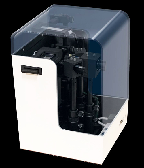
The researchers are hopeful the CytoPAN diagnostic tool (above) can be a valuable resource in developing countries and remote areas where patients face long wait times before receiving a cancer diagnosis. In these areas, diagnoses typically come after advanced symptoms, such as palpable mass lesions and malaise, become present, which can have a negative impact on patientoutcomes. And anatomic pathologists worldwide would benefit greatly from such an advance in cancer diagnostics. (Photo copyright: Jouha Min, Lip Ket Chin Center for Systems Biology, Massachusetts General Hospital.)
“Unfortunately, in many low- and middle-income countries, [breast cancer] diagnosis often takes an extraordinarily long time—up to a few months—due to a lack of specialists and limited laboratory infrastructure,” Hyungsoon Im, PhD, Assistant Professor at Harvard Medical School and one of the researchers involved in the project, told United Press International (UPI).
“From a public health aspect, it is critically important to develop new diagnostic methods that overcome these barriers,” he added.
Because FNA testing is less invasive than surgical biopsy collection, it has fewer complications and is generally considered safe. Thus, it is “feasible to be performed even in resource-limiting settings at much lower costs,” Im told UPI. “This could lead to earlier treatment and accelerate new drug testing in clinical trials.”
CytoPAN Testing and Additional Trials
The researchers tested CytoPAN on 68 breast cancer patients in South Korea.
“To determine the clinical utility of the approach,” they wrote in the published study, “we next conducted a prospective clinical study in which the FNA could be directly compared to conventional pathology results. We enrolled treatment-native patients at the Kyungpook National University Chilgok Hospital (Daegu, South Korea) and who were referred for primary surgery. All patients consented to have a preoperative breast FNA before clinically indicated surgery. The breast masses were visualized by ultrasound or computed tomography, and a coaxial needle was introduced through which FNA samples (CytoPAN) and core biopsies were obtained. Surgical specimens and/or core biopsies were processed by routine pathology and served as the gold standard.”
The CytoPAN platform detected the presence of breast cancer cells with a 100% accuracy, using as few as 50 harvested cells per collected specimen.
The test also successfully identified two key breast cancer biomarkers:
“We are also preparing additional trials in the US and other countries,” Im told UPI. “The success in those trials will (hopefully) accelerate … widespread adoption of the technology.”
The researchers are currently testing CytoPAN on a larger number of patients in Botswana, with funding from the US federal National Institutes of Health (NIH).
According to the American Cancer Society (ACS), approximately 300,000 individuals are diagnosed with breast cancer annually in the US. The Union for International Cancer Control (UICC) states on their website that, globally, there are more than two million new cases of breast cancer diagnosed each year. And more than 600,000 people died from breast cancer worldwide in 2018. A disproportionate number of those deaths occurred in developing countries that have limited resources to diagnose and treat the disease.
Additional Research for Other Applications in Cancer Testing and Pathology
The new CytoPAN technology requires minimal training, according to the researchers, and only costs about $5 per test kit. This is substantially less expensive than the price associated with other tests available on the market, UPI noted.
Though additional research and clinical trials are needed before CytoPAN will be available for widespread clinical use, a cost-effective, relatively non-invasive test that can accurately diagnose cancer within an hour would be transformational for anatomic pathology and, potentially, could save many lives.