This AI platform has the potential to also reduce workload of radiologists, but also of anatomic pathologists and oncologists allowing them to be more productive
When the UK’s National Health Service (NHS) recently tested an artificial intelligence (AI) platform’s ability to analyze mammograms, the AI found early signs of breast cancer that “human doctors” had previously missed, the BBC reported. This level of ability by AI might soon be adapted to aid overworked anatomic pathologists and cancer doctors in the United Kingdom.
Out of 10,000 mammograms MIA analyzed, the AI platform found “tiny signs of breast cancer in 11 women” which had not been spotted during earlier examinations, the BBC noted, adding that the cancers “were practically invisible to the human eye.”
This is a significant development in AI’s role in healthcare. Anatomic pathologists and clinical laboratory leaders will note that ongoing advancements in AI are enabling technology developers to apply their solutions to assessing radiology images, as well as in whole slide imaging used in digital pathology. In the UK, use of AI, the BBC noted, may also help ease doctor’s workloads.
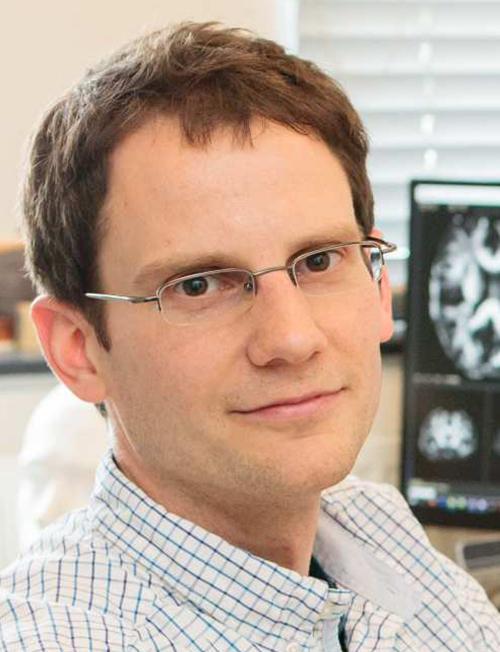
“This is just the beginning of our work with Kheiron,” said Ben Glocker, PhD (above), Professor in Machine Learning for Imaging at Imperial College London and Head of ML Research at Kheiron Medical, in a news release. “We are actively working on new methodologies for the safe deployment and continuous monitoring of MIA to support a US and UK rollout. We are working hard to make sure that as many women as possible will benefit from the use of this new technology within the next year.” AI tools such as MIA may soon take much of the load from anatomic pathologists and radiologists. (Photo copyright: Imperial College London.)
MIA Cloud-based AI Platform
Kheiron was founded in 2016 and MIA was named one of the seven biggest medical breakthroughs in 2023 by ABC News. A study conducted by Imperial College London in 2023 found that MIA “could significantly increase the early detection of breast cancers in a European healthcare setting by up to 13%,” according to an Imperial news release.
“The study was conducted over three phases (two pilot phases and a live roll-out). Overall across the three phases, the AI reader found 24 more cancers than the standard human reading—a 7% relative increase—and resulted in 70 more women recalled (0.28% relative increase),” the news release reported. “Of the additional recalls, six (initial pilot), 13 (extended pilot), and 11 (live use) additional cancers were found, increasing relative cancer detection rate by 13%, 10%, and 5% respectively. [The researchers] found that 83% of the additional cancers detected using MIA in real clinical practice were invasive, showing that MIA can detect cancers where early detection is particularly vital.”
Supported by Microsoft’s Azure Cloud, MIA came together over six years based on training encompassing millions of mammograms worldwide, Healthcare Digital reported.
“AI tools are generally pretty good at spotting symptoms of a specific disease if they are trained on enough data to enable them to be identified. This means feeding the program with as many different anonymized images of those symptoms as possible, from as diverse a range of people as possible,” Sarah Kerruish, Chief Strategy Officer, Kheiron, told Healthcare Digital.
MIA has been trained to “recognize subtle patterns and anomalies” that can point to “cancerous cells even in their earliest stages of development,” Dataconomy reported.
MIA Finds Early Cancer Signs
In the pilot study, MIA examined mammograms from 10,889 women. Each image had previously been reviewed by two radiologists, the BBC reported.
Findings include the following according to Healthcare Digital:
MIA “flagged” all people the physicians previously identified with symptoms.
The AI platform discovered 11 people with cancer the doctors did not identify.
The cancer MIA discovered—and the doctors did not—suggested cancer in early stages.
So, how did the doctors miss the cancer that MIA spotted? Gerald Lip, MD, Clinical Director for Breast Screening in North East Scotland who led the pilot study for the NHS, told Healthcare Digital, “part of the power of AI is it’s not prone to exhaustion or distraction.
“There is an element of fatigue,” he said. “You get disruptions, someone’s coming in, someone’s chatting in the background. There are lots of things that can probably throw you off your regular routine as well. And in those days when you have been distracted, you go, ‘how on earth did I miss that?’ It does happen.”
Lip is also the Chief Investigator in the Mammography Artificial Intelligence Project in the Industrial Center for Artificial Intelligence and Digital Diagnostics in Scotland.
“I see MIA as a friend and an augmentation to my practice,” he told Healthcare Digital. “MIA isn’t perfect. It had no access to patient history so [it] would flag cysts that had already been identified by previous scans and designated harmless.”
AI as a Safety Net
In the 2023 study, researchers from Imperial College London deployed MIA as an extra reader for mammograms of 25,065 women who visited screening sites in Hungary between April 2021 and January 2023, according to a news release.
“Our prospective real-world usage data in Hungary provides evidence for a significant, measurable increase of early breast cancer detection when MIA is used in clinical practice,” said Peter Kecskemethy, PhD, CEO and co-founder of Kheiron Medical, in the news release.
“Our study shows that AI can act as an effective safety net—a tool to prevent subtler signs of cancer from falling through the cracks,” said Ben Glocker, PhD, Professor in Machine Learning for Imaging at Imperial College London and Head of ML Research at Kheiron Medical, in the news release.
More studies are needed before MIA can be used in clinical settings. Nevertheless, use of AI in radiology—specifically mammograms—where the AI tool can identify very small cancers typically undetectable by radiologists, would be a boon to cancer doctors and the patients they treat.
So far, the research suggests that the AI-powered MIA has benefits to deployment in breast cancer screening. Eventually, it may also make impressive contributions to medical diagnosis and patient care, particularly if MIA eventually proves to be effective at analyzing the whole slide images used by anatomic pathologists.
Good behavior in federal prison by the disgraced founder of the now-defunct clinical laboratory company earned her the reduction in her original sentence of 11 years
Elizabeth Holmes, founder of failed clinical laboratory blood analysis company Theranos, continues to serve a lengthy term in prison after being convicted of multiple counts of fraud in 2022. However, now comes news that good behavior at her federal prison has shortened her sentence by nearly two years, according to NBC News.
The latest reduction took Holmes’ release from December 2032 to August 2032 in her “11-plus-year (135 month) prison sentence for wire fraud and conspiracy,” NBC reported, adding that Holmes, though Theranos, “defrauded investors out of hundreds of millions of dollars.”
Holmes entered FPC Bryan, a federal prison camp in Bryan, Texas, to begin serving her term in May 2023.
“Holmes had her sentence computation done within the first 30 days of arriving at Bryan,” Forbes reported. Given Good Conduct Time (GCT), Holmes was given 608 days off calculated from the start of her sentence. “If she were to incur a disciplinary infraction, some of those days can be taken away. Most all prisoners receive 54 days per year of GCT based on the sentence imposed,” Forbes added.
The Federal Bureau of Prisons (BOP) can additionally shave off up to a year through its Residential Drug Abuse Program (RDAP). “To qualify, the prisoner must not have a disqualifying offense, such as terrorism or gun charge, and voluntarily provided information that they had a drug or alcohol problem prior to their arrest. This disclosure has to be done prior to sentencing during the pre-sentence interview and must be also documented in the Presentence Report, a detailed report used by the BOP to determine things like classification and programming for the prisoner,” Forbes noted.
Additionally, the federal First Step Act, which President Trump signed into law in 2018, enables Holmes to “earn up to 365 days off any imposed sentence by participating in prison programming such as a self-improvement classes, a job, or religious activities,” Forbes reported.
Given the opportunities to shave time off her sentence, Holmes may ultimately serve just 66 months of her original 135 month sentence in federal prison.
Elizabeth Holmes (above) taken backstage at TechCrunch Disrupt San Francisco 2014 when Holmes was at the height of her fame and popularity. At this point, Theranos’ Edison blood testing device had not yet been shown to be a fake. But evidence was mounting as clinical laboratory scientists and anatomic pathologists became aware of the technology’s shortcomings. (Photo copyright: Max Morse/Wikimedia Commons.)
Fall of a Silicon Valley Darling
Theranos boasted breakthrough technology and became an almost overnight sensation in Silicon Valley when it burst onto the scene in 2003. Holmes, a then 19-year-old Stanford University dropout, claimed Theranos would “revolutionize the world of blood testing by reducing sample sizes to a single pin prick,” Quartz reported.
The height of the company saw Theranos valued at $9 billion, which came crashing down when the Wall Street Journal reported in 2015 that questionable accuracy and procedures were being followed by the company, CNN reported.
“From the moment Holmes concluded her presentation and stepped off the podium on Monday afternoon, she, her company, and her comments became the number one subject discussed by attendees in the halls between sessions and in the AACC exhibit hall,” Michel wrote, adding, “The executive team and the investors at Theranos have burned through their credibility with the media, the medical laboratory profession, and the public. In the future, the company’s claims will only be accepted if presented with scientific data developed according to accepted standards and reviewed by credible third parties. Much of this data also needs to be published in peer-reviewed medical journals held in highest esteem.”
Ultimately, investors who had jumped in early with financial support for Theranos were defrauded of hundreds of millions of dollars and Holmes was sentenced to 11 years/three months behind bars.
“Theranos had only ever performed roughly a dozen of the hundreds of tests it offered using its proprietary technology, and with questionable accuracy. It also came to light that Theranos was relying on third-party manufactured devices from traditional blood testing companies rather than its own technology,” CNN added.
The company shut down in 2018.
And so, the Elizabeth Holmes saga continues with reductions in her prison sentence for “good behavior.” The irony will likely not be lost on the anatomic pathologists, clinical laboratory scientists, and lab managers who followed the federal trials.
With further study, this research may provide clinical laboratories with a new proteomic biomarker for dementia screenings that identifies risk more than 10 years before symptoms appear
Researchers at the University of Warwick in the UK and Fudan University in Shanghai, China, identified four protein biomarkers in blood that they say can predict dementia up to 15 years before diagnosis. They say these biomarkers may lead to clinical laboratory blood tests that offer alternatives to costly brain scans and lumbar punctures for diagnosis of dementia.
The scientists “used the largest cohort of blood proteomics and dementia to date,” according to a University of Warwick news release. This included taking blood from 52,645 “healthy” people without dementia who participated in the UK Biobank—a population-based study cohort, the new release noted.
“The proteomic biomarkers are [easy] to access and non-invasive, and they can substantially facilitate the application of large-scale population screening,” said neurovegetative disease specialist Jin-tai Yu, MD, PhD, a professor at Fudan University and co-author of the study, in the news release.
“The advent of proteomics offers an unprecedented opportunity to predict dementia onset,” the researchers wrote.
“This is a well-conducted study that adds to what we know about changes in blood that occur very early in diseases that cause dementia, which will be important for early diagnosis in the future,” said Tara Spires-Jones, PhD, in a post from the Science Media Center in the UK. “However,” she added, “it is important to note that these are still scientific research studies and that there are currently no blood tests available for routine use that can diagnose dementia with certainty.
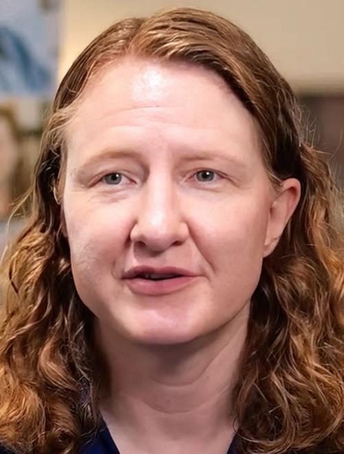
“Based on this study, it does seem likely that blood tests will be developed that can predict risk for developing dementia over the next 10 years, although individuals at higher risk often have difficulty knowing how to respond,” Suzanne Schindler, MD, PhD (above), told Reuters. Schindler, an Associate Professor of Neurology at Washington University in St. Louis, was not involved in the research. Clinical laboratories may soon have a new blood test for dementia. (Photo copyright: VJDementia.)
Predicting Onset of Dementia with 90% Accuracy
The researchers analyzed 52,645 blood samples from the UK Biobank (UKBB). The samples were collected between 2006 and 2010 from healthy individuals who at that time were without dementia.
By March 2023, 1,417 of the study participants had developed Alzheimer’s disease or some other form of dementia. The researchers looked at 1,463 proteins and identified four that were present in high levels among those people:
“Individuals with higher GFAP levels were 2.32 times more likely to develop dementia,” the researchers wrote in Nature Aging. “Notably, GFAP and LTBP2 were highly specific for dementia prediction. GFAP and NEFL began to change at least 10 years before dementia diagnosis.”
When adding known risk factors such as age, sex, and genetics, the researchers said they could predict onset of dementia with 90% accuracy, according to the University of Warwick news release.
“Our findings strongly highlight GFAP as an optimal biomarker for dementia prediction, even more than 10 years before the diagnosis, with implications for screening people at high risk for dementia and for early intervention,” the researchers wrote.
The news release also noted that smaller studies had already identified some of the proteins as potential biomarkers, “but this new research was much larger and conducted over several years.”
Further Validation Needed
Amanda Heslegrave, PhD, of the UK Dementia Research Institute, University College London described the UKBB as “an excellent resource” in the Science Media Center (SMC) post. However, she noted, it’s “a highly curated biobank and may not capture all populations that we need to know the risk for. The new biomarkers identified will need further validation before being used as screening tools.”
Another expert raised additional questions about the University of Warwick/Fudan University study in the SMC post.
“These results may help researchers understand the biological systems involved in the development of dementia,” said David Curtis, MD, PhD, of the UCL Genetics Institute at University College London. “However in my view the strengths of the reported associations are not really strong enough to say that these would form a useful test for predicting who will get dementia in the future.”
Conversely, Curtis pointed to other studies suggesting that phosphorylated tau (p-tau) proteins are better candidates for developing a simple blood test.
P-tau “provides a very good indicator of whether the pathological processes leading to Alzheimer’s disease are present in the brain,” he said. “When effective treatments for Alzheimer’s disease are developed it will be very helpful indeed to have simple blood tests—such as measuring phosphorylated tau—available in order to identify who could benefit.”
At least two blood tests based on the p-tau217 variant—from ALZpath and C2N—are currently available to US clinicians as laboratory developed tests (LDT).
The UK Biobank continues to be used by researchers both in the UK and abroad because of the full sets of data on large numbers of patients over many years. There are few other sources of such data elsewhere in the world. The UK Biobank is a large-scale biomedical database and research resource. It contains de-identified genetic, lifestyle and health information, and biological samples from 500,000 UK participants.
On its website, the UK Biobank states, “It is the most comprehensive and widely-used dataset of its kind and is globally accessible to approved researchers who are undertaking health-related research that is in the public interest, whether they are from academic, commercial, government or charitable settings.”
Thus, clinical laboratory managers and pathologists can expect a continuing stream of published studies that identify biomarkers associated with different health conditions and to see where the data used in these analyses came from the UK’s biobank.
As the cancer registry expands it will increasing become more useful to anatomic pathologists, histopathologists, oncologists, and even clinical laboratories
Oncologists, histopathologists, anatomic pathologists, and other cancer physicians now have a powerful new Wikipedia-style tumor registry to help them with their diagnoses and in educating patients on their specific types of cancer. Clinical laboratory managers may find it useful to understand the value this searchable database, and it can help their staff pathologists as well.
Free to use by both physicians and patients the World Tumor Registry (WTR) is designed “to minimize diagnostic errors by giving doctors a searchable online database of cancers that have been collected and categorized with cellular images collected from around the world,” Pittsburg-Post Gazette reported.
Prompt, accurate cancer diagnoses offer cancer patients the best chance for optimal treatment outcomes. However, many medical professionals around the globe do not have the training and resources to offer superior cancer diagnoses. That deficiency can translate to inferior treatment options and lower survival rates among cancer patients.
To help improve cancer diagnoses, pathologist Yuri E. Nikiforov, MD, PhD, Division Director, Molecular and Genomic Pathology, Vice Chair of the Department of Pathology, and Professor of Pathology, University of Pittsburgh, developed the WTR to provide educational and practical resources for individuals and organizations involved in cancer research.
Officially announced at the United States and Canadian Academy of Pathology (USCAP) annual convention, the WTR is an open-access catalog of digital microscopic images of human cancer types and subtypes.
The lower cost of technology and improved speed of access via the internet are technologies enabling this effort.
“We are creating sort of a Wikipedia for cancer images,” said Alyaksandr V. Nikitski, MD, PhD (above), Research Assistant Professor of Pathology, Division of Molecular and Genomic Pathology at Pittsburg School of Medicine and Administrative Director of the WTR, in an exclusive interview with Dark Daily. “Anyone in the world, if they can access the internet, can look at the well-annotated, diagnostic digital slides of cancer,” said Nikitski. Clinical laboratories may also find this new pathology tool useful. (Photo copyright: Alyaksandr V. Nikitski)
Minimizing Diagnostic Errors
Based in Pittsburgh, the WTR is freely available to anyone for viewing digital pathology slides of known cancer tumors as well as borderline and questionable cases. On the website, individuals can search for pictures of tumors in the registry by diagnosis, specific cohorts, and by microscopic features. Individuals may search further by tumor type and subtype to receive a picture of related tumors.
According to the WTR website, the mission of the nonprofit “is to minimize diagnostic errors, eliminate inequality in cancer recognition, diagnosis, and treatment in diverse populations, and improve outcomes by increasing access to the diagnostic pathology expertise and knowledge of microscopic characteristics of cancers that occur in different geographic, environmental, and socio-economic settings.”
This new comprehensive initiative will eventually encompass cancer images from all over the world.
“Let’s assume that I am a pathologist or a trainee who has little experience, or I don’t have access to collections of atypical tumors,” Nikitski explained. “I can view tumor collections online [in the WTR database] and check how typical and rare tumors look in various geographic regions and environmental settings.”
Once an image of a slide is selected, users will then receive a brief case history of the tumor in addition to such data as the age of the patient, their geographic location, sex, family history of the disease, and the size and stage of the tumor.
Increasing Probability of Correct Diagnosis
Pathologists and clinicians may also predict the probability of a particular diagnosis by searching under the microscopic feature of the database. This feature utilizes an innovative classifier known as PathDxFinder, where users may compare a slide from their lab to slides in the database by certain criteria. This includes:
After completing the questions above, the user presses the “predict diagnosis” button to receive the probability of cancer and most likely diagnosis based on the answers provided in the questionnaire.
WTR Editorial Boards
The WTR represents collections for each type of cancer site, such as lung or breast. A chairperson and editorial board are responsible for reviewing submitted slides before they are placed online. The editorial boards include 20 pathologists who are experts in diagnosing cancer categories, Nikitski explained.
Thousands of identified microscopic whole slide images (WSI) representing various types of cancer are deposited by the editors and other contributors to the project. The editorial board then carefully analyzes and compiles the data before posting the images for public viewing.
The editorial boards are located in five world regions:
Africa and the Middle East
Asia and Oceania
Central and South America
North America and Europe
Northern Asia
Any physicians or pathologists can contribute images to the database, by “simply selecting the editor of their region on the website, writing their name, and asking if they can submit tumor cases,” Nikitski stated.
“We have established a platform that allows pathologists to contact editors who are in the same geographic region,” he added.
Helping Physicians Identify Cancer Types
In a YouTube video, Nikiforov states that the WTR is an “educational nonprofit organization rooted in [the] beliefs that every cancer patient deserves accurate and timely diagnosis as the first and essential step in better treatment and outcomes.”
“We believe this can be achieved only when modern diagnostic tools and technologies are freely available to every physician and pathologist. Only when we understand how microscopic features of cancer vary in different geographic, environmental and ethnic populations, and only by integrating histopathology with clinical immunohistochemical and molecular genetic information for every cancer type,” he stated.
Since patient privacy is important, the database contains only basic data about patients, and all patient information is protected.
Launched in March, there are currently more than 400 thyroid tumor slides available to view in the online database. At the time of the announcement, the WTR platform was planned to be implemented in three phases:
Thyroid cancer (released in March of this year).
Lung cancer and breast cancer (anticipated to be completed by the third quarter of 2026).
Remaining cancers, including brain, soft tissue and bone, colorectal, head and neck, hematolymphoid, female genital, liver, pancreatic, prostate and male genital, skin, urinary system, pediatric, other endocrine cancers, and rare cancers (anticipated to be completed by the end of 2029).
“We believe that this resource will help physicians and pathologists practicing in small or big or remote medical centers to learn how cancer looks under a microscope in their own communities,” Nikiforov said in the video. “We also see WTR as a platform that connects physicians and scientists from different parts of the world who can work together to better understand and treat cancer.”
Catalogs like the World Tumor Registry might potentially create a pool of information that that could be mined by analytical and artificial intelligence (AI) platforms to ferret out new ways to improve the diagnosis of certain types of cancer and even enable earlier diagnoses.
“It is an extremely useful resource,” Nikitski said.
Anatomic pathologists will certainly find it so. And clinical laboratory managers may find the information useful as well when interacting with histopathologists and oncologists.
Clinical studies show that new ‘cell-free’ test can predict cardiovascular disease risk better than standard HDL cholesterol test
Researchers from the National Institutes of Health (NIH) have developed a diagnostic assay that measures how well high-density lipoprotein (HDL)—the so-called “good” cholesterol—is working in the body. And their findings could lead to new clinical laboratory tests that supplement standard HDL level testing to better determine a person’s risk for heart disease.
Cholesterol tests are among the most commonly performed assays by clinical laboratories. A new test that reveals how well HDL is working in the body would certainly boost a medical laboratory’s test requisition volume.
“Measuring HDL function is limited to research labs and isn’t conducive to large-scale testing by routine clinical laboratories. To try to solve that problem, researchers from NHLBI’s Lipoprotein Metabolism Laboratory created a new diagnostic test,” noted an NHLBI news release.
“This is going to quicken the pace of basic research,” said Edward B. Neufeld, PhD, who along with guest researcher Masaki Sato, PhD, developed the test. “It increases the number of samples that you can study. It increases the number of experiments you can do.”
Such a new cholesterol test would quickly become one of the most commonly performed clinical lab tests because just about every American who has a physical gets cholesterol tests as part of that process.
“Other people may modify this or come up with better versions, which is fine with us,” Edward Neufeld, PhD (above), NHLBI Staff Scientist, said in a news release. “We just really wanted to tackle this problem of evaluating HDL function.” Clinical laboratories may soon have a new cholesterol test to supplement standard HDL level testing. (Photo copyright: ResearchGate.)
Faster Answers Needed about HDL
According to the NIH, the goal should go beyond measuring level of HDL as part of a person’s annual physical. What is also needed is finding out whether HDL cholesterol is effectively doing certain tasks, such as removing extra cholesterol from arteries and transporting it to the liver.
The NHLBI’s new cell-free test may make it possible to step up large-scale clinical testing of HDL function, according to the news release. As it stands now, HDL function study has been limited to research labs where testing involves “harvesting cells in the lab [which] can take days to process,” according to NIH Record.
“Most studies to date that have assessed CAD (coronary artery disease) risk by HDL functionality still use the CEC (cellular cholesterol efflux capacity) in vitro assay and are based on the use of radioisotopes (3H-cholesterol) and cultured cells, which is very labor intensive and impractical to do in a clinical laboratory,” the researchers wrote in The Journal of Clinical Investigation. They also pointed out that CEC batch-to-batch variability does not fit clinical laboratories’ need for standardization.
Advantages of NHLBI’s Test
To overcome these barriers, the NHLBI researchers created an HDL-specific phospholipid efflux (HDL-SPE) assay that has certain advantages over current HDL function assessments done in research labs.
According to the NIH, the HDP-SPE assay:
Is easy to replicate in clinical labs.
Is more suited to automation and large samples.
Offers up results in about an hour.
Is a better predictor of cardiovascular disease risk than HDL cholesterol testing for CAD risk.
“We developed a cell-free, HDL-specific phospholipid efflux assay for the assessment of CAD risk on the basis of HDL functionality in whole plasma or serum. One of the main advantages of the HDL-SPE assay is that it can be readily automated, unlike the various CEC assays currently in use,” the authors noted in their paper.
Here is how the test is performed, according to the NIH:
Plasma with HDL is separated from the patient’s blood.
“Plasma is added to donor particles coated with a lipid mixture resembling plaque and a fluorescent-tagged phospholipid” that only HDL can remove.
The fluorescent signal by HDL is then measured.
A bright signal suggests optimal HDL lipid removal function, while a dim light means reduced function.
The test builds on the scientists’ previous findings and data. In creating the new assay they drew on data from:
A study of 50 severe CAD and 50 non-CAD people.
A Japanese study of 70 CAD and 154 non-CAD participants.
Examined association of HDL-SPE with cardiovascular disease in a study of 340 patients and 340 controls.
“We have established the HDL-SPE assay for assessment of the functional ability of HDL to efflux phospholipids. Our combined data consistently show that our relatively simple HDL-SPE assay captures a pathophysiologically relevant parameter of HDL function that is at least equivalent to the CEC assay in its association with prevalent and incident CAD,” the researchers concluded in The Journal of Clinical Investigation.
Test May Be Subject to New FDA Rule
While HDL cardiovascular-related research is moving forward, studies aimed at the therapeutic side need to pick up, NIH noted.
“Someday we may have a drug that modulates HDL and turns out to be beneficial, but right now we don’t have that,” said Alan Remaley MD, PhD, NHLBI Senior Investigator and Head of the Lipoprotein Metabolism Laboratory, in the news release.
It may be years before the HDL-SPE test is used in medical settings, the researchers acknowledged, adding that more studies are needed with inclusion of different ethnicities.
Additionally, in light of the recently released US Food and Drug Administration (FDA) final rule on regulation of laboratory developed tests (LDT), the company licensed to bring the test to market may need to submit the HDL-SPE assay to the FDA for premarket review and clearance. That could lengthen the time required for the developers to comply with the FDA before the test is used by doctors and clinical laboratories in patient care.
New technology could enable genetic scientists to identify antibiotic resistant genes and help physicians choose better treatments for genetic diseases
Genomic scientists at the Icahn School of Medicine at Mount Sinai Medical Center in New York City have developed what they call a “smart tweezer” that enables researchers to isolate a single bacterium from a patient’s microbiome in preparation for genetic sequencing. Though primarily intended for research purposes, the new technology could someday be used by clinical laboratories and microbiologists to help physicians diagnose chronic disease and choose appropriate genetic therapies.
The researchers designed their new technology—called mEnrich-seq—to improve the effectiveness of research into the complex communities of microorganisms that reside in the microbiomes within the human body. The discovery “ushers in a new era of precision in microbiome research,” according to a Mount Sinai Hospital press release.
“Imagine you’re a scientist who needs to study one particular type of bacteria in a complex environment. It’s like trying to find a needle in a large haystack,” said the study’s senior author Gang Fang, PhD (above), Professor of Genetics and Genomic Sciences at Icahn School of Medicine at Mount Sinai Medical Center, in a press release. “mEnrich-seq essentially gives researchers a ‘smart tweezer’ to pick up the needle they’re interested in,” he added. Might smart tweezers one day be used to help physicians and clinical laboratories diagnose and treat genetic diseases? (Photo copyright: Icahn School of Medicine.)
Addressing a Technology Gap in Genetic Research
Any imbalance or decrease in the variety of the body’s microorganisms can lead to an increased risk of illness and disease.
In researching the microbiome, many scientists “focus on studying specific types of bacteria within a sample, rather than looking at each type of bacteria present,” the press release states. The limitation of this method is that a specific bacterium is just one part of a complicated environment that includes other bacteria, viruses, fungi and host cells, each with their own unique DNA.
“mEnrich-seq effectively distinguishes bacteria of interest from the vast background by exploiting the ‘secret codes’ written on bacterial DNA that bacteria use naturally to differentiate among each other as part of their native immune systems,” the press release notes. “This new strategy addresses a critical technology gap, as previously researchers would need to isolate specific bacterial strains from a given sample using culture media that selectively grow the specific bacterium—a time-consuming process that works for some bacteria, but not others. mEnrich-seq, in contrast, can directly recover the genome(s) of bacteria of interest from the microbiome sample without culturing.”
Isolating Hard to Culture Bacteria
To conduct their study, the Icahn researchers used mEnrich-seq to analyze urine samples taken from three patients with urinary tract infections (UTIs) to reconstruct Escherichia coli (E. Coli) genomes. They discovered their “smart tweezer” covered more than 99.97% of the genomes across all samples. This facilitated a comprehensive examination of antibiotic-resistant genes in each genome. They found mEnrich-seq had better sensitivity than standard study methods of the urine microbiome.
They also used mEnrich-seq to selectively examine the genomes of Akkermansia muciniphila (A. muciniphila), a bacterium that colonizes the intestinal tract and has been shown to have benefits for obesity and Type 2 diabetes as well as a response to cancer immunotherapies.
“Akkermansia is very hard to culture,” Fang told GenomeWeb. “It would take weeks for you to culture it, and you need special equipment, special expertise. It’s very tedious.”
mEnrich-seq was able to quickly segregate it from more than 99.7% of A. muciniphila genomes in the samples.
Combatting Antibiotic Resistance Worldwide
According to the press release, mEnrich-seq could potentially be beneficial to future microbiome research due to:
Cost-Effectiveness: It offers a more economical approach to microbiome research, particularly beneficial in large-scale studies where resources may be limited.
Broad Applicability: The method can focus on a wide range of bacteria, making it a versatile tool for both research and clinical applications.
Medical Breakthroughs: By enabling more targeted research, mEnrich-seq could accelerate the development of new diagnostic tools and treatments.
“One of the most exciting aspects of mEnrich-seq is its potential to uncover previously missed details, like antibiotic resistance genes that traditional sequencing methods couldn’t detect due to a lack of sensitivity,” Fang said in the news release. “This could be a significant step forward in combating the global issue of antibiotic resistance.”
More research and clinical trials are needed before mEnrich-seq can be used in the medical field. The Icahn researchers plan to refine their novel genetic tool to improve its efficiency and broaden its range of applications. They also intend to collaborate with physicians and other healthcare professionals to validate how it could be used in clinical environments.
Should all this come to pass, hospital infection control teams, clinical laboratories, and microbiology labs would welcome a technology that would improve their ability to detect details—such as antibiotic resistant genes—that enable a faster and more accurate diagnosis of a patient’s infection. In turn, that could contribute to better patient outcomes.