Skin patch technologies could enable clinical laboratories to monitor patients’ vitals and report to medical professionals in real time
Pathologists and clinical laboratory leaders have read many Dark Daily ebriefings on the development of skin patches over the years that do everything from monitoring fatigue in the military to being a complete lab-on-skin technology. Now, researchers at the University of California San Diego (UCSD) have developed a wearable patch that can monitor cardiovascular signals and other various biochemical levels in the body simultaneously.
The researchers believe there is enormous potential for such a patch in helping patients monitor conditions such as hypertension or diabetes. They also foresee a scenario where the patch could be used in settings where vitals must be constantly monitored. They hope to develop future versions of the patch that can detect more biomarkers within the body.
“This type of wearable would be very helpful for people with underlying medical conditions to monitor their own health on a regular basis,” Lu Yin, a PhD student and co-first author of the study, told New Atlas. “It would also serve as a great tool for remote patient monitoring, especially during the COVID-19 pandemic when people are minimizing in-person visits to the clinic,” she added.
Combining Precision Medicine with Telehealth and the Internet of Things
About the size of a postage stamp and consisting of stretchy polymers that conform to the skin, the UCSD patch monitors blood pressure and contains sensors that measure different biochemical levels in the body, such as:
The sensors are carefully arranged on the patch to eliminate interference between the signals, noted a UCSD press release.
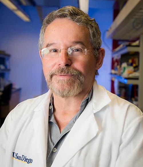
In their published research, the UCSD researchers wrote of their new skin patch monitoring device, “Intertwined with concepts of telehealth, the internet of medical things, and precision medicine, wearable sensors offer features to actively and remotely monitor physiological parameters. Wearable sensors can generate data continuously without causing any discomfort or interruptions to daily activity, thus enhancing the self-monitoring compliance of the wearer, and improving the quality of patient care.” (Photo copyright: University of California San Diego.)
“Each sensor provides a separate picture of a physical or chemical change. Integrating them all in one wearable patch allows us to stitch those different pictures together to get a more comprehensive overview of what’s going on in our bodies,” said Sheng Xu, PhD, Principle Investigator, Xu Research Group at UCSD, Assistant Professor in the Department of NanoEngineering Department, and a co-first author of the study, in the press release.
The UCSD researchers developed their skin patch to monitor specific biomarkers that can affect blood pressure.
“Let’s say you are monitoring your blood pressure and you see spikes during the day and think that something is wrong,” co-first author Juliane Sempionatto, PhD, a postdoctoral researcher at California Institute of Technology (Caltech) and co-first author of the study, told New Atlas. “But a biomarker reading could tell you if those spikes were due to an intake of alcohol or caffeine. This combination of sensors can give you that type of information,” she added.
The blood pressure sensor sits near the center of the patch and consists of a set of small transducers welded to the patch via a conductive link. Voltage applied to the transducers send ultrasound waves through the body which bounce off arteries and create echoes that are detected by the sensor and converted into an accurate blood pressure reading.
The chemical sensor releases the drug pilocarpine into the skin to induce sweat and then measures the chemicals contained in the sweat to provide readings of certain biochemical levels.
The glucose sensor located in the patch emits a mild electrical current to the body that stimulates the release of interstitial fluid and then reads the glucose level in that fluid.
“The novelty here is that we take completely different sensors and merge them together on a single small platform as small as a stamp,” Joseph Wang, D.Sc, SAIC Endowed Chair, Distinguished Professor of NanoEngineering, Director of the Center for Wearable Sensors at UCSD, and co-author of the study told New Atlas. “We can collect so much information with this one wearable and do so in a non-invasive way, without causing discomfort or interruptions to daily activity.” (Photo copyright: University of Southern California San Diego.)
Skin Patch Measurements Closely Match Those of Traditional Devices
Test subjects wore the patch on their neck while performing various combinations of the following tasks:
exercising on a stationary bicycle,
eating a high-sugar meal,
drinking an alcoholic beverage, and
drinking a caffeinated beverage.
The results of the measurements taken from the patch closely matched measurements collected by traditional monitoring devices such as a:
For now, the patch must be connected to an external power source which transmits the reading to a counter-top machine, but the researchers hope to create a wireless version in the future.
“There are opportunities to monitor other biomarkers associated with various diseases,” Sempionatto said in the UCSD press release. “We are looking to add more clinical value to this device.”
Other Similar Skin Patch Monitoring Technologies
Though an important breakthrough, the UCSD’s device is not the first skin patch monitor to be developed.
Multiple research and clinical studies are underway that hope to prove the accuracy and safety of wearable devices at detecting and monitoring certain health conditions. It’s a worthy goal.
Skin patches, such as the one created at UCSD, could enable clinical laboratories to provide value-added service to medical professionals and patients alike. Medical labs could potentially monitor skin patch readings in real-time and notify physicians and patients of changes in biomarkers that require attention.
Further, as this technology is developed, it will likely find a ready market with the latest generation of consumers who are more willing than previous generations to buy their own diagnostic tests for home use. These “next-generation” healthcare consumers have demonstrated their willingness to use Apple watches, Fitbits, and similar wearable devices to monitor their condition during exercise and other health metrics.
Pathologists and clinical laboratory managers should not overlook the potential for robust consumer demand to accelerate development and market adoption of such skin patches.
This is yet another example that dogs can be highly accurate screeners for disease. But are they ready to be included in clinical laboratory diagnostic tests?
Thailand researchers have trained dogs to screen for COVID-19 infections in humans, despite the country’s “spicy and flavorful cuisine,” the AP reported. This is just the latest example of a country using dogs to identify individuals who are infected with the SARS-CoV-2 coronavirus. Clinical laboratory managers and pathologists have seen other examples of dogs being trained to identify different diseases or health conditions.
In fact, dogs have been shown to be highly accurate at spotting disease in humans and the practice is becoming common worldwide. But could dogs achieve the required clinical accuracy and reproducibility in detecting disease for the procedure to be translated into clinical practice?
Smelling Disease as a Clinical Laboratory Diagnostic
Clinical laboratory professionals are quite familiar with the concept of the human body producing volatile chemicals that can serve as biomarkers for disease or illness. Dark Daily has previously reported on multiple breath/aroma-based diagnostic clinical laboratory tests going as far back as 2013.
But it is in the use of dogs to spot COVID-19 infections in humans where this type of breath/aroma-based diagnostic test research is making a notable impact.
“Even if this approach were not warranted as a clinical diagnostic procedure, trained dogs could be deployed at airports, train stations, sporting events, concerts, and other public places to identify individuals who may be positive for SARS-CoV-2, the coronavirus that causes the COVID-19 illness,” we wrote. “Such an approach would make it feasible to ‘screen’ large numbers of people as they are on the move. Those individuals could then undergo a more precise medical laboratory test as confirmation of infections.”
According to the researchers, individuals with a COVID-19 infection emit a unique odor that is present in sweat samples. The six Labrador retrievers used in the research were able to detect the presence of COVID-19 with an impressive 95% accuracy rate in more than 1,000 samples presented to them, the AP reported.
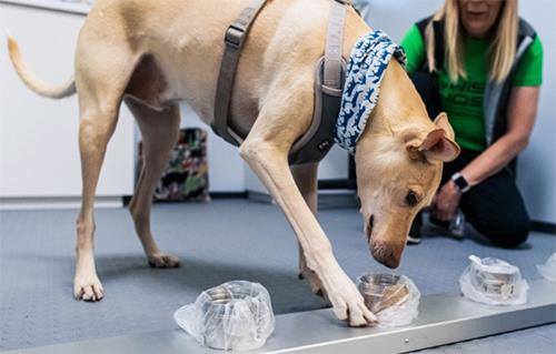
A Labrador Retriever named Bobby (above) sniffs sample of human sweat through containers to detect COVID-19 coronavirus at Veterinary Faculty, Chulalongkorn University in Bangkok. Thailand has deployed a canine virus detection squad to help provide a fast and effective way of identifying people with COVID-19 as the country faces a surge in cases, with clusters found in several crowded slum communities and large markets. Clinical laboratory professionals and pathologists will find it interesting that the dogs are given a sample of sweat, each presented in a unique container. Thus, the dogs never are in the presence of the humans who provided the specimens. (Photo and caption copyright: AP/Sakchai Lalit)
To perform the study, the scientists placed sweat samples in metal containers and allowed the dogs to sniff each sample. If no trace of the infection was present, the dogs simply walked past the container. If the disease was detected in a particular sample, the dogs would sit down in front of the container.
Would Spicy Food Interfere with Dogs’ Ability to Detect COVID-19?
The head of the research team, Professor Kaywalee Chatdarong, PhD, noted that other countries also have been using canines to detect the presence of COVID-19. She did have some concerns that the utilization of dogs for this purpose may not work in Thailand due to their often-spicy cuisine. However, since the samples used were from students and faculty at the university, as well as people from the surrounding area, the cuisine did not seem to affect the study results, the AP reported.
Thailand is facing a surge in COVID-19 cases with recent clusters reported at construction sites, crowded neighborhoods, and large markets. The research team plans to use the canines in mobile units in communities suspected of being hotspots for the disease.
A major plus of using dogs to sniff out the disease from sweat samples is the ability to test people who may not be able to get out of their homes to be tested.
“People can simply put cotton balls underneath their armpits to collect sweat samples and send them to the lab,” Suwanna Thanaboonsombat, a volunteer who collects samples and brings them to the clinical laboratory for testing, told the AP. “And the result is quite accurate.”
According to the US Centers for Disease Control and Prevention (CDC), dogs can become infected with the SARS-CoV-2 coronavirus. However, their chances of transmitting the disease to humans is extremely low. Nevertheless, to ensure the dogs do not become infected with COVID-19 themselves, the researchers designed the sample containers to avoid contact between the samples and the dogs’ noses.
Living Animals Come with Limitations
While dogs can provide a quick and inexpensive method of testing for COVID-19, they do have limitations.
“5 p.m. is their dinner time. When it’s around 4:50, they will start to be distracted. So, you can’t really have them work anymore,” Chatdarong told the AP. “And we can’t have them working after dinner either because they need a nap. They are living animals and we do have to take their needs and emotions into consideration. But for me, they are heroes and heroines.”
Using Dogs to Detect COVID-19 in Other Countries
Last fall, the Helsinki Airport in Finland announced it would use a team of trained dogs to detect the presence of COVID-19 among visitors to the airport to ensure the health and safety of its customers and their families, and to help prevent the spread of SARS-CoV-2 in Finland.
Being tested for the coronavirus at the Helsinki airport in Finland does not require direct contact with a dog. Individuals simply need to swipe their skin with a test wipe and drop the wipe into a cup. The cup is then given to a dog that is working in a separate booth (shown above), which protects both the dog and the dog’s handler from contamination. All tests are processed anonymously and anyone testing positive for COVID-19 is directed to a health information point located at the airport. (Photo copyright: Finavia.)
“We are among the pioneers. As far as we know no other airport has attempted to use canine scent detection on such a large scale against COVID-19,” said Airport Director Ulla Lettijeff in a Finavia press release. “This might be an additional step forward on the way to beating COVID-19.”
In addition to being “man’s best friend,” dogs serve valuable purposes in the medical community. Their strong sense of smell may render them useful in the detection of and fight against illnesses, including COVID-19.
Whether the performance and accuracy of individual dogs can be validated with acceptable quality control (QC) procedures remains to be seen. Medical laboratory managers and pathologists understand the challenges presented with demonstrating accuracy and reproducibility with this method of diagnostic testing. That obstacle has prevented research outcomes from being translated into clinical practice.
New non-invasive test could replace traditional painful spinal taps and clinical laboratory fluid analysis for diagnosis of Parkinson’s disease
Scientists at AXIM Biotechnologies of San Diego have added another specimen that can be collected non-invasively for rapid, point-of-care clinical laboratory testing. This time it is tears, and the diagnostic test is for Parkinson’s disease (PD).
The new assay measures abnormal alpha-synuclein (a-synuclein), a protein that is a biomarker for Parkinson’s, according to an AXIM news release which also said the test is the first rapid test for PD.
“The revolutionary nature of AXIM’s new test is that it is non-invasive, inexpensive, and it can be performed at a point of care. It does not require a lumbar puncture, freezing, or sending samples to a lab. AXIM’s assay uses a tiny tear drop versus a spinal tap to collect the fluid sample and the test can be run at a doctor’s office with quantitative results delivered from a reader in less than 10 minutes,” the news release notes.
“Furthermore, emerging evidence shows that a-synuclein assays have the potential to differentiate people with PD from healthy controls, enabling the potential for early identification of at-risk groups,” the news release continues. “These findings suggest a crucial role for a-synuclein in therapeutic development, both in identifying pathologically defined subgroups of people with Parkinson’s disease and establishing biomarker-defined at-risk cohorts.”
This is just the latest example of a disease biomarker that can be collected noninvasively. Other such biomarkers Dark Daily has covered include:
“With this new assay, AXIM has immediately become a stakeholder in the Parkinson’s disease community, and through this breakthrough, we are making possible new paradigms for better clinical care, including earlier screening and diagnosis, targeted treatments, and faster, cheaper drug development,” said John Huemoeller, CEO, AXIM (above), in a news release. Patients benefit from non-invasive clinical laboratory testing. (Photo copyright: AXIM Biotechnologies.)
Fast POC Test versus Schirmer Strip
AXIM said it moved forward with its novel a-synuclein test propelled by earlier tear-related research that found “a-synuclein in its aggregated form can be detected in tears,” Inside Precision Medicine reported.
But that research used what AXIM called the “outdated” Schirmer Strip method to collect tears. The technique involves freezing tear samples at -80 degrees Celsius (-112 Fahrenheit), then sending them to a clinical laboratory for centrifugation for 30 minutes; quantifying tear protein content with a bicinchoninic acid assay, and detecting a-synuclein using a plate reader, AXIM explained.
Alternatively, AXIM says its new test may be performed in doctors’ offices and offers “quantitative results delivered from a reader in less than 10 minutes.”
“Our proven expertise in developing tear-based diagnostic tests has led to the development of this test in record speed, and I’m extremely proud of our scientific team for their ability to expand our science to focus on such an important focus area as Parkinson’s,” said John Huemoeller, CEO, AXIM in the news release.
“This is just the beginning for AXIM in this arena,” he added. “But I am convinced when pharmaceutical companies, foundations, and neurologists see how our solution can better help diagnose Parkinson’s disease in such an expedited and affordable way, we will be at the forefront of PD research, enabling both researchers and clinicians a brand-new tool in the fight against PD.”
One of those tests was “a lateral flow diagnostic for point-of-care use that measures the level of lactoferrin proteins in tear fluid, which work to protect the surface of the eye. … Axim said that low lactoferrin levels have also been linked to Parkinson’s disease and that the assay can be used alongside its alpha-synuclein test,” Fierce Biotech noted.
“It made sense to try and look at the proteinaceous [consisting of or containing protein] constituents of tear fluid,” Lew told Neurology Live. “Tear fluid is easy to collect. It’s noninvasive, inexpensive. It’s not like when you do a lumbar puncture, which is a much more involved ordeal. There’s risk of contamination with blood (saliva is dirty) issues with blood and collection. [Tear fluid analysis] is much safer and less expensive to do.”
In Biomarkers in Medicine, Lew et al noted why tears make good biomarkers for Parkinson’s disease, including “the interconnections between the ocular [eye] surface system and neurons affected in Parkinson’s disease.”
The researchers also highlighted “recent data on the identification of tear biomarkers including oligomeric α-synuclein, associated with neuronal degeneration in PD, in tears of PD patients” and discussed “possible sources for its release into tears.”
Future Clinical Laboratory Testing for Parkinson’s
Parkinson’s disease is the second most common neurodegenerative disorder after Alzheimer’s. It affects nearly one million people in the US. About 1.2 million people may have it by 2030, according to the Parkinson’s Foundation.
Thus, an accurate, inexpensive, non-invasive diagnostic test that can be performed at the point of care, and which returns clinical laboratory test results in less than 10 minutes, will be a boon to physicians who treat PD patients worldwide.
Clinical laboratory managers and pathologists may want to follow AXIM’s future research to see when the diagnostic test may become available for clinical use.
Founder of now defunct clinical laboratory testing company was supposed to report to prison April 27, but a last-minute legal challenge has delayed that judge’s order
Anatomic pathologists and clinical laboratory leaders who are following the continuing saga of Theranos and Elizabeth Holmes may be interested to learn that the former CEO’s attorneys are making last-minute legal moves to delay her prison sentence while she appeals her guilty verdict. At the same time, Holmes appears to be on a mission to revamp her public image.
Apparently, the twists and turns in Holmes’ never-ending story are not yet over when it comes to Theranos, its maligned clinical laboratory technology, and the company’s convicted founder.
On May 7, The New York Times (NYT) profiled Holmes in a massive, 5,000-word story that attempted to portray her as a flawed businessperson who now prefers a simpler life with her partner and two young children.
“I made so many mistakes and there was so much I didn’t know and understand, and I feel like when you do it wrong, it’s like you really internalize it in a deep way,” disgraced Theranos founder Elizabeth Holmes recently told The New York Times. Anatomic pathologists and clinical laboratory directors impacted by the revelation that Theranos hide the fact that its blood testing technology was faulty may not sympathize with Holmes’ position. (Photo copyright: Stuart Isett/Fortune Global Forum.)
Legal Team Secures Last-Minute Delay in Holmes’ Surrender
Holmes admitted to the news outlet that the deep voice she used in public, along with her black turtleneck sweaters, were part of a character she created.
“I believed it would be how I would be good at business and taken seriously and not taken as a little girl or a girl who didn’t have good technical ideas,” Holmes told the NYT. “Maybe people picked up on that not being authentic, since it wasn’t.”
However, on April 26, the 9th Circuit Court of Appeals stayed her surrender date until that court rules on Holmes’ latest bid to stay free while she appeals her conviction, The Washington Post reported.
Just days earlier on April 10, a district court judge ruled that Holmes would not stay free while her appeal progresses. The 9th Circuit announcement curtailed the district court ruling. It is not known when the 9th Circuit will issue a decision in the matter.
New York Times Reports on Holmes’ Change in Personality
The somewhat odd New York Times profile of Holmes varied between reflections on her past crimes and on her current personal life, where she is known as “Liz.”
“In case you’re wondering, Holmes speaks in a soft, slightly low, but totally unremarkable voice—no hint of the throaty contralto she used while running her blood-testing startup Theranos, now defunct,” the NYT reported.
Holmes still lives in California with her partner, Billy Evans (whose parents own a luxury hotel chain), and their two children: a son who is almost two years old and a daughter born in February. She works at home for a rape-crisis hotline.
Balwani’s Role in Theranos Again Publicly Debated
In the NYT interview, Holmes talked about being raped while a student at Stanford University and about alleged abuse from her Theranos business partner and former lover, Ramesh “Sunny” Balwani.
Balwani, Theranos’ former President and Chief Operating Officer, began his 12-year, 11-month prison sentence on April 20 in a Southern California facility for his role in defrauding Theranos investors, KTVU TV reported. Balwani has also appealed his conviction on the 12 fraud charges.
Holmes reiterated to the NYT past statements she made in court that Balwani allegedly exerted social and sexual control over her when they both worked at Theranos and were in a romantic relationship.
“She lived by entrepreneurial tenets that she said Balwani told her she needed to follow in order to succeed,” the NYT reported. “These included not sleeping for more than five hours, going vegan, getting to the office daily by 5 a.m., no alcohol.”
“[I] deferred to [Balwani] in the areas he oversaw because I believed he knew better than I did,” including on clinical lab activities at Theranos, Holmes said.
Balwani’s attorneys dismissed Holmes’ allegations, as they have in the past.
Clinical laboratory professionals can reasonably make two broad observations from the continuing saga of Theranos and Elizabeth Holmes:
Justice for healthcare crimes is often deferred for those who have influence and money.
Holmes’ image overhaul may be a last-ditch effort to sway public opinion about her, in the event that she receives a new jury trial as a result of her appeal.
Dark Daily will continue to keep you updated on further developments in this case.
Dogs’ acute sense of smell can even surpass effectiveness of some clinical laboratory testing in detecting certain diseases in humans
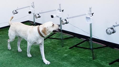
When it comes to COVID-19 testing, a recent Italian study demonstrates that trained dogs can detect SARS-CoV-2 with accuracy comparable to rapid molecular tests used in clinical laboratories. The researchers wanted to determine if dogs could be more effective at screening people for COVID-19 at airports, schools, and other high-traffic environments as a way to detect the coronavirus and reduce the spread of this infectious disease.
Scientists at the State University of Milan in Italy conducted a study that shows dogs can be trained to accurately identify the presence of the COVID-19 infection from both biological samples and by simply smelling an individual.
For their validation study, the Italian team trained three dogs named Nala, Otto, and Helix, “to detect the presence of SARS-CoV-2 in sweat samples from infected people. At the end of the training, the dogs achieved an average sensitivity of 93% and a specificity of 99%, showing a level of accuracy highly consistent with that of the RT-PCR [reverse transcription polymerase chain reaction] used in molecular tests and a moderate to strong reproducibility over time,” Nature reported.
RT-PCR tests are the current gold-standard for SARS-CoV-2 detection. This is yet another example of scientists training dogs to smell a disease with “acceptable” accuracy. This time for COVID-19.
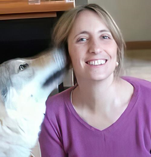
“We only recruited dogs that showed themselves predisposed and positively motivated to carry out this type of activity. One of the fundamental aspects was not to cause stress or anxiety in the subjects used,” Federica Pirrone, PhD (above), Associate Professor, Department of Veterinary Medicine and Animal Sciences, University of Milan, and one of the authors of the study told Lifegate. “Training always takes place using positive reinforcement of a food nature: whether it’s a particularly appetizing morsel, a biscuit, or something that associates the dog’s search with a rewarding prize.” In some instances, dogs have been shown to be as good or more effective at detecting certain diseases than clinical laboratory testing. (Photo copyright: Facebook.)
Dogs More Accurate than Rapid Antigen Testing
Nala and four other dogs (Nim, Hope, Iris and Chaos) were later trained by canine technicians from Medical Detection Dogs Italy (MDDI) to identify the existence of the SARS-CoV-2 virus by directly smelling people waiting in line in pharmacies to get a nasal swab to test for the coronavirus.
Working with their handlers, the five dogs accurately signaled the presence or absence of the virus with 89% sensitivity and 95% specificity. That rate is “well above the minimum required by the WHO [World Health Organization] for rapid swabs for SARS-CoV-2,” according to Nature.
“The results of studies published so far on the accuracy of canine smell in detecting the presence of SARS-CoV-2 in biological samples (e.g., saliva, sweat, urine, trachea-bronchial secretions) from infected people suggest that sniffer dogs might reach percentages of sensitivity and specificity comparable to, or perhaps even higher, than those of RT-PCR,” the scientists wrote in Scientific Reports.
“However, although most of these studies are of good quality, none of them provided scientific validation of canine scent detection, despite this being an important requirement in the chemical analysis practice. Therefore, further applied research in this field is absolutely justified to provide definitive validation of this biodetection method,” the researchers concluded.
Other Studies into Using Dogs for Detecting Disease
Scientists from the Division of Biological and Health Sciences, Department of Agriculture and Livestock at the University of Sonora; and the Canine Training Center Obi-K19, both in Hermosillo, Mexico, conducted the study “as part of a Frontiers of Science Project of the National Council of Science and Technology (CONACYT), in which in addition to analyzing sweat compounds, trained dogs are put to sniff the samples and make detections in people who show symptoms or could be positive for coronavirus,” Mexico Daily Post reported.
The researchers trained four dogs with sweat samples and three dogs with saliva samples of COVID-19 positive patients. The samples were obtained from a health center located in Hermosillo, Sonora, in Mexico. The dogs were restricted to spend five minutes per patient and the researchers calculated the performance of the dogs by measuring sensitivity, specificity, and their 95% confidence intervals (CI).
The researchers concluded that all four of the dogs could detect COVID-19 from either sweat or saliva samples “with sensitivity and specificity rates significantly different from random [sampling] in the field.” According to the Frontiers in Medicine study, the researchers found their results promising because, they said, it is reasonable to expect the detection rate would improve with longer exposure to the samples.
The objective of the Mexican researchers is for the dogs to ultimately reach the sensitivity range requested by WHO for the performance of an antigen test, which is at least 80% sensitivity and 97% specificity. If that goal is achieved, dogs could become important partners in the control of the COVID-19 pandemic, the scientists wrote.
Data obtained so far from these studies indicate that biosensing dogs may represent an effective method of screening for COVID-19 as well as other diseases. More studies and clinical trials are needed before the widespread use of dogs might become feasible. Nevertheless, scientists all over the world are finding that Man’s best friend can be a powerful ally in the fight against the spread of deadly diseases.
In the meantime, the gold standard in COVID-19 testing will continue to be the FDA-cleared assays used by clinical laboratories throughout the United States.