With further study, this research may provide clinical laboratories with a new proteomic biomarker for dementia screenings that identifies risk more than 10 years before symptoms appear
Researchers at the University of Warwick in the UK and Fudan University in Shanghai, China, identified four protein biomarkers in blood that they say can predict dementia up to 15 years before diagnosis. They say these biomarkers may lead to clinical laboratory blood tests that offer alternatives to costly brain scans and lumbar punctures for diagnosis of dementia.
The scientists “used the largest cohort of blood proteomics and dementia to date,” according to a University of Warwick news release. This included taking blood from 52,645 “healthy” people without dementia who participated in the UK Biobank—a population-based study cohort, the new release noted.
“The proteomic biomarkers are [easy] to access and non-invasive, and they can substantially facilitate the application of large-scale population screening,” said neurovegetative disease specialist Jin-tai Yu, MD, PhD, a professor at Fudan University and co-author of the study, in the news release.
“The advent of proteomics offers an unprecedented opportunity to predict dementia onset,” the researchers wrote.
“This is a well-conducted study that adds to what we know about changes in blood that occur very early in diseases that cause dementia, which will be important for early diagnosis in the future,” said Tara Spires-Jones, PhD, in a post from the Science Media Center in the UK. “However,” she added, “it is important to note that these are still scientific research studies and that there are currently no blood tests available for routine use that can diagnose dementia with certainty.
“Based on this study, it does seem likely that blood tests will be developed that can predict risk for developing dementia over the next 10 years, although individuals at higher risk often have difficulty knowing how to respond,” Suzanne Schindler, MD, PhD (above), told Reuters. Schindler, an Associate Professor of Neurology at Washington University in St. Louis, was not involved in the research. Clinical laboratories may soon have a new blood test for dementia. (Photo copyright: VJDementia.)
Predicting Onset of Dementia with 90% Accuracy
The researchers analyzed 52,645 blood samples from the UK Biobank (UKBB). The samples were collected between 2006 and 2010 from healthy individuals who at that time were without dementia.
By March 2023, 1,417 of the study participants had developed Alzheimer’s disease or some other form of dementia. The researchers looked at 1,463 proteins and identified four that were present in high levels among those people:
“Individuals with higher GFAP levels were 2.32 times more likely to develop dementia,” the researchers wrote in Nature Aging. “Notably, GFAP and LTBP2 were highly specific for dementia prediction. GFAP and NEFL began to change at least 10 years before dementia diagnosis.”
When adding known risk factors such as age, sex, and genetics, the researchers said they could predict onset of dementia with 90% accuracy, according to the University of Warwick news release.
“Our findings strongly highlight GFAP as an optimal biomarker for dementia prediction, even more than 10 years before the diagnosis, with implications for screening people at high risk for dementia and for early intervention,” the researchers wrote.
The news release also noted that smaller studies had already identified some of the proteins as potential biomarkers, “but this new research was much larger and conducted over several years.”
Further Validation Needed
Amanda Heslegrave, PhD, of the UK Dementia Research Institute, University College London described the UKBB as “an excellent resource” in the Science Media Center (SMC) post. However, she noted, it’s “a highly curated biobank and may not capture all populations that we need to know the risk for. The new biomarkers identified will need further validation before being used as screening tools.”
Another expert raised additional questions about the University of Warwick/Fudan University study in the SMC post.
“These results may help researchers understand the biological systems involved in the development of dementia,” said David Curtis, MD, PhD, of the UCL Genetics Institute at University College London. “However in my view the strengths of the reported associations are not really strong enough to say that these would form a useful test for predicting who will get dementia in the future.”
Conversely, Curtis pointed to other studies suggesting that phosphorylated tau (p-tau) proteins are better candidates for developing a simple blood test.
P-tau “provides a very good indicator of whether the pathological processes leading to Alzheimer’s disease are present in the brain,” he said. “When effective treatments for Alzheimer’s disease are developed it will be very helpful indeed to have simple blood tests—such as measuring phosphorylated tau—available in order to identify who could benefit.”
At least two blood tests based on the p-tau217 variant—from ALZpath and C2N—are currently available to US clinicians as laboratory developed tests (LDT).
The UK Biobank continues to be used by researchers both in the UK and abroad because of the full sets of data on large numbers of patients over many years. There are few other sources of such data elsewhere in the world. The UK Biobank is a large-scale biomedical database and research resource. It contains de-identified genetic, lifestyle and health information, and biological samples from 500,000 UK participants.
On its website, the UK Biobank states, “It is the most comprehensive and widely-used dataset of its kind and is globally accessible to approved researchers who are undertaking health-related research that is in the public interest, whether they are from academic, commercial, government or charitable settings.”
Thus, clinical laboratory managers and pathologists can expect a continuing stream of published studies that identify biomarkers associated with different health conditions and to see where the data used in these analyses came from the UK’s biobank.
This is yet another example that dogs can be highly accurate screeners for disease. But are they ready to be included in clinical laboratory diagnostic tests?
Thailand researchers have trained dogs to screen for COVID-19 infections in humans, despite the country’s “spicy and flavorful cuisine,” the AP reported. This is just the latest example of a country using dogs to identify individuals who are infected with the SARS-CoV-2 coronavirus. Clinical laboratory managers and pathologists have seen other examples of dogs being trained to identify different diseases or health conditions.
In fact, dogs have been shown to be highly accurate at spotting disease in humans and the practice is becoming common worldwide. But could dogs achieve the required clinical accuracy and reproducibility in detecting disease for the procedure to be translated into clinical practice?
Smelling Disease as a Clinical Laboratory Diagnostic
Clinical laboratory professionals are quite familiar with the concept of the human body producing volatile chemicals that can serve as biomarkers for disease or illness. Dark Daily has previously reported on multiple breath/aroma-based diagnostic clinical laboratory tests going as far back as 2013.
But it is in the use of dogs to spot COVID-19 infections in humans where this type of breath/aroma-based diagnostic test research is making a notable impact.
“Even if this approach were not warranted as a clinical diagnostic procedure, trained dogs could be deployed at airports, train stations, sporting events, concerts, and other public places to identify individuals who may be positive for SARS-CoV-2, the coronavirus that causes the COVID-19 illness,” we wrote. “Such an approach would make it feasible to ‘screen’ large numbers of people as they are on the move. Those individuals could then undergo a more precise medical laboratory test as confirmation of infections.”
According to the researchers, individuals with a COVID-19 infection emit a unique odor that is present in sweat samples. The six Labrador retrievers used in the research were able to detect the presence of COVID-19 with an impressive 95% accuracy rate in more than 1,000 samples presented to them, the AP reported.
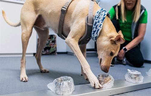
A Labrador Retriever named Bobby (above) sniffs sample of human sweat through containers to detect COVID-19 coronavirus at Veterinary Faculty, Chulalongkorn University in Bangkok. Thailand has deployed a canine virus detection squad to help provide a fast and effective way of identifying people with COVID-19 as the country faces a surge in cases, with clusters found in several crowded slum communities and large markets. Clinical laboratory professionals and pathologists will find it interesting that the dogs are given a sample of sweat, each presented in a unique container. Thus, the dogs never are in the presence of the humans who provided the specimens. (Photo and caption copyright: AP/Sakchai Lalit)
To perform the study, the scientists placed sweat samples in metal containers and allowed the dogs to sniff each sample. If no trace of the infection was present, the dogs simply walked past the container. If the disease was detected in a particular sample, the dogs would sit down in front of the container.
Would Spicy Food Interfere with Dogs’ Ability to Detect COVID-19?
The head of the research team, Professor Kaywalee Chatdarong, PhD, noted that other countries also have been using canines to detect the presence of COVID-19. She did have some concerns that the utilization of dogs for this purpose may not work in Thailand due to their often-spicy cuisine. However, since the samples used were from students and faculty at the university, as well as people from the surrounding area, the cuisine did not seem to affect the study results, the AP reported.
Thailand is facing a surge in COVID-19 cases with recent clusters reported at construction sites, crowded neighborhoods, and large markets. The research team plans to use the canines in mobile units in communities suspected of being hotspots for the disease.
A major plus of using dogs to sniff out the disease from sweat samples is the ability to test people who may not be able to get out of their homes to be tested.
“People can simply put cotton balls underneath their armpits to collect sweat samples and send them to the lab,” Suwanna Thanaboonsombat, a volunteer who collects samples and brings them to the clinical laboratory for testing, told the AP. “And the result is quite accurate.”
According to the US Centers for Disease Control and Prevention (CDC), dogs can become infected with the SARS-CoV-2 coronavirus. However, their chances of transmitting the disease to humans is extremely low. Nevertheless, to ensure the dogs do not become infected with COVID-19 themselves, the researchers designed the sample containers to avoid contact between the samples and the dogs’ noses.
Living Animals Come with Limitations
While dogs can provide a quick and inexpensive method of testing for COVID-19, they do have limitations.
“5 p.m. is their dinner time. When it’s around 4:50, they will start to be distracted. So, you can’t really have them work anymore,” Chatdarong told the AP. “And we can’t have them working after dinner either because they need a nap. They are living animals and we do have to take their needs and emotions into consideration. But for me, they are heroes and heroines.”
Using Dogs to Detect COVID-19 in Other Countries
Last fall, the Helsinki Airport in Finland announced it would use a team of trained dogs to detect the presence of COVID-19 among visitors to the airport to ensure the health and safety of its customers and their families, and to help prevent the spread of SARS-CoV-2 in Finland.
Being tested for the coronavirus at the Helsinki airport in Finland does not require direct contact with a dog. Individuals simply need to swipe their skin with a test wipe and drop the wipe into a cup. The cup is then given to a dog that is working in a separate booth (shown above), which protects both the dog and the dog’s handler from contamination. All tests are processed anonymously and anyone testing positive for COVID-19 is directed to a health information point located at the airport. (Photo copyright: Finavia.)
“We are among the pioneers. As far as we know no other airport has attempted to use canine scent detection on such a large scale against COVID-19,” said Airport Director Ulla Lettijeff in a Finavia press release. “This might be an additional step forward on the way to beating COVID-19.”
In addition to being “man’s best friend,” dogs serve valuable purposes in the medical community. Their strong sense of smell may render them useful in the detection of and fight against illnesses, including COVID-19.
Whether the performance and accuracy of individual dogs can be validated with acceptable quality control (QC) procedures remains to be seen. Medical laboratory managers and pathologists understand the challenges presented with demonstrating accuracy and reproducibility with this method of diagnostic testing. That obstacle has prevented research outcomes from being translated into clinical practice.
Hello primary diagnosis of digital pathology images via artificial intelligence! Goodbye light microscopes!
Digital pathology is poised to take a great leap forward. Within as few as 12 months, image analysis algorithms may gain regulatory clearance in the United States for use in primary diagnosis of whole-slide images (WSIs) for certain types of cancer. Such a development will be a true revolution in surgical pathology and would signal the beginning of the end of the light microscope era.
A harbinger of this new age of digital pathology and automated image analysis is a press release issued last week by Ibex Medical Analytics of Tel Aviv, Israel. The company announced that its Galen artificial intelligence (AI)-powered platform for use in the primary diagnosis of specific cancers will undergo an accelerated review by the Food and Drug Administration (FDA).
FDA’s ‘Breakthrough Device Designation’ for Pathology AI Platform
Ibex stated that “The FDA’s Breakthrough Device Designation is granted to technologies that have the potential to provide more effective treatment or diagnosis of life-threatening diseases, such as cancer. The designation enables close collaboration with, and expedited review by, the FDA, and provides formal acknowledgement of the Galen platform’s utility and potential benefit as well as the robustness of Ibex’s clinical program.”
“All surgical pathologists should recognize that, once the FDA begins to review and clear algorithms capable of using digital pathology images to make an accurate primary diagnosis of cancer, their daily work routines will be forever changed,” stated Robert L. Michel, Editor-in-Chief of Dark Daily and its sister publication The Dark Report. “Essentially, as FDA clearance is for use in clinical care, pathology image analysis algorithms powered by AI will put anatomic pathology on the road to total automation.
“Clinical laboratories have seen the same dynamic, with CBCs (complete blood counts) being a prime example. Through the 1970s, clinical laboratories employed substantial numbers of hematechnologists [hematechs],” he continued. “Hematechs used a light microscope to look at a smear of whole blood that was on a glass slide with a grid. The hematechs would manually count and record the number of red and white blood cells.
“That changed when in vitro diagnostics (IVD) manufacturers used the Coulter Principle and the Coulter Counter to automate counting the red and white blood cells in a sample, along with automatically calculating the differentials,” Michel explained. “Today, only clinical lab old-timers remember hematechs. Yet, the automation of CBCs eventually created more employment for medical technologists (MTs). That’s because the automated instruments needed to be operated by someone trained to understand the science and medicine involved in performing the assay.”
Primary Diagnosis of Cancer with an AI-Powered Algorithm
Surgical pathology is poised to go down a similar path. Use of a light microscope to conduct a manual review of glass slides will be supplanted by use of digital pathology images and the coming next generation of image analysis algorithms. Whether these algorithms are called machine learning, computational pathology, or artificial intelligence, the outcome is the same—eventually these algorithms will make an accurate primary diagnosis from a digital image, with comparable quality to a trained anatomic pathologist.
How much of a threat is automated analysis of digital pathology images? Computer scientist/engineer Ajit Singh, PhD, a partner at Artiman Ventures and an authority on digital pathology, believes that artificial intelligence is at the stage where it can be used for primary diagnosis for two types of common cancer: One is prostate cancer, and the other is dermatology.
On June 17, Ajit Singh, PhD (above), Partner at Artiman Ventures, will lead a special webinar and roundtable discussion for all surgical pathologists and their practice administrators on the coming arrival of artificial intelligence-powered algorithms to aid in the primary diagnosis of certain cancers. Regulatory approval for such solutions may happen by the end of this year. Such a development would accelerate the transition from light microscopes to a fully digital pathology workflow. Singh is shown above addressing the 2018 Executive War College. (Photo copyright: The Dark Report.)
“It is now possible to do a secondary read, and even a first read, in prostate cancer with an AI system alone. In cases where there may be uncertainty, a pathologist can review the images. Now, this is specifically for prostate cancer, and I think this is a tremendous positive development for diagnostic pathways,” he added.
Use of Digital Pathology with AI-Algorithms Changes Diagnostics
Pathologists who are wedded to their light microscopes will want to pay attention to the impending arrival of a fully digital pathology system, where glass slides are converted to whole-slide images and then digitized. From that point, the surgical pathologist becomes the coach and quarterback of an individual patient’s case. The pathologist guides the AI-powered image analysis algorithms. Based on the results, the pathologist then orders supplementary tests appropriate to developing a robust diagnosis and guiding therapeutic decisions for that patient’s cancer.
In his interview with The Dark Report, Singh explained that the first effective AI-powered algorithms in digital pathology will be developed for prostate cancer and skin cancer. Both types of cancer are much less complex than, say, breast cancer. Moreover, the AI developers have decades of prostate cancer and melanoma cases where the biopsies, diagnoses, and downstream patient outcomes create a rich data base from which the algorithms can be trained and tuned.
This webinar is organized as a roundtable discussion so participants can interact with the expert panelists. The Chair and Moderator is Ajit Singh, PhD, Adjunct Professor at the Stanford School of Medicine and Partner at Artiman Ventures.
The panelists (above) represent academic pathology, community hospital pathology, and the commercial sector. They are:
Because the arrival of automated analysis of digital pathology images will transform the daily routine of every surgical pathologist, it would be beneficial for all pathology groups to have one or more of their pathologists register and participate in this critical webinar.
The roundtable discussion will help them understand how quickly AI-powered image analysis is expected be cleared for use by the FDA in such diseases as prostate cancer and melanomas. Both types of cancers generate high volumes of case referrals to the nation’s pathologists, so potential for disruption to long-standing client relationships, and the possible loss of revenue for pathology groups that delay their adoption of digital pathology, can be significant.
On the flip side, community pathology groups that jump on the digital pathology bandwagon early and with the right preparation will be positioned to build stronger client relationships, increase subspecialty case referrals, and generate additional streams of revenue that boost partner compensation within their group.
Also, because so many pathologists are working remotely, Dark Daily has arranged special group rates for pathology practices that would like their surgical pathologists to participate in this important webinar and roundtable discussion on AI-powered primary diagnosis of pathology images. Inquire at info@darkreport.com or call 512-264-7103.