As this therapeutic approach gains regulatory approval, clinical laboratory tests to determine condition of patient’s gut microbiota and monitor therapy will be needed
Some developments in the clinical laboratory industry are less about diagnostic tests and more about novel approaches to therapy. Such is the case with a new carbon bead technology developed by researchers from University College London (UCL) and the Royal Free Hospital intended to remove harmful bacteria toxins from the gut before they leak to the liver. The macroporous beads, which come in small pouches, are delivered orally and could be utilized in the future to treat a number of diseases.
Why is this relevant? Once a new treatment is accepted for clinical use, demand increases for a clinical laboratory test that confirms the therapy will likely work and to monitor its progress.
In collaboration with Yaqrit, a UK-based life sciences company that develops treatments for chronic liver disease, the UCL and Royal Free Hospital scientists engineered the carbon beads—known as CARBALIVE—to help restore gut health. They measured the technology’s impact on liver, kidney, and brain function in both rats and mice.
“The influence of the gut microbiome on health is only just beginning to be fully appreciated,” said Rajiv Jalan, PhD, Professor of Hepatology at UCL in a press release. “When the balance of the microbiome is upset, ‘bad’ bacteria can proliferate and out-compete the ‘good’ bacteria that keeps the gut healthy.
“One of the ways [the ‘bad’ bacteria] do this is by excreting endotoxin, toxic metabolites, and cytokines that transform the gut environment to make it more favorable to them and hostile to good bacteria,” he continued. “These substances, particularly endotoxin, can trigger gut inflammation and increase the leakiness of the gut wall, resulting in damage to other organs such as the liver, kidneys, and brain.”
“I have high hopes that the positive impact of these carbon beads in animal models will be seen in humans, which is exciting not just for the treatment of liver disease but potentially any health condition that is caused or exacerbated by a gut microbiome that doesn’t work as it should,” said Rajiv Jalan, PhD (above), Professor of Hepatology, University College London, in a press release. “This might include conditions such as irritable bowel syndrome (IBS), for example, which is on the rise in many countries.” Though not a clinical laboratory diagnostic test, new therapies like CARBALIVE could be a boon to physicians treating patients with IBS and other gastrointestinal conditions.
Developing the Carbon Beads
The team discovered CARBALIVE is effective in the prevention of liver scarring and injury in animals with cirrhosis when ingested daily for several weeks. They also found a reduced mortality rate in test animals with acute-on-chronic-liver-failure (ACLF).
After achieving success with CARBALIVE in animals, the researchers tested the technology on 28 cirrhosis patients. The carbon beads proved to be safe for humans and had inconsequential side effects.
“In cirrhosis, a condition characterized by scarring of the liver, it is known that inflammation caused by endotoxins can exacerbate liver damage,” Jalan explained. “Part of the standard treatment for cirrhosis is antibiotics aimed at controlling bad bacteria, but this comes with the risk of antibiotic resistance and is only used in late-stage disease.”
The beads, which are smaller than a grain of salt, contain an exclusive physical structure that absorbs large and small molecules in the gut. They are intended to be taken with water at bedtime as harmful bacteria is more likely to circulate through the body at night which could result in damage. The carbon beads do not kill bacteria, which decreases the risk of antibiotic resistance. They eventually pass through the body as waste.
“They work by absorbing the endotoxins and other metabolites produced by ‘bad’ bacteria in the gut, creating a better environment for the good bacteria to flourish and helping to restore microbiome health,” said Michal Kowalski, M.Sc.Eng, Director and VP of Operations at Yaqrit, in the UCL news release.
“This prevents these toxins from leaching into other areas of the body and causing damage, as they do in cirrhosis,” he added. “The results in animal models are very positive, with reduction in gut permeability, liver injury, as well as brain and kidney dysfunction.”
Additional Research
The researchers plan to perform further clinical trials in humans to determine if the carbon beads are effective at slowing the progression of liver disease. If the benefits that were observed in lab animals prove to be compelling in humans, the technology may become an invaluable tool for the treatment of liver disease and other diseases associated with poor microbiome health in the future.
According to the American Liver Foundation, 4.5 million adults in the US have been diagnosed with liver disease. However, it is estimated that 80 to 100 million adults have some form of fatty liver disease and are unaware of it. Liver disease was the 12th leading cause of death in the US in 2020 with 51,642 adults perishing from the disease that year.
According to BMC Public Health, globally there were 2.05 million new cases of liver cirrhosis diagnosed in 2019. In that year, 1.47 million people around the world died from the disease.
More research and clinical studies are needed before this novel technology can be used clinically. When and if that happens, the demand for clinical laboratory tests that measure microbiome deficiencies and monitor patient progress during therapy will likely be high.
“On-a-chip” devices continue to advance and medical laboratories will be natural repositories for patient data as the technology continues to improve
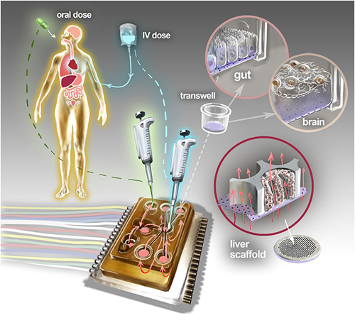
Dark Daily has predicted that the future of clinical laboratory testing will include highly complex multi-analyte test panels. The biomarkers, however, could number in the hundreds or thousands. So, it’s interesting to see new research by a Massachusetts Institute of Technology (MIT) team currently developing a multi-biomarker organ test device for clinical purposes.
Motivated by the costly failure of animal testing efforts to develop drug safety and efficacy in humans, the MIT research engineers created a microfluidic platform technology they dubbed “physiome-on-a-chip,” or more colloquially, “body-on-a-chip.” Their goal is to identify drug reaction in different cell groups within the body (in vivo).
They acknowledged contributions of in vitro microphysiological systems (MPSs), AKA “organ-on-a-chip” (OOC) systems. They note, however, in their paper published in Scientific Reports, that more complex systems that interconnect and receive data from multiple MPSs are needed due to increasing limitations arising from drugs’ “lack of efficacy” rather than toxicity.
“Here we describe the development and implementation of multi-MPS platforms, AKA physiome-on-a-chip, supporting four-way, seven-way, and 10-way MPS interactions for several weeks,” the MIT engineers wrote.
Though MIT’s new technology needs further research and development time, as well as clinical trials, this type of chip design and its ability to scale is a positive development and progress toward Dark Daily’s prediction. Once finalized, it could be adopted in medical laboratories for many types of diagnostic testing purposes.
Researchers Motivated to Improve Drug Efficacy
According to an MIT news release, “MIT engineers have developed new technology that could be used to evaluate new drugs and detect possible side effects before the drugs are tested in humans. Using a microfluidic platform that connects engineered tissues from up to 10 organs, the researchers can accurately replicate human organ interactions for weeks at a time, allowing them to measure the effects of drugs on different parts of the body.”
The “body-on-a-chip” technology, MIT says, is aimed at determining how drugs may affect one organ while also having side effects on others.
“Some of these effects are really hard to predict from animal models because the situations that lead to them are idiosyncratic. With our chip, you can distribute a drug and then look for the effects on other tissues and measure the exposure and how it is metabolized,” said Linda Griffith, PhD, Professor of Teaching Innovation at MIT’s School of Engineering, and a senior author of the study, in the news release.
According to MIT, factors affecting the effectiveness of pharmaceuticals may include:
Genetics;
Environment;
Personal lifestyles; and,
Interactions with other drugs.
TechCrunch called the study “unprecedented,” pointing to the platform’s connection of so many tissues and the technology’s ability to keep them stable for weeks.
“An advantage of our platform is that we can scale it up or down and accommodate a lot of different configurations,” Linda Griffith, PhD, MIT Professor, MIT School of Engineering, told Science Daily. “I think the field is going to go through a transition where we start to get more information out of a three-organ or four-organ system, and it will start to become cost-competitive because the information you’re getting is so much more valuable.” (Photo copyright: MacArthur Foundation.)
How “Body-on-a-Chip” Works
“Body-on-a-chip” is about the size of a tablet computer and links 10 organ types, including: liver, lung, gut, endometrium, brain, heart, pancreas, kidney, skin, and skeletal muscle.
Using microfluidic platform technology, the researchers placed one- to two-million cells from human tissue samples into the device and then pushed fluid through the chip to resemble blood flow, the Daily Mail reported, adding that MIT’s MPS platform design features:
Compartments made from a plastic block;
Passages for fluid to move (as a circulatory system does) between the compartments;
A water reservoir to limit fluid evaporation; and,
Ability to monitor flow of molecular exchanges and drug distribution.
Essentially, using the MIT device, a drug can be introduced to one organ, processed normally, and then passed to other organs for processing and use in other ways, TechCrunch summarized.
The physiome-on-a-chip system (above schematic) comprises bioengineered devices that nurture many interconnected 3D MPSs representing specified functional behaviors of each organ of interest, designed to capture essential features of in vivo physiology based on quantitative systems models tailored for individual applications such as drug fate or disease modeling. This technology could eventually be utilized for clinical laboratory and anatomic pathology testing. (Image and caption copyright: Victor O. Leshyk/Scientific Reports.)
Drug Delivery, Effects on Multiple Tissues Noted in MIT Study
The MIT researcher engineers reported these findings and accomplishments:
Delivering a drug to the gastrointestinal tissue;
Replicating digesting a drug;
Observing as a drug was transported to other tissues and metabolized;
Measuring a drug’s path; and,
Noting effects of a drug on different tissues and how drugs break down.
“The huge potential of MPS technology is revealed by connecting multiple organ chips in an integrated system for in vitropharmacology. This study beautifully illustrates that multi-MPS ‘physiome-on-a-chip’ approaches, which combine the genetic background of human cells with physiologically relevant tissue-to-media volumes, allow accurate prediction of drug pharmacokinetics and drug absorption, distribution, metabolism, and excretion,” said Kevin Healy, PhD, Professor of Bioengineering and Materials Science and Engineering, at University of California Berkeley in the MIT news release. Healy was not involved in the research.
Unique Device Design
In addition to making it possible to study so many different tissue types, the device design, according to MIT, is unique for these reasons:
Its open microfluidic system, rather than a closed system, means the lid can be removed to manipulate tissue samples;
Instead of external pumps common in closed systems, the MIT team used “on-board pumps” to control flow of liquid between the organs; and,
The pumps used enabled larger engineered tissues, such as those from tumors in an organ, to be assessed.
The MIT engineers next plan to focus on specific organs—including the brain, liver, and gastrointestinal tissue—to model Parkinson’s disease, Digital Trends reported.
As healthcare providers and medical laboratories adopt precision medicine, MIT’s contributions are both timely and important. The ability to accommodate many different configurations in one platform is impressive, and something Dark Daily has been anticipating.
Obesity may be one of several health conditions and diseases where the human microbiome can be harnessed for diagnostic and therapeutic uses
Microbiologists could soon be the front lines in the nation’s fight against obesity and possibly other chronic diseases. New research underway at Vanderbilt University could lead to a host of new clinical laboratory tests that use engineered microbes.
This research is revealing how the human microbiome can be the source of new biomarkers for diagnostic tests and therapeutic drugs. In fact, early research findings point to the possibility that pathologists and clinical laboratories may eventually use the human microbiome in their daily work.
Engineering Bacteria to Battle Obesity
The human microbiome has remained largely unstudied. One reason why this is true is that it has been difficult to recreate, in the laboratory, the optimal conditions to allow these microbes to grow and thrive just as they do in the human body. However, as researchers continue to make new discoveries about this community of micro-organisms, there is optimism that elements of the human microbiome can be used to develop novel medical laboratory tests. (more…)