Immunotherapy device could also enable clinical laboratories to receive in vivo biomarker data wirelessly
Researchers from Rice University in Houston and seven other states in the US are working on a new oncotherapy sense-and-respond implant that could dramatically improve cancer outcomes. Called Targeted Hybrid Oncotherapeutic Regulation (THOR), the technology is intended primarily for the delivery of therapeutic drugs by monitoring specific cancer biomarkers in vivo.
Through a $45 million federal grant from the Advanced Research Projects Agency for Health (ARPA-H), the researchers set out to develop an immunotherapy implantable device that monitors a patient’s cancer and adjusts antibody treatment dosages in real time in response to the biomarkers it measures.
It’s not a far stretch to envision future versions of the THOR platform also being used diagnostically to measure biomarker data and transmit it wirelessly to clinical laboratories and anatomic pathologists.
ARPH-A is a federal funding agency that was established in 2022 to support the development of high-impact research to drive biomedical and health breakthroughs. THOR is the second program to receive funding under its inaugural Open Broad Agency Announcement solicitation for research proposals.
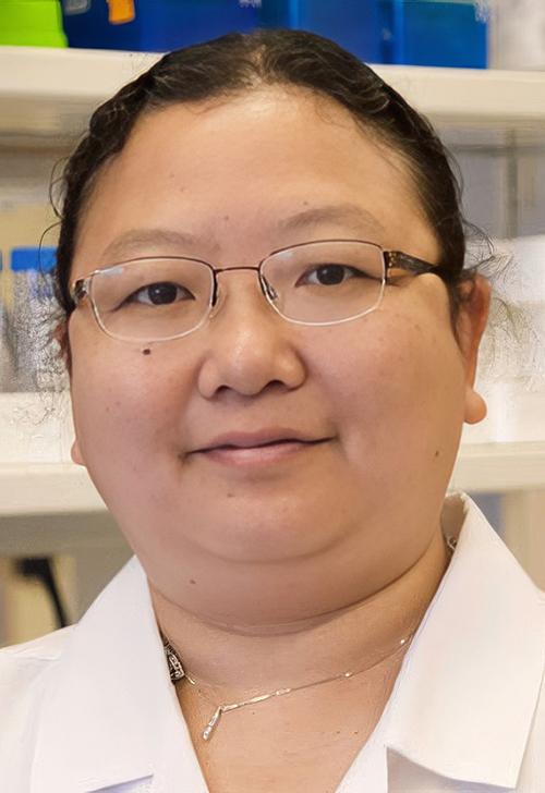
“By integrating a self-regulated circuit, the THOR technology can adjust the dose of immunotherapy reagents based on a patient’s responses,” said Weiyi Peng, MD, PhD (above), Assistant Professor of Biology and Biochemistry at the University of Houston and co-principal investigator on the research, in a UH press release. “With this new feature, THOR is expected to achieve better efficacy and minimize immune-related toxicity. We hope this personalized immunotherapy will revolutionize treatments for patients with peritoneal cancers that affect the liver, lungs, and other organs.” If anatomic pathologists and clinical laboratories could receive biometric data from the THOR device, that would be a boon to cancer diagnostics. (Photo copyright: University of Houston.)
Antibody Therapy on Demand
Omid Veiseh, PhD, Associate Professor of Bioengineering at Rice University and principal investigator on the project, described the THOR device as a “living drug factory” inside the body. The device is a rod-like gadget that contains onboard electronics and a wireless rechargeable battery. It is three inches long and has a miniaturized bioreactor that contains human epithelial cells that have been engineered to produce immune modulating therapies.
“Instead of tethering patients to hospital beds, IV bags, and external monitors, we’ll use a minimally invasive procedure to implant a small device that continuously monitors their cancer and adjusts their immunotherapy dose in real time,” said Veiseh in a Rice University press release. “This kind of ‘closed-loop therapy’ has been used for managing diabetes, where you have a glucose monitor that continuously talks to an insulin pump.
But for cancer immunotherapy, it’s revolutionary.”
The team believes the THOR device will have the ability to monitor biomarkers and produce an antibody on demand that will trigger the immune system to fight cancer locally. They hope the sensor within THOR will be able to monitor biomarkers of toxicity for the purpose of fine-tuning therapies to a patient immediately in response to signals from a tumor.
“Today, cancer is treated a bit like a static disease, which it’s not,” Veiseh said. “Clinicians administer a therapy and then wait four to six weeks to do radiological measurements to see if the therapy is working. You lose quite a lot of time if it’s not the right therapy. The tumor may have evolved into a more aggressive form.”
The THOR device lasts 60 days and can be removed after that time. It is designed to educate the immune system to recognize a cancer and prevent it from recurring. If the cancer is not fully eradicated after the first implantation, the patient can be implanted with THOR again.
Use of AI in THOR Therapy
The researchers plan to spend the next two and a half years building prototypes of the THOR device, testing them in rodents, and refining the list of biomarkers to be utilized in the device. Then, they intend to take an additional year to establish protocols for the US Food and Drug Administration’s (FDA) good manufacturing practices requirements, and to test the final prototype on large animals. The researchers estimate the first human clinical trials for the device will begin in about four years.
“The first clinical trial will focus on refractory recurrent ovarian cancer, and the benefit of that is that we have an ongoing trial for ovarian cancer with our encapsulated cytokine ‘drug factory’ technology,” said Veiseh in the UH press release.
The group is starting with ovarian cancer because research in this area is lacking and it will provide the opportunity for THOR to activate the immune system against ovarian cancer, which is typically challenging to fight with immunotherapy approaches. If successful in ovarian cancer, the researchers hope to test THOR in other cancers that metastasize within the abdomen, such as:
All control and decision-making will initially be performed by a healthcare provider based on signals transmitted by THOR using a computer or smartphone. However, Veiseh sees the device ultimately being powered by artificial intelligence (AI) algorithms that could independently make therapeutic decisions.
“As we treat more and more patients [with THOR], the devices are going to learn what type of biomarker readout better predicts efficacy and toxicity and make adjustments based on that,” he predicted. “Between the information you have from the first patient versus the millionth patient you treat, the algorithm is just going to get better and better.”
Moving Forward
In addition to UH and Rice University, scientists working on the project come from several institutions, including:
More research and clinical trials are needed before THOR can be used in the clinical treatment of cancer patients. If the device reaches the commercialization stage, Veiseh plans to either form a new company or license the technology to an existing company for further development.
“We know that the further we advance it in terms of getting that human data, the more likely it is that this could then be transferred to another entity,” he told Precision Medicine Online.
Pathologists and clinical laboratories will want to monitor the progress of the THOR technology’s ability to sense changes in cancer biomarkers and deliver controlled dosages of antibiotic treatments.
Similar health monitoring devices have been popular with chronic disease patients and physicians who treat them; this technology may give clinical laboratories a new diagnostic tool
There is an ever-increasing number of companies working to develop lab testing technologies that would be used outside of the traditional clinical laboratory. One such example is Nutromics, an Australia-based medical technology company which recently announced it has raised US $14 million to fund its new lab-on-a-patch platform, according to a company press release.
Nutromics’ lab-on-a-patch device “uses DNA sensor technology to track multiple targets in the human body, including disease biomarkers and hard-to-dose drugs,” according to MobiHealthNews. Notably, Nutromics’ technology uses interstitial fluid as the sample source.
Nutromics raised $4 million last year to support a manufacturing facility and an initial human clinical trial of its “continuous molecular monitoring (CMM) platform technology that is able to track multiple targets in the human body via a single wearable sensor. The platform provides real-time, continuous molecular-level insights for remote patient monitoring and hospital-at-home systems,” MobiHealthNews reported.
“We are aiming to cause a paradigm shift in diagnostic healthcare by essentially developing a lab-on-a-patch. A lack of timely and continuous diagnostic insights can strongly impact outcomes when dealing with critical disease states. With this strategic industry and VC (venture capital) investment in us, we see more confidence in our technology and hope to accelerate our growth,” said entrepreneur and chemical engineer Peter Vranes (above), co-founder and CEO of Nutromics, in a press release. Clinical laboratory leaders have watched similar biometric monitoring devices come to fruition. (Photo copyright: Nutromics.)
.
How Nutromics’ Lab-on-a-Patch Works
“Our technology is, in fact, two technologies coming together—a marker and needle. What that does is give us access to fluid under your skin called interstitial fluid. If you’re going to measure something continuously, that’s a really good fluid [to measure],” Vranes told Outcomes Rocket.
Vranes calls the system’s aptamer-based sensor platform technology the “jewel in the crown.” An aptamer is a short sequence of artificial DNA or RNA that binds a specific target molecule. Nutromics’ aptamer sensor, Vranes said, enables targeting of analytes, unlike continuous glucose monitors (CGMs).
“[CGMs] are limited to metabolites—things that are already in the body like glucose and lactate. We’re not limited to those. We can do a whole range of different targets. And what that gives us is a ‘blue ocean’ opportunity to go in and solve problems in areas that other technologies just can’t solve,” Vranes said.
Nutromics plans to develop multiple aptamer-based sensors that measure a variety of analytes in interstitial fluid, Medtech Insight noted.
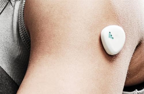
Nutromics’ wearable DNA sensor lab-on-a-patch technology (above) enables monitoring of multiple targets, including disease biomarkers and some medications, MobiHealthNews explained. The wearable patch contains microneedles that painlessly access interstitial fluid under the skin. Collected data is wirelessly transmitted to a software application and integrates with consumer health software and provider platforms, according to Nutromics. Medical laboratories could have a role in collecting this data and adding it other test results from patients using the wearable patch. (Photo copyright: Nutromics.)
Initial Launch Will Include Antibiotic Monitoring
Nutromics expects to initially launch therapeutic monitoring of vancomycin, a glycopeptide antibiotic medication used to treat various bacterial infections. The company says 60% of doses for this prescription antibiotic are not within therapeutic range.
“Interstitial fluid originates in the blood and then leaks out of capillaries to bring nutrients to cells in the body’s tissues. Because interstitial fluid is in direct communication with the cells, it should have information about the tissues themselves beyond what can be measured from testing the blood,” said Mark Prausnitz, PhD, Regents Professor and J. Erskine Love Jr. Chair, Georgia Tech School of Chemical and Biomolecular Engineering, in a 2020 news release announcing results of human trials of microneedle-based ISF sampling.
“We sampled interstitial fluid from 21 human participants and identified clinically relevant and sometimes distinct biomarkers in interstitial fluid when compared to companion plasma samples based on mass spectrometry analysis,” the scientists wrote.
Clinical laboratory leaders and pathologists will find it useful to monitor the development of diagnostics for use outside the lab. Nutromics is an example of a company developing wearable health technology that painlessly gathers data for lab tests to be conducted in point-of-care and near-patient settings.
Tiny sensors with Bluetooth technology that measure useful biomarkers may eliminate need for invasive blood draws used for clinical laboratory tests
What if a baby’s pacifier could be used to measure electrolyte levels in newborns? An international research team has developed just such a device, and it has the potential to reduce invasive blood collections required to provide specimens for clinical laboratory testing of critical biomarkers. At the same time, this device may allow continuous monitoring of electrolyte levels with wireless alerts to caregivers.
Typical blood draws for NICU babies can cause information gaps as they are usually only performed twice a day. This can be problematic in cases where more frequent monitoring of these biomarkers is required to monitor the infant’s condition.
“We know that premature babies have a better chance of survival if they get a high quality of care in the first month of birth,” said Jong-Hoon Kim, PhD, Associate Professor at the WSU School of Electrical Engineering and Computer Science, in a WSU news release. “Normally, in a hospital environment, they draw blood from the baby twice a day, so they just get two data points. This device is a non-invasive way to provide real-time monitoring of the electrolyte concentration of babies.”
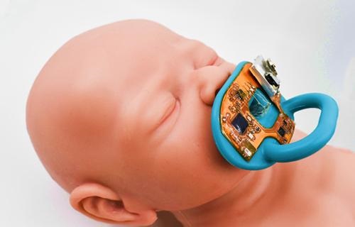
The smart pacifier (above) developed by researchers at the Washington State University School of Electrical Engineering and Computer Science—in collaboration with scientists in two South Korean institutions—provides continuous monitoring of sodium and potassium ion levels. This can help detect and prevent potentially dangerous dehydration issues in NICU babies without invasive blood draws for traditional clinical laboratory testing. (Photo copyright: University of Washington.)
How the Smart Pacifier Works
The miniature system developed by the WSU researchers utilizes a typical, commercially available pacifier outfitted with ion-selective sensors, flexible circuits, and microfluidic channels that monitor salivary electrolytes. These flexible, microfluidic channels attract the saliva when the pacifier is in the infant’s mouth which enables continuous and efficient saliva collection without the need for any type of pumping system. The gathered data is relayed wirelessly to caregivers using Bluetooth technology.
When the researchers tested their smart pacifier on infants, they discovered that the results captured from the device were comparable to information obtained from normal blood draws and standard clinical laboratory tests. Kim noted in the press release that technology currently in use to test infant saliva for electrolytes tend to be bulky, rigid devices that require a separate sample collection.
“You often see NICU pictures where babies are hooked up to a bunch of wires to check their health conditions such as their heart rate, the respiratory rate, body temperature, and blood pressure,” said Kim in the press release. “We want to get rid of those wires.”
The researchers intend to make the components for the device more affordable and recyclable. They also plan to perform testing for their smart pacifier on larger test groups to prove efficacy and hope the gadget will help make NICU treatment less disruptive for infant patients.
Going as far back as 2013, Dark Daily has covered research into the use of sensors placed in wearables and disposables to detect and monitor health issues.
“It should be noted that the ability to put reliable diagnostic sensors in disposables like diapers has been around for almost a decade and does not seem to have caught on with either caregivers or the public,” said Robert Michel, Editor-in-Chief of Dark Daily and its sister publication, The Dark Report. “Because the researchers who developed the pacifier are attempting to solve a problem for NICU babies, this solution might find acceptance.”
This is another example of how researchers are thinking outside the box as to how to measure critical biomarkers without the need to send a specimen to the core clinical laboratory and wait hours—sometimes overnight—for results.
This new technology could replace needle biopsies and allow physicians to detect rejection of transplanted organs earlier, saving patients’ lives
Anatomic pathologists
may be reading fewer biopsy reports for patients with organ transplants in the
future. That’s thanks to a new technology that may be more sensitive to and
capable of detecting organ rejection earlier than traditional needle biopsies.
When clinicians can detect organ transplant rejection
earlier, patients survive longer. Unfortunately, extensive organ damage may
have already occurred by the time rejection is detected through a traditional
needle biopsy. This led a group of researchers at Emory University School of Medicine to
search for a better method for detecting organ rejection in patients with transplants.
The Emory researchers describe the method and technology
they devised in a paper published in Nature Biomedical
Engineering, titled, “Non-Invasive Early Detection of Acute Transplant
Rejection Via Nanosensors of Granzyme B Activity.” The new technology could
make it easier for clinicians to detect when a patient’s body is rejecting a
transplanted organ at an earlier time than traditional methods.
This technology also provides a running measure of processes,
so clinicians have more powerful tools for deciding on the most appropriate
dosage of immunosuppressant
drugs.
“Right now, most tests are aimed at organ dysfunction, and
sometimes they don’t signal there is a problem until organ function is below 50
percent,” Andrew
Adams, MD, PhD Co-Principal Investigator and an Associate Professor of Surgery
at Emory University School of Medicine, in a Georgia
Institute of Technology news release.
How the Technology Works
The method that Adams and his colleagues tested involves the
detection of granzyme B,
a serine protease
often found in the granules of natural killer cells
(NK cells) and cytotoxic
T cells. “Before any organ damage can happen, T cells have to produce granzyme
B, which is why this is an early detection method,” said Gabe Kwong, PhD, Assistant
Professor in the Wallace H. Coulter Department of Biomedical Engineering at
Georgia Tech and Emory University, in the news release.
The new technology is made up of sensor nanoparticles in the
shape of a ball with iron oxide in the middle. Amino acids stick out of the
ball like bristles. Each amino acid has a fluorescent molecule attached to the
tip.
The nanoparticles are injected into the patient. Their size
prevents them from gathering in the patient’s tissue or from being flushed out
through the kidneys. They are designed to accumulate in the tissue of the
transplanted organ.
If the T cells in the transplanted organ begin to produce
granzyme B, the amino acids break away from the nanoparticles, releasing the
fluorescent molecules attached to their tips. Those molecules are small enough
to be processed through the kidneys and can be detected in the patient’s urine.
Pathologists Play Crucial Role on Transplant Teams
Anatomical pathologists (histopathologists in the UK) are key
members of transplant teams for many reasons, including their ability to assess
biopsies. The current method for detecting organ transplant rejection involves
needle biopsies. It is considered the gold standard.
However, according to a paper published in the International
Journal of Organ Transplantation Medicine: “Although imaging studies
and laboratory findings are important and helpful in monitoring of the
transplanted liver, in many circumstances they are not sensitive enough. For
conditions such as rejection of the transplant, liver histology remains the
gold-standard test for the diagnosis of allograft dysfunction. Therefore,
histopathologic assessments of allograft liver
biopsies have an important role in managing patients who have undergone liver
transplantation.”
There are two main problems with needle biopsies. The first,
as mentioned above, is that they don’t always catch the rejection soon enough.
The second is that the needle may cause damage to the transplanted organ.
“The biggest risk of a biopsy is bleeding and injury to the transplanted organ,” noted Andrew Adams, MD, PhD (above), Co-Principal Investigator and an Associate Professor of Surgery at Emory University School of Medicine, in the Georgia Tech news release. “Then there’s the possibility of infection. You’re also just taking a tiny fraction of the transplanted organ to determine what’s going on with the whole organ, and you may miss rejection or misdiagnose it because the needle didn’t hit the right spot,” he added.
And, according to Kwong, even though biopsies are the gold
standard, the results represent one moment in time. “The biopsy is not
predictive. It’s a static snapshot. It’s like looking at a photo of people in
mid-jump. You don’t know if they’re on their way up or on their way down. With
a biopsy, you don’t know whether rejection is progressing or regressing.”
Future Directions of Emory’s Research
The research conducted by Adams and Kwong, et al, is in its
early stages, and the new technology they created won’t be ready to be used on patients
for some time. Nevertheless, there’s reason to be excited.
Nanoparticles are not nearly as invasive as a needle biopsy.
Thus, risk of infection or damaging the transplanted organ is much lower. And Emory’s
technology would allow for much earlier detection, as well as giving clinicians
a better way to adjust the dose of immunosuppressant drugs the patient takes.
“Adjusting the dose is very difficult but very important
because heavy immunosuppression increases occurrence of infections and patients
who receive it also get cancer more often,” said Kwong. The new technology
provides a method of measuring biological activity rates, which would give
clinicians a clearer picture of what’s happening.
The Emory team’s plan is to enhance the new sensors to
detect at least one other major cause of transplant rejection—antibodies. When
a patient’s body rejects a transplanted organ, it produces antibodies to
neutralize what it sees as a foreign entity.
“Antibodies kill their target cells through similar types of
enzymes. In the future, we envision a single sensor to detect both types of
rejection,” said Kwong.
Adams adds, “This method could be adapted to tease out
multiple problems like rejection, infection, or injury to the transplanted
organ. The treatments for all of those are different, so we could select the
proper treatment or combination of treatments and also use the test to measure
how effective treatment is.”
This line of research at Emory University demonstrates how
expanding knowledge in a variety of fields can be combined in new ways. As this
happens, medical laboratories not only get new biomarkers that can be
clinically useful without the need for invasive procedures like needle biopsies,
but these same biomarkers can guide the selection of more effective therapies.
Innovative web-based educational formats might add value to training initiatives for pathology residents and fellows and medical laboratory workers
In the final months of 2013, the regulatory fight between gene testing company 23andMe.com and the Food and Drug Administration (FDA) generated national headlines. In that encounter, 23andMe.com blinked and ceased offering health-related genetic tests to consumers.
However, the company continues to work to position itself as a major player in genetic testing and genetic medicine. In the second half of 2013, for example, 23andMe.com initiated a business campaign to position itself as a source of on-line information about genetics for educational purposes. (more…)