Goal is to demonstrate how whole human genome sequencing of newborns can deliver important diagnostic findings associated with 250 genetic conditions
Clinical laboratory testing and genetics are moving closer to the delivery room than ever before. In the largest study of its kind in North America, genomic scientists plan to supplement traditional screening for inherited diseases—traditionally performed on a blood sample taken shortly after birth—with whole genome sequencing (WGS) on 100,000 newborns in New York City during their first five years of life, LifeSciencesIntelligence reported.
Conducted by genetic scientists at NewYork-Presbyterian (NYP) and Columbia University, in collaboration with genetic company GeneDx, a wholly-owned subsidiary of health intelligence company Sema4 (NASDAQ:SMFR), the genetic research study, called GUARDIAN (Genomic Uniform-screening Against Rare Diseases In All Newborns), will screen newborn babies for 250 rare diseases that are generally not tested for.
The GUARDIAN program will “drive earlier diagnosis and treatment to improve the health of the babies who participate, generate evidence to support the expansion of newborn screening through genomic sequencing, and characterize the prevalence and natural history of rare genetic conditions,” according to a Sema4 news release.
“The appetite for this is growing. The awareness of this is growing. We all see it as inevitable,” medical geneticist Robert Green, MD, at Brigham and Women’s Hospital and Harvard Medical School told USA Today. “We are grossly underutilizing the life-saving benefits of genetics and we have to get past that.” Clinical laboratory leaders understand the value of early detection of disease and subsequent early treatment. (Photo copyright: Harvard Medical School.)
Improving Health of Babies Through Early Detection of Disease
GUARDIAN aims to use WGS to identify conditions at birth that can affect long-term health and subsequently enhance treatment options and possibly prevent disability or death.
The 250 different diseases GUARDIAN will be screening for typically strike young children. They are mostly rare conditions that:
have an onset before five years of age,
have a greater than 90% probability of the condition developing based on the genetic result,
have effective approaches and treatments that are already available, and/or
have a well-established natural history of the condition.
“We’re entering the therapeutic era and leaving the diagnostic era,” Paul Kruszka, MD, Chief Medical Officer at GeneDx told USA Today. “This potentially has the opportunity to change the way we practice medicine, especially in rare disease.”
Some Parents Reluctant to Agree to Genetic Testing
Green and his research team first began analyzing the genetic sequences of newborns back in 2013. They believe the costs of performing infant WGS is worthwhile because it can improve lives. However, Green also recognizes that some parents are reluctant to agree to this type of genetic testing due to concerns regarding privacy and the fear of discovering their baby may have an illness.
“You’ve gone through all this pregnancy and you’re sitting there with a healthy baby (and I’m) offering you the opportunity to find out something that’s devastating and terrifying,” he told USA Today. “How fun is that?”
Green continued. “We can respect people who don’t want to know, but also respect people who do want to know. Some families will say ‘I treasure the precious ignorance.’ Others will say ‘If I could have known, I would have poured my heart and soul into clinical trials or spent more time with the child when she was healthy.’”
WGS Screening Identifies Undiagnosed Illnesses in Newborn’s Family
The scientists also found that performing WGS in newborns can detect diseases in the infants as well as unknown illnesses in the families of those babies. According to Kruszka, many parents often seek a diagnosis for a rare disease present in their children for several years. Since many common diseases develop as a result of certain combinations of genes, if illnesses are diagnosed at birth, it could extradite the treatment process, prevent complications, and provide better health outcomes for patients.
“We are relentlessly focused on accelerating the adoption and use of genomic information to impact the lives of as many people as possible, particularly newborns and children,” said Katherine Stueland, President and CEO, Sema4, in the Sema4 news release. “As the first commercial laboratory to launch a rapid whole genome sequencing offering, to address broad unmet needs for early diagnosis, participation in this study is an important step forward for healthcare and in delivering on our goal to sequence once, analyze forever.”
The study is open to all babies in New York City who are born in a health system that participates in the GUARDIAN program, regardless of their race, income, or health insurance coverage.
“The results from this study will help us understand the true impact sequencing at birth can have on newborns and their families in comparison to the current standard of care, particularly as we’ll evaluate clinical outcomes in addition to the psychosocial effect on families,” said Kruszka in the Sema4 news release.
Anything that improves the health of newborn babies is a good thing. Regardless of the cost, if DNA analysis can give newborns and their families a better chance at detecting inherited diseases early while clinical laboratory treatment could make a difference, it is worth pursuing.
Columbia University’s MediSCAPE enables surgeons to examine tissue structures in vivo and a large-scale clinical trial is planned for later this year
Scientists at Columbia University in New York City have developed a high-speed 3D microscope for diagnosis of cancers and other diseases that they say could eventually replace traditional biopsy and histology “with real-time imaging within the living body.”
The technology is designed to enable in situ tissue analysis. Known as MediSCAPE, the microscope is “capable of capturing images of tissue structures that could guide surgeons to navigate tumors and their boundaries without needing to remove tissues and wait for pathology results,” according to a Columbia University news story.
“The way that biopsy samples are processed hasn’t changed in 100 years, they are cut out, fixed, embedded, sliced, stained with dyes, positioned on a glass slide, and viewed by a pathologist using a simple microscope. This is why it can take days to hear news back about your diagnosis after a biopsy,” said Hillman in the Columbia news story.
“Our 3D microscope overcomes many of the limitations of prior approaches to enable visualization of cellular structures in tissues in the living body. It could give a doctor real-time feedback about what type of tissue they are looking at without the long wait,” she added in I News.
Hillman’s team previously used the technology—originally dubbed SCAPE for “Swept Confocally Aligned Planar Excitation” microscopy—to capture 3D images of neurological activity in living samples of worms, fish, and flies. In their recent study, the researchers tested the technology with human kidney tissue, a human volunteer’s tongue, and a mouse with pancreatic cancer.
“This was something I didn’t expect—that I could actually look at structures in 3D from different angles,” said nephropathologist and study co-author Shana M. Coley, MD, PhD (above), Director, Transplant Translational Research and Multiplex Imaging Center at Arkana Laboratories, in the Columbia news story. At the time of the Columbia study, Coley was an assistant professor at Columbia University and a renal pathologist at the Columbia University Medical Center. “We found many examples where we would not have been able to identify a structure from a 2D section on a histology slide, but in 3D we could clearly see its shape. In renal pathology in particular, where we routinely work with very limited amounts of tissue, the more information we can derive from the sample, the better for delivering more effective patient care,” she added. (Photo copyright: Arkana Laboratories.)
How MediSCAPE Works
Unlike traditional 3D microscopes that use a laser to scan tiny spots of a tissue sample and then assemble those points into a 3D image, the MediSCAPE 3D microscope “illuminates the tissue with a sheet of light—a plane formed by a laser beam that is focused in a special way,” I News reported.
The MediSCAPE microscope thus captures 2D slices which are rapidly stacked into 3D images at a rate of more than 10 volumes per second, according to I News.
“One of the first tissues we looked at was fresh mouse kidney, and we were stunned to see gorgeous structures that looked a lot like what you get with standard histology,” said optical systems engineer and the study’s lead author, Kripa Patel, PhD, in the Columbia news story. “Most importantly, we didn’t add any dyes to the mouse—everything we saw was natural fluorescence in the tissue that is usually too weak to see.
“Our microscope is so efficient that we could see these weak signals well,” she continued, “even though we were also imaging whole 3D volumes at speeds fast enough to rove around in real time, scanning different areas of the tissue as if we were holding a flashlight.”
A big advantage of the technology, Hillman noted, is the ability to scan living tissue in the body.
“Understanding whether tissues are staying healthy and getting good blood supply during surgical procedures is really important,” she said in the Columbia news story. “We also realized that if we don’t have to remove (and kill) tissues to look at them, we can find many more uses for MediSCAPE, even to answer simple questions such as ‘what tissue is this?’ or to navigate around precious nerves. Both of these applications are really important for robotic and laparoscopic surgeries, where surgeons are more limited in their ability to identify and interact with tissues directly.”
Clinical Trials and FDA Clearance
Early versions of the SCAPE microscopes were too large for practical use by surgeons, so Columbia post-doctoral research scientist Wenxuan Liang, PhD, co-author of the study, helped the team develop a smaller version that would fit into an operating room.
Later this year, the researchers plan to launch a large-scale clinical trial, I News reported. The Columbia scientists hope to get clearance from the US Food and Drug Administration (FDA) to develop a commercialized version of the microscope.
“They will initially seek permission to use it for tumor screening and guidance during operations—a lower and easier class of approval—but ultimately, they hope to be allowed to use it for diagnosis,” Liang wrote.
Charles Evans, PhD, research information manager at Cancer Research UK, told I News, “Using surgical biopsies to confirm a cancer diagnosis can be time-consuming and distressing for patients. And ensuring all the cancerous tissue is removed during surgery can be very challenging unaided.”
He added, “more work will be needed to apply this technique in a device that’s practical for clinicians and to demonstrate whether it can bring benefits for people with cancer, but we look forward to seeing the next steps.”
Will the Light Microscope be Replaced?
In recent years, research teams at various institutions have been developing technologies designed to enhance or even replace the traditional light microscope used daily by anatomic pathologists across the globe.
And digital scanning algorithms for creating whole-slide images (WSIs) that can be analyzed by pathologists on computer screens are gaining in popularity as well.
Such developments may spark a revolution in surgical pathology and could signal the beginning of the end of the light microscope era.
Surgical pathologists should expect to see a steady flow of technologically advanced systems for tissue analysis to be submitted to the FDA for pre-market review and clearance for use in clinical settings. The light microscope may not disappear overnight, but there are a growing number of companies actively developing different technologies they believe can diagnose either or both tissue and digital images of pathology slides with accuracy comparable to a pathologist.
The proof-of-concept experiment showed data can be encoded in DNA and retrieved using automated systems, a development that may have positive significance for clinical laboratories
It may seem far-fetched, but computer scientists and research groups have worked for years to discover if it is possible to store data on Deoxyribonucleic acid (DNA). Now, Microsoft Research (MR) and the University of Washington (UW) have achieved just that, and the implications of their success could be far-reaching.
Clinical pathologists are increasingly performing genetic DNA sequencing in their medical laboratories to identify biomarkers for disease, help clinicians understand their patients’ risk for a specific disease, and track the progression of a disease. The ability to store data in DNA would take that to another level and could have an impact on diagnostic pathology. Pathologist familiar with DNA sequencing may find a whole new area of medical service open to them.
The MR/UW researchers recently demonstrated a fully automated system that encoded data into DNA and then recovered the information as digital data. “In a simple proof-of-concept test, the team successfully encoded the word ‘hello’ in snippets of fabricated DNA and converted it back to digital data using a fully automated end-to-end system,” Microsoft stated in a news release.
DNA’s Potential Storage Capacity and Why We Need It
Thus far, the challenge of using DNA for data storage has
been that there wasn’t a way to easily code and retrieve the information. That,
however, seems to be changing quite rapidly. Several major companies have
invested heavily in research, with consumer offerings expected soon.
At Microsoft Research, ‘consumer interest’ in genetic testing has driven the research into using DNA for data storage. “As People get better access to their own DNA, why not also give them the ability to read any kind of data written in DNA?” asked Doug Carmean, an Architect at Microsoft, during an interview with Wired.
Scientists are interested in using DNA for data storage because
humanity is creating more data than ever before, and the pace is accelerating.
Currently, most of that data is stored on tape, which is inexpensive, but has
drawbacks. Tape degrades and has to be replaced every 10 years or so. But DNA,
on the other hand, lasts for thousands of years!
“DNA won’t degrade over time like cassette tapes and CDs, and it won’t become obsolete,” Yaniv Erlich, PhD, Chief Science Officer at MyHeritage, an online genealogy platform located in Israel, and Associate Professor, Columbia University, told Science Mag.
Tape also takes up an enormous amount of physical space compared to DNA. One single gram of DNA can hold 215 petabytes (roughly one zettabyte) of data. Wired puts the storage capacity of DNA into perspective: “Imagine formatting every movie ever made into DNA; it would be smaller than the size of a sugar cube. And it would last for 10,000 years.”
Researchers at the University of Washington claim, “All the movies, images, emails and other digital data from more than 600 basic smartphones (10,000 gigabytes) can be stored in the faint pink smear of DNA at the end of this test tube.” (Photo and caption copyright: Tara Brown/University of Washington.)
Victor Zhirnov, Chief Scientist at Semiconductor Research Corporation says the worries over storage space aren’t simply theoretical. “Today’s technology is already close to the physical limits of scaling,” he told Wired, which stated, “Five years ago humans had produced 4.4 zettabytes of data; that’s set to explode to 160 zettabytes (each year!) by 2025. Current infrastructure can handle only a fraction of the coming data deluge, which is expected to consume all the world’s microchip-grade silicon by 2040.”
MIT Technology Review agrees, stating, “Humanity is creating information at an unprecedented rate—some 16 zettabytes every year. And this rate is increasing. Last year, the research group IDC calculated that we’ll be producing over 160 zettabytes every year by 2025.”
Heavy Investment by Major Players
The whole concept may seem like something out of a science
fiction story, but the fact that businesses are investing real dollars into it
is evidence that DNA for data storage will likely be a reality in the near
future. Currently, there are a couple of barriers, but work is commencing to
overcome them.
First, the cost of synthesizing DNA in a medical laboratory
for the specific purpose of data storage must be cheaper for the solution to
become viable. Second, the sequencing process to read the information must also
become less expensive. And third is the problem of how to extract the data
stored in the DNA.
In a paper published in ASPLOS ‘16, the MR/UW scientists wrote: “Today, neither the performance nor the cost of DNA synthesis and sequencing is viable for data storage purposes. However, they have historically seen exponential improvements. Their cost reductions and throughput improvements have been compared to Moore’s Law in Carlson’s Curves … Important biotechnology applications such as genomics and the development of smart drugs are expected to continue driving these improvements, eventually making data storage a viable application.”
Automation appears to be the final piece of the puzzle. Currently,
too much human labor is necessary for DNA to be used efficiently as data
storage.
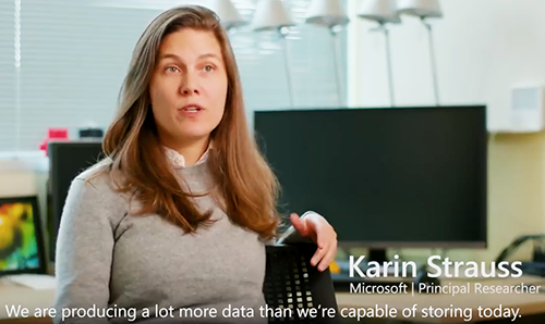
“Our ultimate goal is to put a system into production that, to the end user, looks very much like any other cloud storage service—bits are sent to a datacenter and stored there and then they just appear when the customer wants them,” said Microsoft principal researcher Karin Strauss (above), in the Microsoft news release. “To do that, we needed to prove that this is practical from an automation perspective.” Click here to watch a Microsoft Research video on the DNA storage process. (Photo copyright: Microsoft Research/YouTube.)
It may take some time before DNA becomes a viable medium for
data storage. However, savvy pathology laboratory managers should be aware of,
and possibly prepared for, this coming opportunity.
While it’s unlikely the average consumer will see much
difference in how they save and retrieve data, medical laboratories with the
ability to sequence DNA may find themselves very much in demand because of
their expertise in sequencing DNA and interpreting gene sequences.
Studies show consumer genealogy databases are much broader than is generally known. If your cousins are in such a database, it’s likely you are too
Recent news stories highlighted crime investigators who used the DNA data in consumer genetic genealogy databases to solve cold cases. Though not widely known, such uses of direct-to-consumer DNA databases is becoming more commonplace, which might eventually lead to requests for clinical laboratories to assist in criminal investigations involving DNA data.
Case in point: investigators found the Golden State Killer, a serial killer/rapist/burglar who terrorized multiple California counties over a dozen years in the 1970s to 1980s, after uploading a DNA sample from the crime scene to GEDmatch, an open-data genomics database that features tools for genealogy research. They made the arrest after discovering a distant relative’s DNA in the genealogy database and matching it to the suspect, CBS News revealed in a 60 Minutes Overtime online report.
These and other investigators are using a technique called familial DNA testing (AKA, DNA Profiling), which enables them to use genetic material from relatives to solve crimes.
Clinical laboratories oversee DNA databases. Could DNA databases—developed and managed over years by medical laboratories for patient care—be subpoenaed by law enforcement investigating crimes?
The question raises many issues for society and for labs, including privacy responsibilities and appropriate use of genetic information. On the other hand, the genetic genie is already out of the bottle.
Leveraging Familia DNA to Solve Crimes a New Trend
“The solving of the Golden State Killer case opened this method up as a possibility, and other crime labs are taking advantage of it. Clearly, a trend has started,” Ruth Dickover, PhD, Director of Forensic Science, University of California, Davis, told the Los Angeles Times.
Indeed, the use of familial DNA testing is moving forward. The Verge reported 19 cold case samples have been identified in recent familial DNA testing and public database searches. It also said two new published studies may propel the technique further.
One study, published in the journal Science, suggests nearly every American of European ancestry may soon be identified through familial DNA testing.
The other study, published in Cell, shows that a person’s relatives can be detected when forensic DNA data are compared with consumer genetic databases.
Noah Rosenberg, PhD (above left), Professor of Population Genetics and Society Biology at Stanford University, is shown above working with Jaehee Kim, PhD (right), a Postdoctoral Research Fellow in Biology, on math that could be used to track down relatives in genealogy databases based on forensic DNA. “This could be a way of expanding the reach of forensic genetics, potentially for solving even more cold cases. But at the same time, it could be exposing participants in those databases to forensic searches they might not have anticipated,” he told Wired. (Photo copyright: Stanford University/L.A. Cicero.)
15 Million People Already in Genealogy Databases
Researchers at Columbia University in New York and Hebrew University of Jerusalem told Science they were motivated by the recent trend of investigations leveraging third-party consumer genomics services to find criminals. But they perceived a gap.
“The big limitation is coverage. And even if you find an individual it requires complex analysis from that point,” Yaniv Erlich, PhD, Associate Professor at Columbia and Chief Science Officer at MyHeritage, told The Verge. MyHeritage is an online genealogy platform.
Others offering consumer genetic testing and family history exploration include 23andMe and Ancestry. As of April 2018, more than 15 million people have participated in direct-to-consumer genetic testing, the researchers noted.
The study aimed to find the likelihood that a person can be identified using a long-range familial search. It included these steps and findings:
Statistical analysis of 1.28 million people in the MyHeritage database;
Pairs of people with “identity-by-descent” were removed to avoid bias, such as first cousins and closer relationships;
Researchers aimed at finding a third cousin or closer relatives for each person in the database;
60% of the 1.28 million people were matched with a third cousin or closer relative.
“We project that about 60% of the searches for individuals of European-descent will result in a third cousin or closer match, which can allow their identification using demographic identifiers. Moreover, the technique could implicate nearly any US individual of European descent in the near future,” the researchers wrote.
In an interview with Wired, Erlich added, “The takeaway is it doesn’t matter if you’ve been tested or not tested. You can be identified because the databases already cover such large fractions of the US—at least for European ancestry.”
Matching Forensic and Consumer Genetic Data
Meanwhile, the study published in Cell by researchers at Stanford University, University of California, Davis, and the University of Michigan also suggests investigators could compare forensic DNA samples with consumer genetic databases to find people related to criminals.
That study found:
30% to 32% of people in a forensic database could be related to a child or parent in a consumer database;
35% to 36% could be tied to a sibling.
These studies reveal that genetic data and familial DNA testing can help law enforcement find suspects, which is a good thing for society. But people who uploaded DNA data to some direct-to-consumer databases may find themselves caught up in searches they do not know about. So may their cousins.
Dark Daily recently covered other similar studies that showed it takes just one person’s DNA to reveal genetic information on an entire family. (See, “The Problems with Ancestry DNA Analyses,” October 18, 2018.) These developments in the use of DNA databases to identify criminals should be an early warning to clinical laboratories building databases of genetic information that, at some future point, law enforcement agencies might want access to those databases as part of ongoing criminal investigations.
Goal is to enable gene sequencing data to reside in EMRs, which would provide pathologists and clinical lab professionals with an opportunity to add value
More federal grant money is available to speed up research designed to make it possible to incorporate genome information into the electronic medical record (EMR). This is a development that can have both positive and negative consequences for clinical laboratories and anatomic pathology groups.
The National Institutes of Health (NIH) is awarding more than $48.6 million in grants to researchers seeking to better understand the clinical implications of genomic information and determine the best ways to deliver news to patients when their genetic data indicates they may be predisposed to certain diseases or medical conditions.
The grants are administered by the National Human Genome Research Institute (NHGRI) and represent the third phase of the Electronic Medical Records and Genomics (eMERGE) program. This is a national consortium working to move genomics research closer to clinical application by identifying the potential medical effects of rare genomic variants in about 100 clinically-relevant genes. (more…)