Multiple studies have shown that people with darker skin pigmentation run a higher risk of being misdiagnosed and undertreated than patients with lighter skin due to inaccurate oxygen level readings
Now, scientists at multiple institutions are working to improve the basic pulse oximeter’s design by making it capable of measuring multiple biomarkers, as well as addressing long-standing inaccuracies in the device when used on people with darker skin pigmentation.
This ongoing research demonstrates how new technologies are enabling innovators to add useful functions to standard, well-accepted devices.
Valencia Koomson, PhD (above), Associate Professor, Electrical and Computer Engineering, and head of the Advanced Integrated Circuits and Systems Lab at Tufts University, has developed a pulse oximeter that measures oxygenation in tissue, rather than in blood. Her approach could ensure patients with darker skin pigmentation will be accurately diagnosed at the point-of-care. Though generally not used in clinical laboratory settings, medical technologists will be interested to learn of these new innovations in pulse oximeters. (Photo copyright: Tufts University.)
Measuring Tissues Instead of Blood
The pulse oximeter—a device that attaches to a person’s finger—uses red and infrared light to measure blood oxygen saturation (SpO2) and display pulse rate.
Studies in 2022 that looked into how hospitals administered oxygen to different patients found that inconsistent pulse oximeter readings could cause caregivers to administer less oxygen than is actually needed to people with darker skin pigmentation.
This is because melanin in the skin can interfere with “absorption of light used to measure oxygenated blood in a person’s finger,” according to a National Science Foundation (NSF) news story. Such inaccurate pulse oximeter readings can lead to “inaccurate readings and poorer treatment outcomes” for people with dark skin tones, the NSF wrote.
“Addressing this problem will require innovation in pulse oximeter design and revised regulatory standards,” said Valencia Koomson, PhD, Associate Professor, Electrical and Computer Engineering, Tufts University, Medford, Massachusetts, in the NSF news story.
Koomson, who leads the Advance Integrated Circuit and Systems Lab at Tufts, has developed a prototype pulse oximeter device, which NSF explained, measures oxygenation in biological tissues instead of blood.
“My lab’s work on pulse oximeter devices will provide an alternative technology to address many confounding factors that affect pulse oximeter accuracy, including skin pigmentation, motion artifact, and others,” Koomson said.
National Public Radio (NPR) said Koomson’s device has built-in “technology that can measure a person’s skin tone.”
“We can send more light if there’s a higher level of melanin present, so that melanin doesn’t become a confounding factor that obscures our results,” Koomson told NPR.
Another Pulse Oximeter Redesign
Another new approach to pulse oximetry was developed at Brown University in Providence, Rhode Island.
Rutendo Jakachira, Research Assistant, School of Engineering, and a PhD student in physics, turned to new optical techniques to address the challenge of oxygen saturation levels in dark skin tones, according to a Brown University news release.
Jakachira and Kimani Toussaint, PhD, Professor of Engineering and Senior Associate Dean in the School of Engineering, say they have created possibly the first LED-based light source to emit radially polarized light.
When the LED passes light through a person’s finger, the device calculates the amount of light the hemoglobin in the blood absorbed, NPR explained.
“We did a preliminary study on about five people, and although it was a small study, the results are promising,” said Jakachira, who plans a larger study and clinical trial.
Study Suggests Patients with Darker Skin May Have Received Delayed COVID-19 Care
The researchers analyzed electronic health record (EHR) data from 43,753 patients at Sutter Health in Sacramento, California, who had SpO2 measurements done between January 2020 and February 2022, and 8,735 patients seen for COVID-19 between July 2020 and February 2021 in the hospital’s emergency department.
In their AJE paper, they wrote, “We investigated whether or not pulse oximetry systematically underestimated oxygen saturation in patients who identified as NHB [non-Hispanic Black/African-American] as compared with NHW [non-Hispanic White] counterparts. We also assessed whether or not differences in oxygen saturation measurement affected hospital admission, care delivered, or return to the hospital post discharge among patients with COVID-19.
“We found evidence of differential pulse oximeter measurement error in NHB individuals, resulting in nonrandom overestimation of blood oxygenation as compared with NHW individuals. NHB individuals were also more likely to have hypoxemia [abnormally low oxygen levels in the blood] not detected by pulse oximetry.
“For NHB patients presenting in the ED with COVID-19, we found that overestimation of oxygen saturation was associated with underestimation of the need for admission and underestimation of the need for treatment with dexamethasone and supplemental oxygen. Additionally, we observed associated delays in dexamethasone initiation and initiation of oxygen supplementation.
“There are also broader implications beyond COVID-19, as differential pulse oximeter accuracy has the potential to exacerbate disparities for any condition that relies upon blood oxygenation measurement to inform clinical decision-making.”
Importance of Accurate Readings
Developing pulse oximeters that are accurate for all people, regardless of skin tone, is clearly an important breakthrough. Medical laboratory leaders and pathologists recognize that SpO2 data—along with clinical laboratory test results—are critical for successful diagnostics and treatment. Thus, new technologies that add useful functions to well-accepted devices are positive developments and worth watching.
Two studies show the accuracy of perception-based systems in detecting disease biomarkers without needing molecular recognition elements, such as antibodies
Researchers from multiple academic and research institutions have collaborated to develop a non-conventional machine learning-based technology for identifying and measuring biomarkers to detect ovarian cancer without the need for molecular identification elements, such as antibodies.
Traditional clinical laboratory methods for detecting biomarkers of specific diseases require a “molecular recognition molecule,” such as an antibody, to match with each disease’s biomarker. However, according to a Lehigh University news release, for ovarian cancer “there’s not a single biomarker—or analyte—that indicates the presence of cancer.
“When multiple analytes need to be measured in a given sample, which can increase the accuracy of a test, more antibodies are required, which increases the cost of the test and the turnaround time,” the news release noted.
Unveiled in two sequential studies, the new method for detecting ovarian cancer uses machine learning to examine spectral signatures of carbon nanotubes to detect and recognize the disease biomarkers in a very non-conventional fashion.
“Carbon nanotubes have interesting electronic properties,” said Daniel Heller, PhD (above), in the Lehigh University news release. “If you shoot light at them, they emit a different color of light, and that light’s color and intensity can change based on what’s sticking to the nanotube. We were able to harness the complexity of so many potential binding interactions by using a range of nanotubes with various wrappings. And that gave us a range of different sensors that could all detect slightly different things, and it turned out they responded differently to different proteins.” This method differs greatly from traditional clinical laboratory methods for identifying disease biomarkers. (Photo copyright: Memorial Sloan-Kettering Cancer Center.)
Perception-based Nanosensor Array for Detecting Disease
In the Science Advances paper, the researchers described their development of “a perception-based platform based on an optical nanosensor array that leverages machine learning algorithms to detect multiple protein biomarkers in biofluids.
“Perception-based machine learning (ML) platforms, modeled after the complex olfactory system, can isolate individual signals through an array of relatively nonspecific receptors. Each receptor captures certain features, and the overall ensemble response is analyzed by the neural network in our brain, resulting in perception,” the researchers wrote.
“This work demonstrates the potential of perception-based systems for the development of multiplexed sensors of disease biomarkers without the need for specific molecular recognition elements,” the researchers concluded.
In the Nature Biomedical Engineering paper, the researchers described a fined-tuned toolset that could accurately differentiate ovarian cancer biomarkers from biomarkers in individuals who are cancer-free.
“Here we show that a ‘disease fingerprint’—acquired via machine learning from the spectra of near-infrared fluorescence emissions of an array of carbon nanotubes functionalized with quantum defects—detects high-grade serous ovarian carcinoma in serum samples from symptomatic individuals with 87% sensitivity at 98% specificity (compared with 84% sensitivity at 98% specificity for the current best [clinical laboratory] screening test, which uses measurements of cancer antigen 125 and transvaginal ultrasonography,” the researchers wrote.
“We demonstrated that a perception-based nanosensor platform could detect ovarian cancer biomarkers using machine learning,” said Yoona Yang, PhD, a postdoctoral research associate in Lehigh’s Department of Chemical and Biomolecular Engineering and co-first author of the Science Advances article, in the news release.
How Perception-based Machine Learning Platforms Work
According to Yang, perception-based sensing functions like the human brain.
“The system consists of a sensing array that captures a certain feature of the analytes in a specific way, and then the ensemble response from the array is analyzed by the computational perceptive model. It can detect various analytes at once, which makes it much more efficient,” Yang said.
“SWCNTs have unique optical properties and sensitivity that make them valuable as sensor materials. SWCNTS emit near-infrared photoluminescence with distinct narrow emission bands that are exquisitely sensitive to the local environment,” the researchers wrote in Science Advances.
“Carbon nanotubes have interesting electronic properties,” said Daniel Heller, PhD, Head of the Cancer Nanotechnology Laboratory at Memorial Sloan Kettering Cancer Center and Associate Professor in the Department of Pharmacology at Weill Cornell Medicine of Cornell University, in the Lehigh University news release.
“If you shoot light at them, they emit a different color of light, and that light’s color and intensity can change based on what’s sticking to the nanotube. We were able to harness the complexity of so many potential binding interactions by using a range of nanotubes with various wrappings. And that gave us a range of different sensors that could all detect slightly different things, and it turned out they responded differently to different proteins,” he added.
The researchers put their technology to practical test in the second study. The wanted to learn if it could differentiate symptomatic patients with high-grade ovarian cancer from cancer-free individuals.
The research team used 269 serum samples. This time, nanotubes were bound with a specific molecule providing “an extra signal in terms of data and richer data from every nanotube-DNA combination,” said Anand Jagota PhD, Professor, Bioengineering and Chemical and Biomolecular Engineering, Lehigh University, in the news release.
This year, 19,880 women will be diagnosed with ovarian cancer and 12,810 will die from the disease, according to American Cancer Society data. While more research and clinical trials are needed, the above studies are compelling and suggest the possibility that one day clinical laboratories may detect ovarian cancer faster and more accurately than with current methods.
New nanotechnology device is significantly faster than typical rapid detection clinical laboratory tests and can be manufactured to identify not just COVID-19 at point of care, but other viruses as well
Researchers at the University of Central Florida (UCF) announced the development of an optical sensor that uses nanotechnology to identify viruses in blood samples in seconds with an impressive 95% accuracy. This breakthrough underscores the value of continued research into technologies that create novel diagnostic tests which offer increased accuracy, faster speed to answer, and lower cost than currently available clinical laboratory testing methods.
The innovative UCF device uses nanoscale patterns of gold that reflect the signature of a virus from a blood sample. UCF researchers claim the device can determine if an individual has a specific virus with a 95% accuracy rate. Different viruses can be identified by using their DNA sequences to selectively target each virus.
According to a UCF Today article, the University of Central Florida research team’s device closely matches the accuracy of widely-used polymerase chain reaction (PCR) tests. Additionally, the UCF device provides nearly instantaneous results and has an accuracy rate that’s a marked improvement over typical rapid antigen detection tests (RADT).
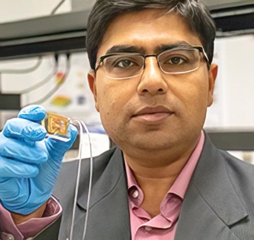
Debashis Chanda, PhD (above), holds up the nanotechnology biosensor he and his team at the University of Central Florida developed that can detect viruses in a blood sample in seconds with 95% accuracy and without the need for pre-preparation of the blood sample. Chanda is professor of physics at the NanoScience Technology Center and the College of Optics and Photonics (CREOL) at UCF. Should this detection device prove effective at instantly detecting viruses at the point of care, clinical laboratories worldwide could have a major new tool in the fight against not just COVID-19, but all viral pathogens. (Photo copyright: University of Central Florida.)
Genetic Virus Detection on a Chip
“The sensitive optical sensor, along with the rapid fabrication approach used in this work, promises the translation of this promising technology to any virus detection, including COVID-19 and its mutations, with high degree of specificity and accuracy,” Debashis Chanda, PhD, told UCF Today. Chanda is professor of physics at the NanoScience Technology Center at UCF and one of the authors of the study. “Here, we demonstrated a credible technique which combines PCR-like genetic coding and optics on a chip for accurate virus detection directly from blood.”
The team tested their device using samples of the Dengue virus that causes Dengue fever, a tropical disease spread by mosquitoes. The device can detect viruses directly from blood samples without the need for sample preparation or purification. This feature enables the testing to be timely and precise, which is critical for early detection and treatment of viruses. The chip’s capability also can help reduce the spread of viruses.
No Pre-processing or Sample Preparation Needed for Multi-virus Testing
The scientists confirmed their device’s effectiveness with multiple tests using varying virus concentration levels and solution environments, including environments with the presence of non-target virus biomarkers.
“A vast majority of biosensors demonstrations in the literature utilize buffer solutions as the test matrix to contain the target analyte,” Chanda told UCF Today. “However, these approaches are not practical in real-life applications because complex biological fluids, such as blood, containing the target biomarkers are the main source for sensing and at the same time the main source of protein fouling leading to sensor failure.”
The researchers believe their device can be easily adapted to detect other viruses and are optimistic about the future of the technology.
“Although there have been previous optical biosensing demonstrations in human serum, they still require off-line complex and dedicated sample preparation performed by skilled personnel—a commodity not available in typical point-of-care applications,” said Abraham Vazquez-Guardado, PhD, a Postdoctoral Fellow at Northwestern University who worked on the study, in the UCS Today article. “This work demonstrated for the first time an integrated device which separated plasma from the blood and detects the target virus without any pre-processing with potential for near future practical usages.”
More research and additional studies are needed to develop the University of Central Florida scientists’ technology and prove its efficacy. However, should the new chip prove viable for point-of-care testing, it would give clinical laboratories and microbiologists an ability to test blood samples without any advanced preparation. Combined with the claims for the device’s remarkable accuracy, that could be a boon not only for COVID-19 testing, but for testing other types of viruses as well.