

How Automated Tools Increase Collected Revenue, Produce More Claims, and Require Less Labor: Unleashing the Power of AI and Machine Learning in Coding/Billing/Collections
How Automated Tools Increase Collected Revenue, Produce More Claims, and Require Less Labor: Unleashing the Power of AI and Machine Learning in Coding/Billing/Collections by Dean Paluch...Diagnosing Ovarian Cancer Using Perception-based Nanosensors and Machine Learning
Two studies show the accuracy of perception-based systems in detecting disease biomarkers without needing molecular recognition elements, such as antibodies
Researchers from multiple academic and research institutions have collaborated to develop a non-conventional machine learning-based technology for identifying and measuring biomarkers to detect ovarian cancer without the need for molecular identification elements, such as antibodies.
Traditional clinical laboratory methods for detecting biomarkers of specific diseases require a “molecular recognition molecule,” such as an antibody, to match with each disease’s biomarker. However, according to a Lehigh University news release, for ovarian cancer “there’s not a single biomarker—or analyte—that indicates the presence of cancer.
“When multiple analytes need to be measured in a given sample, which can increase the accuracy of a test, more antibodies are required, which increases the cost of the test and the turnaround time,” the news release noted.
The multi-institutional team included scientists from Memorial Sloan Kettering Cancer Center, Weill Cornell Medicine, the University of Maryland, the National Institutes of Standards and Technology, and Lehigh University.
Unveiled in two sequential studies, the new method for detecting ovarian cancer uses machine learning to examine spectral signatures of carbon nanotubes to detect and recognize the disease biomarkers in a very non-conventional fashion.

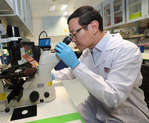
“Carbon nanotubes have interesting electronic properties,” said Daniel Heller, PhD (above), in the Lehigh University news release. “If you shoot light at them, they emit a different color of light, and that light’s color and intensity can change based on what’s sticking to the nanotube. We were able to harness the complexity of so many potential binding interactions by using a range of nanotubes with various wrappings. And that gave us a range of different sensors that could all detect slightly different things, and it turned out they responded differently to different proteins.” This method differs greatly from traditional clinical laboratory methods for identifying disease biomarkers. (Photo copyright: Memorial Sloan-Kettering Cancer Center.)
Perception-based Nanosensor Array for Detecting Disease
The researchers published their findings from the two studies in the journals Science Advances, titled, “A Perception-based Nanosensor Platform to Detect Cancer Biomarkers,” and Nature Biomedical Engineering, titled, “Detection of Ovarian Cancer via the Spectral Fingerprinting of Quantum-Defect-Modified Carbon Nanotubes in Serum by Machine Learning.”
In the Science Advances paper, the researchers described their development of “a perception-based platform based on an optical nanosensor array that leverages machine learning algorithms to detect multiple protein biomarkers in biofluids.
“Perception-based machine learning (ML) platforms, modeled after the complex olfactory system, can isolate individual signals through an array of relatively nonspecific receptors. Each receptor captures certain features, and the overall ensemble response is analyzed by the neural network in our brain, resulting in perception,” the researchers wrote.
“This work demonstrates the potential of perception-based systems for the development of multiplexed sensors of disease biomarkers without the need for specific molecular recognition elements,” the researchers concluded.
In the Nature Biomedical Engineering paper, the researchers described a fined-tuned toolset that could accurately differentiate ovarian cancer biomarkers from biomarkers in individuals who are cancer-free.
“Here we show that a ‘disease fingerprint’—acquired via machine learning from the spectra of near-infrared fluorescence emissions of an array of carbon nanotubes functionalized with quantum defects—detects high-grade serous ovarian carcinoma in serum samples from symptomatic individuals with 87% sensitivity at 98% specificity (compared with 84% sensitivity at 98% specificity for the current best [clinical laboratory] screening test, which uses measurements of cancer antigen 125 and transvaginal ultrasonography,” the researchers wrote.
“We demonstrated that a perception-based nanosensor platform could detect ovarian cancer biomarkers using machine learning,” said Yoona Yang, PhD, a postdoctoral research associate in Lehigh’s Department of Chemical and Biomolecular Engineering and co-first author of the Science Advances article, in the news release.
How Perception-based Machine Learning Platforms Work
According to Yang, perception-based sensing functions like the human brain.
“The system consists of a sensing array that captures a certain feature of the analytes in a specific way, and then the ensemble response from the array is analyzed by the computational perceptive model. It can detect various analytes at once, which makes it much more efficient,” Yang said.
The “array” the researchers are referring to are DNA strands wrapped around single-wall carbon nanotubes (DNA-SWCNTs).
“SWCNTs have unique optical properties and sensitivity that make them valuable as sensor materials. SWCNTS emit near-infrared photoluminescence with distinct narrow emission bands that are exquisitely sensitive to the local environment,” the researchers wrote in Science Advances.
“Carbon nanotubes have interesting electronic properties,” said Daniel Heller, PhD, Head of the Cancer Nanotechnology Laboratory at Memorial Sloan Kettering Cancer Center and Associate Professor in the Department of Pharmacology at Weill Cornell Medicine of Cornell University, in the Lehigh University news release.
“If you shoot light at them, they emit a different color of light, and that light’s color and intensity can change based on what’s sticking to the nanotube. We were able to harness the complexity of so many potential binding interactions by using a range of nanotubes with various wrappings. And that gave us a range of different sensors that could all detect slightly different things, and it turned out they responded differently to different proteins,” he added.
The researchers put their technology to practical test in the second study. The wanted to learn if it could differentiate symptomatic patients with high-grade ovarian cancer from cancer-free individuals.
The research team used 269 serum samples. This time, nanotubes were bound with a specific molecule providing “an extra signal in terms of data and richer data from every nanotube-DNA combination,” said Anand Jagota PhD, Professor, Bioengineering and Chemical and Biomolecular Engineering, Lehigh University, in the news release.
This year, 19,880 women will be diagnosed with ovarian cancer and 12,810 will die from the disease, according to American Cancer Society data. While more research and clinical trials are needed, the above studies are compelling and suggest the possibility that one day clinical laboratories may detect ovarian cancer faster and more accurately than with current methods.
—Donna Marie Pocius
Related Information:
Perception-Based Nanosensor Platform Could Advance Detection of Ovarian Cancer
Perception-Based Nanosensor Platform to Detect Cancer Biomarkers
Machine Learning Nanosensor Platform Detects Early Cancer Biomarkers
Researchers Use Machine Learning to Identify Thousands of New Marine RNA Viruses in Study of Interest to Microbiologists and Clinical Laboratory Scientists
Screening and analysis of ocean samples also identified a possible missing link in how the RNA viruses evolved
An international team of scientists has used genetic screening and machine learning techniques to identify more than 5,500 previously unknown species of marine RNA viruses and is proposing five new phyla (biological groups) of viruses. The latter would double the number of RNA virus phyla to 10, one of which may be a missing link in the early evolution of the microbes.
Though the newly-discovered viruses are not currently associated with human disease—and therefore do not drive any current medical laboratory testing—for virologists and other microbiologists, “a fuller catalog of these organisms is now available to advance scientific understanding of how viruses evolve,” said Dark Daily Editor-in-Chief Robert Michel.
“While scientists have cataloged hundreds of thousands of DNA viruses in their natural ecosystems, RNA viruses have been relatively unstudied,” wrote four microbiologists from Ohio State University (OSU) who participated in the study in an article they penned for The Conversation.
The OSU study authors included:
- Ahmed Zayed, PhD, research scientist, Dept. of Microbiology, OSU.
- Matthew Sullivan, PhD, Professor of Microbiology and Director of the Center of Microbiome Science at OSU.
- Guillermo Dominguez Huerta, PhD, scientific consultant at the Sullivan Lab, Department of Microbiology, OSU.
- James Wainaina, PhD, postdoctoral scholar, Dept. of Microbiology, OSU.
Zayed was lead author of the study and Sullivan led the OSU research team.
The researchers published their findings in the journal Science, titled, “Cryptic and Abundant Marine Viruses at the Evolutionary Origins of Earth’s RNA Virome.”

RNA versus DNA Viruses
In contrast to the better-understood DNA virus, an RNA virus contains RNA instead of DNA as its genetic material, according to Samanthi Udayangani, PhD, in an article she penned for Difference Between. Examples of RNA viruses include:
One major difference, she explains, is that RNA viruses mutate at a higher rate than do DNA viruses.
The OSU scientists identified the new species by analyzing a database of RNA sequences from plankton collected during a series of ocean expeditions aboard a French schooner owned by the Tara Ocean Foundation.
“Plankton are any aquatic organisms that are too small to swim against the current,” the authors explained in The Conversation. “They’re a vital part of ocean food webs and are common hosts for RNA viruses.”
The team’s screening process focused on the RNA-dependent RNA polymerase (RdRp) gene, “which has evolved for billions of years in RNA viruses, and is absent from other viruses or cells,” according to the OSU news story.
“RdRp is supposed to be one of the most ancient genes—it existed before there was a need for DNA,” Zayed said.
The RdRp gene “codes for a particular protein that allows a virus to replicate its genetic material. It is the only protein that all RNA viruses share because it plays an essential role in how they propagate themselves. Each RNA virus, however, has small differences in the gene that codes for the protein that can help distinguish one type of virus from another,” the study authors explained.
The screening “ultimately identified over 44,000 genes that code for the virus protein,” they wrote.
Identifying Five New Phyla
The researchers then turned to machine learning to organize the sequences and identify their evolutionary connections based on similarities in the RdRp genes.
“The more similar two genes were, the more likely viruses with those genes were closely related,” they wrote.
The technique classified many of the sequences within the five previously known phyla of RNA viruses:
But the researchers also identified five new phyla—including two dubbed “Taraviricota” and “Arctiviricota”—that “were particularly abundant across vast oceanic regions,” they wrote. Taraviricota is named after the Tara expeditions and Arctiviricota gets its name from the Arctic Ocean.
They speculated that Taraviricota “might be the missing link in the evolution of RNA viruses that researchers have long sought, connecting two different known branches of RNA viruses that diverged in how they replicate.”
In addition to the five new phyla, the researchers are proposing at least 11 new classes of RNA viruses, according to the OSU story. The scientists plan to issue a formal proposal to the International Committee on Taxonomy of Viruses (ICTV), the body responsible for classification and naming of viruses.
Studying RNA Viruses Outside of Disease Environments
“As the COVID-19 pandemic has shown, RNA viruses can cause deadly diseases. But RNA viruses also play a vital role in ecosystems because they can infect a wide array of organisms, including microbes that influence environments and food webs at the chemical level,” wrote the four study authors in The Conversation. “Mapping out where in the world these RNA viruses live can help clarify how they affect the organisms driving many of the ecological processes that run our planet. Our study also provides improved tools that can help researchers catalog new viruses as genetic databases grow.”
This remarkable study, which was partially funded by the US National Science Foundation, will be most intriguing to virologists and microbiologists. However, clinical laboratories also should be interested in the fact that the catalog of known viruses has just expanded by 5,500 types of RNA viruses.
—Stephen Beale
Related Information:
Cryptic and Abundant Marine Viruses at the Evolutionary Origins of Earth’s RNA Virome
There’s More to RNA Viruses than Diseases
Differences Between DNA and RNA Viruses
Ocean Water Samples Yield Treasure Trove of RNA Virus Data
Global Survey of Marine RNA Viruses Sheds Light on Origins and Abundance of Earth’s RNA Virome
Scientists Find Trove of over 5,000 New Viruses Hidden in Oceans
Virologists Identify More than 5,000 New Viruses in the Ocean
Proof of Concept Study Demonstrates Machine Learning and AI Can Identify Cancer Cells Based on pH Levels; May Have Applications in Surgical Pathology
The new method employs a pH sensitive dye and AI algorithms to ‘distinguish between cells originating from normal and cancerous tissue, as well as among different types of cancer’ the researchers said
Might a pH-sensitive dye in tandem with an image analysis solution soon be used to identify cancerous cells within blood samples as well within tissue? Recent research indicates that could be a possibility. If further studies and clinical trials confirm this capability, then anatomic pathologists could gain another valuable tool to use in diagnosing cancers and other types of disease.
Currently, surgical pathologists use a variety of hematoxylin and eosin stains (H/E) to bring out useful features in cells and cell structures. So, staining tissue on glass slides is a common practice. Now, thanks to machine learning and artificial intelligence, anatomic pathologists may soon have a similar tool for spotting cancer cells within both tissue and blood samples.
Researchers at the National University of Singapore (NUS) have developed a method for identifying cancer that uses a pH sensitive dye called bromothymol blue. The dye reacts to various levels of acidity in cancer cells by turning colors. “The pH inside cancer cells tends to be higher than that of healthy cells. This phenomenon occurs at the very early phases of cancer development and becomes amplified as it progresses,” Labroots reported.
In “Machine Learning Based Approach to pH Imaging and Classification of Single Cancer Cells,” published in the journal APL Bioengineering, the NUS researchers wrote, “Here, we leverage a recently developed pH imaging modality and machine learning-based single-cell segmentation and classification to identify different cancer cell lines based on their characteristic intracellular pH. This simple method opens up the potential to perform rapid noninvasive identification of living cancer cells for early cancer diagnosis and further downstream analyses.”
According to an NUS news release, the bromothymol blue dye is “applied onto patients’ cells” being held ex vivo in cell culture dishes. The dye’s color changes depending on the acidity level of the cancer cells it encounters. Microscopic images of the now-visible cancers cells are taken, and a machine-learning algorithm analyzes the images before generating a report for the anatomic pathologist.
The NUS researchers claim the test can provide answers in about half an hour with 95% accuracy, Labroots reported.
“The ability to analyze single cells is one of the holy grails of health innovation for precision medicine or personalized therapy. Our proof-of-concept study demonstrates the potential of our technique to be used as a fast, inexpensive and accurate tool for cancer diagnosis,” said Lim Chwee Teck, PhD, NUS Society Professor and Director of NUS’ Institute for Health Innovation and Technology, in the NUS news release.
The novel technique for differentiating cancer cells from non-cancerous cells being developed at the National University of Singapore (NUS) could eventually become useful in detecting cancer cells in tissue samples, either obtained from tumor biopsies or blood samples. “As the number of cells in these samples can be in millions or even billions, the ability to detect the very few cancer cells among the others will be useful for clinicians,” NUS Society Professor and Director of NUS’ Institute for Health Innovation and Technology, Lim Chwee Teck, PhD (above) told The Straits Times. (Photo copyright: The Straits Times.)
AI Cell Analysis versus Laborious Medical Laboratory Steps
By developing an AI-driven method, Professor Lim and the NUS team sought to improve upon time-consuming techniques for identifying cells that traditionally involve using florescent probes, nanoparticles, and labeling steps, or for cells to be fixed or terminated.
“Unlike other cell analysis techniques, our approach uses simple, inexpensive equipment, and does not require lengthy preparation and sophisticated devices. Using AI, we are able to screen cells faster and accurately,” Professor Lim told Labroots. “Furthermore, we can monitor and analyze living cells without causing any toxicity to the cells or the need to kill them.”
The new technique may have implications for cancer detection in tumor tissue as well as in liquid biopsies.
“We are also exploring the possibility of performing the real-time analysis on circulating cancer cells suspended in blood,” Professor Lim said in the NUS news release. “One potential application for this would be in liquid biopsy where tumor cells that escaped from a primary tumor can be isolated in a minimally-invasive fashion from bodily fluids such as blood.”
Diagnosing Cancer in Real Time
The NUS’ method requires more research and clinical studies before it could become an actual tool for anatomic pathologists and other cancer diagnosticians. Additionally, the NUS researchers acknowledged that the focus on only four cell lines (normal cells, benign breast tumor cells, breast cancer cells, and pancreatic cancer cells) limited their study, as did lack of comparison with conventional florescent pH indicators.
Still, the NUS scientists are already planning more studies to advance their concept to different stages of cell malignancy. They envision a “real-time” version of the technique to enable recognition of cells and fast separation of those that need to be referred to clinical laboratories for molecular testing and/or genetic sequencing.
Medical laboratory leaders may want to follow the NUS study. An inexpensive AI-driven method that can accurately detect and classify cancer cells based on pH within the cells is provocative and may be eventually become integrated with other cancer diagnostics.
—Donna Marie Pocius
Related Information
Machine Learning-Based Approach to pH Imaging and Classification of Single Cancer Cells
Machine Learning Can Identify Cancerous Cells by Their Acidity
AI Test Distinguishes Cancer Cells from Healthy Ones Based on Acidity Levels
