Development of the Critical Values system redefined what STAT means in clinical laboratory testing turnaround times
Where did the concept of critical values and having clinical laboratories report them to referring physicians originate? How did the concept blossom into a standard practice in laboratory medicine? Given the importance of critical values, a lookback into how this aspect of laboratory medicine was developed is helpful to understand how and why this has become an essential element in the practice of medicine and an opportunity for labs to add value in patient care.
According to Stanford Medicine, critical/panic values are defined as “values that are outside the normal range to a degree that may constitute an immediate health risk to the individual or require immediate action on the part of the ordering physician.”
What you’ll read below is an insider’s account of the “birth of critical values reporting.”
According to Lundberg, an unaccompanied man was brought to the hospital in a coma and an examination revealed a laceration to his scalp. The patient was admitted to the neurosurgical unit where clinical laboratory tests were performed, including a complete blood count (CBC) analysis, urinalysis, and serum electrolytes. All the test results came back normal except the patient’s serum glucose (blood sugar level) which was 6 mg% in concentration.
“The hard-copy laboratory results were returned to the ward of origin within two hours of receipt of the specimens in the laboratory. However, the results were not noticed by the house officers who were busy with several other seriously ill patients. Ward personnel also failed to communicate the lab results to the responsible physicians,” Lundberg wrote.
When hospital staff did finally notice the test result the next morning glucose was immediately administered to the patient, but it was too late to prevent irreversible brain damage. The man soon passed away.
Following this incident, the hospital developed a “Critical Value Recognition and Reporting System.” The system generated new numbers that were termed “Panic Values.”
However, “critics complained that good doctors should never panic, so the name was changed to Critical Values,” Lundberg explained.
When any of these critical test values were out of the norm, “we required the responsible laboratory person to quickly verify the result and use the telephone (long before laboratory computers) to personally notify a responsible individual (no messages left) who agreed to find a physician who could quickly act on the result. All was documented with times and names,” he wrote.
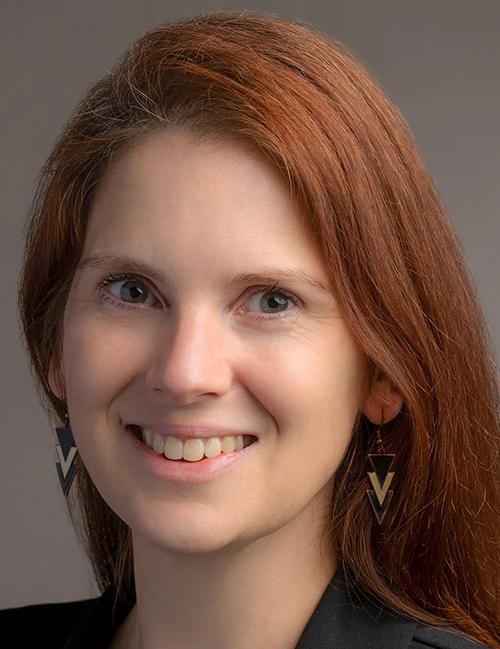
“We understand that when a physician wants something, he/she wants it, no matter what. Well, in this patient-focused approach, the physician cannot have it, except as offered by the patient-focused approach, based on TAT [turnaround times of clinical laboratory tests],” wrote George Lundberg, MD (above), President and Chair of the Board of Directors of the Lundberg Institute, and Clinical Professor of Pathology at Northwestern University in an article he penned for the National Medical Journal of India (Photo copyright: Dark Intelligence Group. Shows Dr. Lundberg in 2011 addressing the Executive War College on Diagnostics, Clinical Laboratory, and Pathology Management.)
New Clinical Laboratory Standards
Recognition of the urgency to adopt new hospital standards related to certain clinical laboratory test results came swiftly. In 1972, Lundberg was invited to publish an article explaining the new Critical Value Recognition and Reporting System in Medical Laboratory Observer.
According to Lundberg, “most laboratory tests that are done do not need to be done; the results are either negative, normal, or show no change from a prior result. But some are crucial.”
The original set of Critical Values included the following testing results:
The list of values were later expanded to include “vital values.” These values describe lab results for which “action” is important, but where timing is less urgent. Examples of vital values include:
Lundberg and his colleagues went on to redefine what constitutes a laboratory test and what renders a test successful. They discussed laboratory procedures with committees of clinicians, lab personnel and patients, and reorganized hematology, chemistry, and toxicology based on the turnaround time (TAT) of tests.
“We ‘started the clock’—any and all days/times 24×7—when a specimen arrived at some place within the laboratory, and stopped the clock when a final result was available somewhere in the laboratory,” Lundberg wrote in NMJI. “We categorized all tests as: less than one hour, less than four hours, less than 24 hours, and more than 24 hours, guaranteed, 24×7. As a trade-off, we abolished the concept of ‘STAT’ orders … NO EXCEPTIONS. The rationale of each TAT was the speed with which a result was needed to render proper medical care that mattered to the welfare of the patient, and, of course, that was technically possible.”
Since then, very little has changed for the Critical Values System over the past 50 years. The majority of values added have fallen under the “Vital” category and not the “Critical” category. Today, most health systems and clinical laboratories create their own internal processes and procedures regarding which values need to be reported immediately (critical), which values are not urgent (vital), and how those results should be handled.
Clinical laboratories nationwide could follow Yale’s example and enact programs to bring much needed lab services to traditionally underserved communities
Ever since the COVID-19 pandemic drove up demand for telehealth medical services, mobile clinical laboratories have grown in popularity as well, especially among residents of remote and traditionally underserved communities. Now, several divisions of Yale University are getting in on the trend.
“Using a van retrofitted with laboratory-grade diagnostic equipment, the mobile clinic will employ SalivaDirect—a saliva-based COVID-19 PCR test developed at YSPH—to facilitate on-site testing with a turnaround time of two to three hours,” Yale Daily News reported.
Funded by a federal grant, the initial goal was to provide 400 free COVID-19 tests, but the program has exceeded that number. By April 10, the mobile lab had been deployed more than 60 times, appearing at events and pop-up sites throughout various communities in Connecticut, including regular stops at the WHEAT Food Pantry of West Haven.
“[The clinical laboratory-in-a-van] is a brilliant way to reduce the barriers to testing, instead taking the lab to communities who may be less likely—or unable—to access the necessary clinic or labs,” microbiologist Anne Wyllie, PhD, a research scientist who helped develop the PCR test deployed by the mobile lab told Yale Daily News. Wyllie works in the Department of Epidemiology of Microbial Diseases at Yale School of Public Health. “We are actively working with our community partners to identify how we can best serve their communities,” she added. (Photo copyright: Yale School of Medicine.)
Mobile Lab’s Capabilities
Collecting samples, processing, and delivering same-day COVID-19 results was the initial goal but that plan has expanded, Yale School of Medicine noted in a news release.
“Same-day onsite delivery of test results is an added benefit for communities and individuals without access to Wi-Fi or the ability to receive private health information electronically,” Yale added.
The mobile van is staffed with trained clinical laboratory technicians as well as community health navigators who provide both healthcare information and proper follow-up connections as needed for patients who receive positive COVID-19 results. The van runs off power from outdoor electrical outlets at each location and currently serves historically underserved populations in Hartford, Middlesex, Fairfield, New Haven, and New London counties, Yale noted.
“The van allows us to bring our services, as well as healthcare information, directly to communities where they are needed,” said Angelique Levi, MD, Associate Professor, Vice Chair and Director of Pathology Reference Services, and CLIA Laboratory Medical Director in the Department of Pathology at Yale University School of Medicine in a news release.
Launch of a High Complexity Molecular Lab on Wheels
YPL and YSPH collaborated to make the mobile lab a reality. YSPH created the saliva-based COVID-19 test and YPL “provided clinical validation necessary to get the testing method ready for emergency use authorization by the US Food and Drug Administration,” Yale noted.
“YPL recognized the need to be closer to the front lines of patient care and that retrofitting a fully licensed, high complexity molecular laboratory into a consumer-sized van was the right next step,” Chen Liu, MD, PhD, Chair of the Department of Pathology at Yale School of Medicine, noted in a Yale news release. This “gives us options to efficiently deliver accurate diagnostic information when and where it’s needed,” he added.
Throughout the COVID-19 pandemic, the Connecticut Department of Public Health, the City of New Haven, and various community organizations partnered with YPL, YSPH, and the SalivaDirect team to offer free SARS-CoV-2 testing to the public at two different sites in New Haven.
Principal investigators Levi and microbiologist Anne Wyllie, PhD, who helped develop the PCR test deployed by the mobile, lab led the Yale lab-in-a-van research project.
Flambeau Diagnostics, a biomedical company that specializing in mobile lab testing, worked with the Yale team to design and implement the mobile lab van.
“According to Wyllie, the new YSPH and YPL initiative utilizes one of the former Flambeau vans that had been retrofitted for clinical testing,” a Yale news release noted.
Kat Fajardo, Laboratory Manager at Yale University, added custom pieces of equipment to ensure seamless PCR testing. One was a Magnetic Induction Cycler (Mic) measuring only six by six inches. The Mic allowed for measurement of 46 biological specimens, while it’s diminutive size freed up space on the van’s countertop. This allowed lab techs to process specimens concurrently while also providing COVID-19 testing, according to a Yale news release.
Additionally, the van has a Myra portable robotic liquid handler which is “designed to automate the process of moving clinical specimens between vials,” the news release notes.
“What we wanted to do is run high complexity testing in the van, with a reduced timeframe, and then be able to get the results out to the patients as soon as we possibly could,” Fajardo stated.
Exploring the Mobile Laboratory’s Potential
According to a news release, YPL and YSPH consult with community partners to select locations for the mobile lab to visit. These partners include:
APT Foundation (New Haven County, in addition to others.
Although the van was initially used to provide SalivaDirect COVID-19 testing to vulnerable populations, YPL is working with its partners in those communities to identify other testing needs beyond COVID.
The Yale team is considering additional offerings and support such as the addition of a social worker as well as expanding lung health awareness beyond COVID-19 to other respiratory diseases. Also under consideration:
Vaccinations including for COVID-19 and Hepatitis B, and
Health education and materials for harm reduction and STI prevention, a Yale news release noted.
Yale’s Laboratory-in-a-Van program is a consumer-facing effort that is bringing much needed clinical lab services to traditionally underserved communities in Connecticut. Clinical laboratories throughout the nation could do the same with remote or homebound patients who cannot reach critical care.
New guidelines also advise people to limit their vitamin D supplementation to recommended daily doses
Clinical laboratories may eventually receive fewer doctors’ orders for vitamin D testing thanks to new guidelines released by the Endocrine Society. The new Clinical Practice Guideline advises against “unnecessary testing for vitamin D levels.” It also urges healthy people, and those 75-years of age or younger, to avoid taking the vitamin at levels above the daily recommended amounts, according to a news release.
Even though the Endocrine Society does recommend vitamin D supplements for certain groups, it advises individuals to hold off on routine testing. That’s because there appears to be uncertainty among ordering clinicians about what to do for patients based on their vitamin D test results.
“When clinicians measure vitamin D, they’re forced to decide what to do about it. That’s where questions about the levels come in. And that’s a big problem. So, what this panel is saying is ‘Don’t screen,’” Clifford Rosen, MD, Director of Clinical and Translational Research and Senior Scientist, Maine Medical Center Research Institute at the University of Maine, told Medscape Medical News.
“We have no data that there’s anything about screening that allows us to improve quality of life. Screening is probably not worthwhile in any age group,” he added.
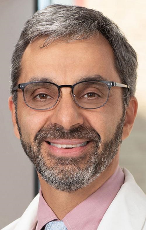
“This guideline refers to people who are otherwise healthy, and there’s no clear indication for vitamin D, such as people with already established osteoporosis. This guideline is not relevant to them,” the author of the Endocrine Society guideline, Anastassios G. Pittas, MD (above), Professor of Medicine at Tufts University School of Medicine in Boston, told Medscape Medical News. This new guideline could result in doctors ordering fewer vitamin D tests from clinical laboratories. (Photo copyright: Tufts University.)
Vitamin D Screening Not Recommended for Certain Groups
The Endocrine Society’s new clinical guidelines advise healthy adults under 75 years of age to refrain from taking vitamin D supplements that exceed US Institute of Medicine—now the National Academy of Medicine (NAM)—recommendations.
Additionally, these updated guidelines:
Recommend vitamin D supplements at levels above NAM recommendations to help lower risks faced by children 18 years and younger, adults 75 and older, pregnant women, and people with prediabetes.
Suggest daily, lower-dose vitamin D (instead of non-daily, higher-dose of the vitamin) for people 50 years and older who have “indications for vitamin D supplementation or treatment.”
Advise “against routine testing for 25-hydroxyvitamin D [aka, calcifediol] levels” in all the above groups “since outcome-specific benefits based on these levels have not been identified. This includes 25-hyrdoxyvitamin D screening in people with dark complexion or obesity.”
One exception to the guideline applies to people with already established osteoporosis, according to the guideline’s author endocrinologist Anastassios G. Pittas, MD, Chief of Endocrinology, Diabetes and Metabolism; Co-Director, Tuft’s Diabetes and Lipid Center; and Professor of Medicine at Tufts University School of Medicine in Boston.
Vitamin D’s Link to Disease Studied
During a panel discussion at the Endocrine Society’s annual meeting, members acknowledged that many studies have shown relationships between serum concentrations of 25-hydroxy vitamin D (25(OH)D) and physical disorders including those of musculoskeletal, metabolic, and cardiovascular systems. Still, they questioned the link of vitamin D supplementation and testing with disease prevention.
“There is paucity of data regarding definition of optimal levels and optimal intake of vitamin D for preventing specific diseases. … What we really need are large-scale clinical trials and biomarkers so we can predict disease outcome before it happens,” said Panel Chair Marie Demay, MD, Endocrinologist, Massachusetts General Hospital, and Professor of Medicine, Harvard Medical School, Boston, Medscape Medical News reported.
Meanwhile, in their Journal of Clinical Endocrinology and Metabolism paper, the researchers note that use of supplements (1,000 IU or more per day) increased from 0.3% to 18.2%, according to the National Health and Nutrition Examination Survey (NHANES) conducted by the National Center for Health Statistics (NCHS), CDC, for the years 1999-2000 and 2013-2014.
“The use of 25(OH)D testing in clinical practice has also been increasing; however, the cost effectiveness of widespread testing has been questioned, especially given the uncertainty surrounding the optimal level of 25(OH)D required to prevent disease,” the authors wrote.
“Thus, the panel suggests against routine 25(OH)D testing in all populations considered,” the researchers stated at the Endocrine Society annual meeting.
Other Groups Weigh-in on Vitamin D Testing
Pathologists and medical laboratory leaders may recall the explosion in vitamin D testing starting about 20 years ago. Vitamin D testing reimbursed by Medicare Part B “increased 83-fold” during the years 2000 to 2010, according to data cited in an analysis by the American Academy of Family Physicians (AAFP).
Also, the US Preventive Services Task Force (USPSTF) said in a statement that there is not enough information to “recommend for or against” testing for vitamin D deficiency.
“No organization recommends population-based screening for vitamin D deficiency, and the American Society for Clinical Pathology recommends against it,” the USPSTF noted.
Clinical Laboratories Can Get the Word Out
The vitamin D debate has been going on for a while. And the latest guidance from the Endocrine Society may cause physicians and patients to stop ordering vitamin D tests as part of annual physicals or in routine screenings.
Medical laboratories can provide value by ensuring physicians and patients have the latest information about vitamin D test orders, reports, and interpretation.
Infection control teams and clinical laboratory managers may want to look at this new product designed to improve the diagnosis and treatment of sepsis
Accurate and fast diagnosis of sepsis for patients arriving in emergency departments is the goal of a new product that was just cleared by the federal Food and Drug Administration (FDA). It is also the newest example of how artificial intelligence (AI) continues to find its way into pathology and clinical laboratory medicine.
Sepsis is one of the deadliest killers in US hospitals. That is why there is interest in the recent action by the FDA to grant marketing authorization for an AI-powered sepsis detection software through the agency’s De Novo Classification Request. The DNCR “provides a marketing pathway to classify novel medical devices for which general controls alone, or general and special controls, provide reasonable assurance of safety and effectiveness for the intended use, but for which there is no legally marketed predicate device,” the FDA’s website states.
Unlike a single analyte assay that is run in a clinical laboratory, Prenosis’ AI/ML software uses 22 diagnostic and predictive parameters, along with ML algorithms, to analyze data and produce a clinically actionable answer on sepsis.
It is important for clinical laboratory managers and pathologists to recognize that this diagnostic approach to sepsis brings together a number of data points commonly found in a patient’s electronic health record (EHR), some of which the lab generated and others the lab did not generate.
“Sepsis is a serious and sometimes deadly complication. Technologies developed to help prevent this condition have the potential to provide a significant benefit to patients,” said Jeff Shuren, MD, JD, Director of the FDA’s Center for Devices and Radiological Health, in a statement. “The FDA’s authorization of the Prenosis Sepsis ImmunoScore software establishes specific premarket and post-market requirements for this device type.” Clinical laboratory EHRs contain some of the data points Prenosis’ diagnostic software uses. (Photo copyright: US Food and Drug Administration.)
How it Works
To assist doctors diagnose sepsis, the ImmunoScore software is first integrated into the patient’s hospital EHR. From there, it leverages 22 parameters including:
White blood cell count to produce a score that informs caregivers of the patient’s risk for sepsis within 24 hours, MedTech Dive reported.
Instead of requiring a doctor or nurse to look at each parameter separately, the SaMD tool uses AI “to evaluate all those markers at once”, CNBC noted. It then produces a risk score and four discrete risk stratification categories (low, medium, high, and very high) which correlate to “a patient’s risk of deterioration” represented by:
By sharing these details—a number from one to 100 for each of the 22 diagnostic and predictive parameters—Sepsis ImmunoScore helps doctors determine which will likely contribute most to the patient’s risk for developing sepsis, MedTech Dive reported.
“A lot of clinicians don’t trust AI products for multiple reasons. We are trying very hard to counter that skepticism by making a tool that was validated by the FDA first, and then the second piece is we’re not trying to replace the clinician,” Bobby Reddy Jr., PhD, Prenosis co-founder and CEO, told MedTech Dive.
Big Biobank and Blood Sample Data
Prenosis, which says its goal is the “enabling [of] precision medicine in acute care” developed Sepsis ImmunoScore using the company’s own biobank and a dataset of more than 100,000 blood samples from more than 25,000 patients.
AI algorithms drew on this biological/clinical dataset—the largest in the world for acute care patients suspected of having serious infections, according to Prenosis—to “elucidate patterns in rapid immune response.”
“It does not work without data, and the data started at Carle,” said critical care specialist Karen White, MD, PhD, Carle Foundation Hospital, St. Louis, MO, in the news release. “The project involved a large number of physicians, research staff, and internal medicine residents at Carle who helped recruit patients, collect data, and samples,” she said.
Opportunity for Clinical Laboratories
Sepsis is a life-threatening condition based on an “extreme response to an infection” that affects nearly 1.7 million adults in the US each year and is responsible for 350,000 deaths, according to US Centers for Disease Control and Prevention (CDC) data.
A non-invasive diagnostic tool like Sepsis ImmunoScore will be a boon to emergency physicians and the patients they treat. Now that the FDA has authorized the SaMD diagnostic tool to go to market, it may not be long before physicians can use the information it produces to save lives.
Clinical laboratory managers inspired by the development of Sepsis ImmunoScore may want to look for similar ways they can take certain lab test results and combine them with other data in an EHR to create intelligence that physicians can use to better treat their patients. The way forward in laboratory medicine will be combining lab test results with other relevant sets of data to create clinically actionable intelligence for physicians, patients, and payers.
Ten year collaboration between Google and Harvard may lead to a deeper understanding of the brain and new clinical laboratory diagnostics
With all our anatomic pathology and clinical laboratory science, we still do not know that much about the structure of the brain. But now, scientists at Harvard University and Google Research studying the emerging field of connectomics have published a highly detailed 3D reconstruction of human brain tissue that allows visualization of neurons and their connections at unprecedented nanoscale resolutions.
Further investigation of the nano-connections within the human brain could lead to novel insights about the role specific proteins and molecules play in the function of the brain. Though it will likely be years down the road, data derived from this study could be used to develop new clinical laboratory diagnostic tests.
The data to generate the model came from Google’s use of artificial intelligence (AI) algorithms to color-code Harvard’s electron microscope imaging of a cubic millimeter of neural tissue—equivalent to a half-grain of rice—that was surgically removed from an epilepsy patient.
“That tiny square contains 57,000 cells, 230 millimeters of blood vessels, and 150 million synapses, all amounting to 1,400 terabytes of data,” according to the Harvard Gazette, which described the project as “the largest-ever dataset of human neural connections.”
“A terabyte is, for most people, gigantic, yet a fragment of a human brain—just a minuscule, teeny-weeny little bit of human brain—is still thousands of terabytes,” said neuroscientist Jeff W. Lichtman, MD, PhD, Jeremy R. Knowles Professor of Molecular and Cellular Biology, whose Lichtman Lab at Harvard University collaborated on the project with researchers from Google. The two labs have been working together for nearly 10 years on this project, the Harvard Gazette reported.
Lichtman’s lab focuses on the emerging field of connectomics, defined “as understanding how individual neurons are connected to one another to form functional networks,” said neurobiologist Wei-Chung Allen Lee, PhD, Assistant Professor of Neurology, Harvard Medical School, in an interview with Harvard Medical News. “The goal is to create connectomes—or detailed structural maps of connectivity—where we can see every neuron and every connection.” Lee was not involved with the Harvard/Google Research study.
“The human brain uses no more power than a dim incandescent light bulb, yet it can accomplish feats still not possible with the largest artificial computing systems,” wrote Google Research scientist Viren Jain, PhD (above), in a blog post. “To understand how requires a level of understanding more profound than knowing what part of the brain is responsible for what function. The field of connectomics aims to achieve this by precisely mapping how each cell is connected to others.” Google’s 10-year collaboration with Harvard University may lead to new clinical laboratory diagnostics. (Photo copyright: Google Research.)
Study Data and Tools Freely Available
Along with the Science paper, the researchers publicly released the data along with analytic and visualization tools. The study noted that the dataset “is large and incompletely scrutinized,” so the scientists are inviting other researchers to assist in improving the model.
“The ability for other researchers to proofread and refine this human brain connectome is one of many ways that we see the release of this paper and the associated tools as not only the culmination of 10 years of work, but the beginning of something new,” wrote Google Research scientist Viren Jain, PhD, in a blog post that included links to the online resources.
One of those tools—Neuroglancer—allows any user with a web browser to view 3D models of neurons, axons, synapses, dendrites, blood vessels, and other objects. Users can rotate the models in xyz dimensions.
Users with the requisite knowledge and skills can proofread and correct the models by signing up for a CAVE (Connectome Annotation Versioning Engine) account.
Researchers Found Several Surprises
To perform their study, Lichtman’s team cut the neural tissue into 5,000 slices, each approximately 30 nanometers thick, Jain explained in the blog post. They then used a multibeam scanning electron microscope to capture high-resolution images, a process that took 326 days.
Jain’s team at Google used AI tools to build the model. They “stitched and aligned the image data, reconstructed the three dimensional structure of each cell, including its axons and dendrites, identified synaptic connections, and classified cell types,” he explained.
Jain pointed to “several surprises” that the reconstruction revealed. For example, he noted that “96.5% of contacts between axons and their target cells have just one synapse.” However, he added, “we found a class of rare but extremely powerful synaptic connections in which a pair of neurons may be connected by more than 50 individual synapses.”
In their Science paper, the researchers suggest that “these powerful connections are not the result of chance, but rather that these pairs had a reason to be more strongly connected than is typical,” Jain wrote in the blog post. “Further study of these connections could reveal their functional role in the brain.”
Mysterious Structures
Another anomaly was the presence of “axon whorls,” as Jain described them, “beautiful but mysterious structures in which an axon wraps itself into complicated knots.”
Because the sample came from an epilepsy patient, Jain noted that the whorls could be connected to the disease or therapies or could be found in all brains.
“Given the scale and complexity of the dataset, we expect that there are many other novel structures and characteristics yet to be discovered,” he wrote. “These findings are the tip of the iceberg of what we expect connectomics will tell us about human brains.”
The researchers have a larger goal to create a comprehensive high-resolution map of a mouse’s brain, Harvard Medical News noted. This would contain approximately 1,000 times the data found in the 1-cubic-millimeter human sample.
Dark Daily has been tracking the different fields of “omics” for years, as research teams announce new findings and coin new areas of science and medicine to which “omics” is appended. Connectomics fits that description.
Though the Harvard/Google research is not likely to lead to diagnostic assays or clinical laboratory tests any time soon, it is an example of how advances in technologies are enabling researchers to investigate smaller and smaller elements within the human body.
Findings could lead to new biomarkers clinical laboratories would use for identifying cancer in patients and monitoring treatments
As DNA “dark matter” (the DNA sequences between genes) continues to be studied, researchers are learning that so-called “junk DNA” (non-functional DNA) may influence multiple health conditions and diseases including cancer. This will be of interest to pathologists and clinical laboratories engaged in cancer diagnosis and may lead to new non-invasive liquid biopsy methods for identifying cancer in blood draws.
This technique could enable non-invasive monitoring of cancer treatment and cancer diagnosis, Technology Networks noted.
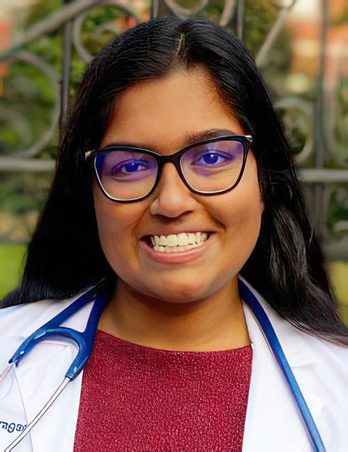
“Our study shows that ARTEMIS can reveal genomewide repeat landscapes that reflect dramatic underlying changes in human cancers,” said study co-leader Akshaya Annapragada (above), an MD/PhD student at the Johns Hopkins University School of Medicine, in a news release. “By illuminating the so-called ‘dark genome,’ the work offers unique insights into the cancer genome and provides a proof-of-concept for the utility of genomewide repeat landscapes as tissue and blood-based biomarkers for cancer detection, characterization, and monitoring.” Clinical laboratories may soon have new biomarkers for the detection of cancer. (Photo copyright: Johns Hopkins University.)
Detecting Early Lung, Liver Cancer
Artemis is a Greek word meaning “hunting goddess.” For the Johns Hopkins researchers, ARTEMIS also describes a technique “to analyze junk DNA found in tumors” and which float in the bloodstream, Financial Times explained.
“It’s like a grand unveiling of what’s behind the curtain,” said geneticist Victor Velculescu, MD, PhD, Professor of Oncology and co-director of the Cancer Genetics and Epigenetics Program at Johns Hopkins Kimmel Cancer Center, in the news release.
“Until ARTEMIS, this dark matter of the genome was essentially ignored, but now we’re seeing that these repeats are not occurring randomly,” he added. “They end up being clustered around genes that are altered in cancer in a variety of different ways, providing the first glimpse that these sequences may be key to tumor development.”
ARTEMIS could “lead to new therapies, new diagnostics, and new screening approaches for cancer,” Velculescu noted.
Repeats of DNA Sequences Tough to Study
For some time technical limitations have hindered analysis of repetitive genomic sequences by scientists.
“Genetic changes in repetitive sequences are a hallmark of cancer and other diseases, but characterizing these has been challenging using standard sequencing approaches,” the study authors wrote in their Science Translational Medicine paper.
“We developed a de novok-mer (short sequences of DNA)-finding approach called ARTEMIS to identify repeat elements from whole-genome sequencing,” the researchers wrote.
The scientists put ARTEMIS to the test in laboratory experiments.
The first analysis involved 1,280 types of repeating genetic elements “in both normal and tumor tissues from 525 cancer patients” who participated in the Pan-Cancer Analysis of Whole Genomes (PCAWG), according to Technology Networks, which noted these findings:
A median of 807 altered elements were found in each tumor.
About two-thirds (820) had not “previously been found altered in human cancer.”
Second, the researchers explored “genomewide repeat element changes that were predictive of cancer,” by using machine learning to give each sample an ARTEMIS score, according to the Johns Hopkins news release.
The scoring detected “525 PCAWG participants’ tumors from the healthy tissues with a high performance” overall Area Under the Curve (AUC) score of 0.96 (perfect score being 1.0) “across all cancer types analyzed,” the Johns Hopkins’ release states.
Liquid Biopsy Deployed
The scientists then used liquid biopsies to determine ARTEMIS’ ability to noninvasively diagnose cancer. Researchers used blood samples from:
ARTEMIS classified patients with lung cancer with an AUC of 0.82.
ARTEMIS detected people with liver cancer, as compared to others with cirrhosis or viral hepatitis, with a score of AUC 0.87.
Finally, the scientists used their “ARTEMIS blood test” to find the origin of tumors in patients with cancer. They reported their technique was 78% accurate in discovering tumor tissue sources among 12 tumor types.
“These analyses reveal widespread changes in repeat landscapes of human cancers and provide an approach for their detection and characterization that could benefit early detection and disease monitoring of patients with cancer,” the researchers wrote in Science Translational Medicine.
Large Clinical Trials Planned
Velculescu said more research is planned, including larger clinical trials.
“While still at an early stage, this research demonstrates how some cancers could be diagnosed earlier by detecting tumor-specific changes in cells collected from blood samples,” Hattie Brooks, PhD, Research Information Manager, Cancer Research UK (CRUK), told Financial Times.
Should ARTEMIS prove to be a viable, non-invasive blood test for cancer, it could provide pathologists and clinical laboratories with new biomarkers and the opportunity to work with oncologists to promptly diagnosis cancer and monitor patients’ response to treatment.