New funding will help Vitestro expand clinical validation, scale manufacturing, and prepare its autonomous blood-draw technology for broader hospital and laboratory adoption.
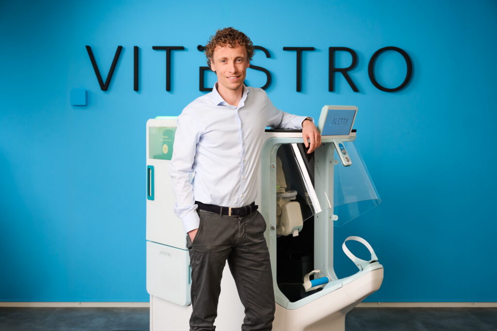
Vitestro has secured $70 million in oversubscribed Series B financing to accelerate development and commercialization of its Aletta Autonomous Robotic Phlebotomy Device (ARPD), a platform designed to automate routine blood collection in clinical settings.
In March 2025, The Dark Reportreported on Vitestro’s development of the ARPD—one of the last manual steps in the laboratory testing workflow. The company had recently received CE mark approval in Europe and reported clinical trial results showing a 95% first-stick success rate and strong patient acceptance, positioning the technology as a potential solution to phlebotomy staffing shortages and pre-analytical variability in clinical laboratories.
For clinical laboratory leaders facing persistent staffing shortages and rising specimen volumes, the investment highlights growing industry interest in automating one of healthcare’s most common clinical procedures.
“Closing our Series B financing reflects strong conviction in our mission to establish a new standard in autonomous robotic venous access and diagnostic blood collection,” said Toon Overbeeke, chief executive officer and co-founder of Vitestro. “Diagnostic blood collection remains the highest-volume invasive medical procedure globally, with billions of procedures performed annually.” (Photo credit: Vitestro)
Vitestro plans to use the capital to advance the next generation of its Aletta platform, conduct additional clinical studies, and scale manufacturing as the company prepares for broader commercial rollout in Europe and eventual entry into the United States through the FDA’s de novo regulatory pathway.
The Aletta system combines multimodal imaging, robotics, and artificial intelligence to autonomously identify veins, guide needle insertion, and collect blood samples with high precision. The platform is designed to perform routine diagnostic blood draws, potentially helping laboratories standardize collection quality while reducing human-dependent variability.
For laboratory executives, the technology could offer a new strategy to stabilize phlebotomy operations as workforce shortages persist. By automating routine blood draws, robotic platforms like Aletta may allow organizations to improve workflow predictability, support existing staff, and maintain patient throughput in high-volume outpatient settings.
A forthcoming issue of The Dark Report will feature interviews with Vitestro executives and provide deeper analysis of what autonomous robotic phlebotomy could mean for clinical laboratories, including its potential impact on staffing, workflow efficiency, and specimen quality.
Biobattery might one day power clinical laboratory testing devices designed to function in vivo to measure and wirelessly report certain biomarkers
Clinical laboratories may one day regularly process biomarker data sent by ingested medical devices from inside the human body, such as the colon and intestines. But powering such devices remains a challenge for developers. Now, researchers at Binghamton University in New York have developed a biobattery that derives its power based on pH reactions when it comes in contact with acids inside the gut.
The battery uses “bacteria to create low levels of electricity that can power sensors and Wi-Fi connections as part of the Internet of Things,” according to a Binghamton University news release.
The biobattery uses microbial fuel cells with spore-forming bacteria for power and it remains inactive until it reaches the small intestine.
Ingestible devices, such as wireless micro cameras, are being utilized more frequently to investigate a myriad of activities that occur in vivo. But traditional batteries that power ingestible diagnostic gadgets can be potentially harmful and are less reliable.
In addition, the small intestine in humans is typically between 10 and 18 feet in length and it folds several times to fit the abdomen. Thus, the inside area can be very difficult to reach for diagnostic purposes.
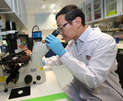
“There are some regions in the small intestine that are not reachable, and that is why ingestible cameras have been developed to solve this issue,” said Seokheun “Sean” Choi, PhD (above), Professor of Electrical and Computer Engineering at Binghamton University, in a news release. “They can do many things, such as imaging and physical sensing, even drug delivery. The problem is power. So far, the electronics are using primary batteries that have a finite energy budget and cannot function for the long term.” As these technologies develop, clinical laboratories may play a role in collecting biomarker data from these devices interpretation by physicians. (Photo copyright: Binghamton University/Jonathan Cohen.)
How Binghamton Researchers Developed Their Biobattery
The dime-sized fuel cell assembly is then sealed with a piece of Kapton tape, which can withstand temperatures from -500 to 750 degrees Fahrenheit. When the tape is removed, moisture mixes with a chemical germinant that causes the bacteria to begin manufacturing spores.
The biobattery generates around 100 microwatts per square centimeter of power density, but it can take up to an hour to germinate completely. After one hour, the energy generated from the device can power an LED light, a small clock, or a digital hygrometer, as well as a micro camera for in vivo use.
“We wanted to make these bio-batteries for portable, storable, and on-demand power generation capabilities,” Choi said in the news release.
“The problem is, how can we provide the long-term storage of bacteria until used? And if that is possible, then how would you provide on-demand battery activation for rapid and easy power generation? And how would you improve the power?” Choi added.
Heating the fuel cell decreased the time it took to reach full power to 20 minutes, and increasing the humidity resulted in higher electrical output.
Potential for Long-term Power Storage
In addition, after a week of being stored at room temperature, the activated battery had only lost 2% of its power. The researchers also believe that the device could function properly in an inactivate state for up to 100 years, provided there is enough moisture to activate the bacteria after the Kapton tape is removed.
“The overall objective is to develop a microbial fuel cell that can be stored for a relatively long period without degradation of bio-catalytic activity, and also can be rapidly activated by absorbing moisture from the air,” said Choi in the news release.
More research and studies are needed to confirm the biobattery performs properly and is feasible for general use. This experimentation would require both animal and human testing, along with biocompatibility studies.
“I think this is a good start,” Choi added. “Hopefully, we can make a commercial product using these ideas.”
If the biobattery can power an ingestible medical device for a reasonable period of time, then this invention may be able to power a clinical laboratory testing device that could function in vivo to measure and wirelessly report certain biomarkers inside the body.
Meet ‘PECOTEX,’ a newly-invented cotton thread with up to 10 sensors that is washable. Its developers hope it can help doctors diagnosis disease and enable patients to monitor their health conditions
Wearable biosensors continue to be an exciting area of research and product development. The latest development in wearable biosensors comes from a team of scientists led by Imperial College London. This team created a conductive cotton thread that can be woven onto T-shirts, textiles, and face masks and used to monitor key biosignatures like heart rate, respiratory rate, and ammonia levels.
Clinical laboratory managers and pathologists should also take note that this wearable technology also can be used to diagnose and track diseases and improve the monitoring of sleep, exercise, and stress, according to an Imperial College London news release.
Should this technology make it into daily use, it might be an opportunity for clinical laboratories to collect diagnostic and health-monitoring data to add to the patient’s full record of lab test results. In turn, clinical pathologists could use that data to add value when consulting with referring physicians and their patients.
“Our research opens up exciting possibilities for wearable sensors in everyday clothing,” said Firat Güder, PhD, Principal Investigator and Chief Engineer at Güder Research Group at Imperial College London, in a news release. “By monitoring breathing, heart rate, and gases, they can already be seamlessly integrated, and might even be able to help diagnose and monitor treatments of disease in the future.” (Photo copyright: Wikipedia.)
Ushering in New Generation of Wearable Health Sensors
The researchers dubbed their new sensor thread PECOTEX. It’s a polystyrene sulfonate-modified cotton conductive thread that can incorporate more than 10 sensors into cloth surfaces, costs a mere 15 cents/meter (slightly over 39 inches), and is machine washable.
“PECOTEX is high-performing, strong, and adaptable to different needs,” stated Firat Güder, PhD, Principal Investigator and Chief Engineer at Güder Research Group, Imperial College London, in the press release.
“It’s readily scalable, meaning we can produce large volumes inexpensively using both domestic and industrial computerized embroidery machines,” he added.
The material is less breakable and more conductive than conventional conductive threads, which allows for more layers to be embroidered on top of each other to develop more complex sensors. The embroidered sensors retain the intrinsic values of the cloth items, such as wearability, breathability, and the feel on the skin. PECOTEX is also compatible with computerized embroidery machines used in the textile industry.
The researchers embroidered the sensors into T-shirts to track heart activity, into a face mask to monitor breathing, and into other textiles to monitor gases in the body like ammonia which could help detect issues with liver and kidney function, according to the news release.
“The flexible medium of clothing means our sensors have a wide range of applications,” said Fahad Alshabouna, a PhD candidate at Imperial College’s Department of Bioengineering and lead author of the study in the news release. “They’re also relatively easy to produce which means we could scale up manufacturing and usher in a new generation of wearables in clothing.”
Uses for PECOTEX Outside of Healthcare
The team plans on exploring new applications for PECOTEX, such as energy storage, energy harvesting, and biochemical testing for personalized medicine. They are also seeking partners for commercialization of the product.
“We demonstrated applications in monitoring cardiac activity and breathing, and sensing gases,” Fahad added. “Future potential applications include diagnosing and monitoring disease and treatment, monitoring the body during exercise, sleep, and stress, and use in batteries, heaters, and anti-static clothing.”
Wearable healthcare devices have enormous potential to perform monitoring for diagnostic, therapeutic, and rehabilitation purposes and support precision medicine.
Further studies and clinical trials need to occur before PECOTEX will be ready for mass consumer use. Nevertheless, it could lead to new categories of inexpensive, wearable sensors that can be integrated into everyday clothes to provide data about an individual’s health and wellbeing.
If this technology makes it to clinical use, it could provide an opportunity for clinical laboratories to collect diagnostic data for patient records and help healthcare professionals track their patients’ medical conditions.
Understanding requirements of digital pathology workflow matters as regulatory and reimbursement elements align toward wider adoption beyond 2023. Upcoming Dark Daily webinar May 10 to cover infrastructure requirements
Nearly all pathology residents and fellows, as well as many histologists and other medical students, have been trained using digital images and, therefore, digital pathology tools. This resounds as a major and important development now working in tandem with recent coding decisions and regulatory recommendations that may combine to advance digital pathology to a significant tipping point.
As Dark Daily’s sister publication, The Dark Report, has described in great detail over the past several years, the trend toward digital pathology implementation started in the mid-2000s. Much has been learned through trial and error that may make the practical path forward clearer for those still on the sidelines.
Digital pathology infrastructure and information technology (IT) requirements are better known after years of research at academic centers throughout the United States—but only for those closest to the action. Two examples are University of Southern California (USC) on the West Coast and Memorial Sloan Kettering Cancer Center (MSKCC) on the East Coast.
During a free 60-minute educational webinar on May 10, W. Dean Wallace, MD, (far left) of University of Southern California (USC) and Orly Ardon, PhD, MBA, (immediate left) of Memorial Sloan Kettering Cancer Center (MSKCC) will explain digital pathology infrastructure, IT, and lessons learned through firsthand experiences. The webinar is sponsored by Hamamatsu, and continuing education credit is available for listening.(Photo copyrights: USC and MSKCC.)
Seven Advantages of Early Adoption of Whole Slide Imaging and Digital Pathology
Many pathologists know that academic centers throughout the U.S. have been the first to adopt and use digital pathology scanners and systems. Early work in what have become custom digital pathology ecosystems has enabled academic pathology groups to:
Learn how to implement, validate, and design workflows that include digital pathology systems and computational pathology.
Determine how physical environments need to change for slide scanners, achieving quality images, maximizing scanner utility, and expanding scanning capabilities in medium- and high-throughput laboratories.
Contract with pharmaceutical companies and drug developers to read digital images in support of drug research and clinical trials.
Understand how digital pathology applies for various use cases, including primary diagnosis, frozen section diagnosis, consultations, second opinions, and telepathology.
Successfully spread pathologist technical and professional support across multiple laboratory locations and remote customers.
Learn best practices for conducting tumor boards and peer reviews of pathology cases.
Validate and verify new hematoxylin and eosin (H&E) stains.
Hospital and Lab Leaders Have Questions About Digital Pathology Requirements
As a result of early adopter projects, digital pathology infrastructure and IT requirements are better understood and documented for a variety of use cases, according to W. Dean Wallace, MD, Professor of Pathology at the Keck School of Medicine of USC. Wallace specializes in pulmonary and renal pathology with a strong interest in informatics, as well as radiology and pathology correlation, and he warns of the danger of implementing an “incomplete digital pathology system.”
This webinar is for hospital and health system leaders, as well as independent pathology groups and reference lab executives, who want to know:
Key workflow aspects of the components needed in a digital pathology service.
Common limitations of commercial digital pathology products.
How to structure a digital pathology implementation team.
A goal-based approach to developing a business case for digital pathology implementation.
Wallace and Orly Ardon, PhD, MBA, Director of Digital Pathology Operations at MSKCC, will lead the call and take questions during the webinar’s live Q&A segment.
Questions About Digital Pathology Implementation
At MSKCC, teams have scanned and archived more than six million histology slides and are prospectively scanning all in-house H&E slides.
“There is a lot of interest out there for digital pathology implementation,” Ardon told Dark Daily, “not only the AI-machine learning opportunities that are enabled with digital slides, but how do we even start a basic digital pathology journey. Institutions and labs don’t realize how many factors they have to think about before they start scanning the first slide.”
“People have limited understanding of the complexities of the business case,” Wallace added. “Do you want to go with a full 100% deployment or a targeted deployment? Do you want to get digital pathology to support tumor boards? By introducing scanners into the tumor board workflow, you can actually cause more problems than you are solving if you are not careful.
“The other aspect of it is the actual technical deployments. You need to begin with careful analysis of functions or services to support,” Wallace said, adding the soft costs of digital pathology can take lab and pathology administrators by surprise.
Ardon and Wallace will present their insights and experiences during the webinar, which has been sponsored by Hamamatsu. Those interested can learn more and register at Dark Dailyhere. P.A.C.E. credit is available for this program through the American Society for Clinical Laboratory Science (ASCLS).
On the Horizon: Incentives and Further Alignment Toward Digital Pathology Adoption
Dark Daily’s new webinar is timely. Earlier this year, the Centers for Medicare and Medicaid Services (CMS) entered what has been called a “tryout” period to gather data about the use of new, digital-pathology-related Current Procedural Terminology (CPT) codes in clinical laboratories and anatomic pathology groups. (See coverage in The Dark Report.)
Some believe the efforts of CMS, clinical labs, and pathology groups will result in new reimbursable codes, reimbursement values, and other incentives for using digital pathology (starting sometime in 2024)—if analysis shows use of digital pathology is as widespread as numerous publications would seem to indicate.
The CPT coding development coincides with recent discussions within the federal Clinical Laboratory Improvement Advisory Committee (CLIAC) about sweeping recommendations to allow continued remote work once the COVID-19 Public Health Emergency ends on May 11 and recognize digital data as a vital component of diagnostic specimens. (See coverage in The Dark Report.)
CLIAC’s recommendations may translate into a running start for modernizing the Clinical Laboratory Improvement Amendments of 1988 (CLIA). CLIA as it is written currently is dated and needs to account for new and emerging technologies, such as digital pathology, medical laboratory industry sources have said for years. (See a recent Dark Report – Dark Daily webinar.)
These developments, as they further align with actions by the U.S. Food and Drug Administration (FDA), could unleash swells of interest in onboarding whole slide scanners and digital pathology tools. Remote workflows became a priority during the COVID-19 pandemic, and it appears they will continue for a period as the Public Health Emergency unwinds, according to the FDA.
Watch Digital Pathology Implementation Strategies
Most executives at hospitals and health systems, private pathology practices, and independent reference labs are on the sidelines watching how digital pathology in research and clinical practice is unfolding.
However, as the pathology field integrates data science and computational pathology, forward-looking hospital and lab leaders can expect greater momentum toward advanced technologies, such as digital pathology tools.
Register here to participate in the upcoming webinar, “Digital Pathology Implementation Strategies.”
—Liz Carey
This content was developed through independent research and interviews by The Dark Intelligence Group, with support from Hamamatsu Photonics K.K., a provider of whole slide imaging systems and related technology such as optical sensors, light sources, and complex instrument systems that use them. Hamamatsu did not participate in the article’s development. Learn more about Hamamatsu at https://nanozoomer.hamamatsu.com/us/en.html.
Some lab experts advise that clinical laboratories and pathology practices should also plan on delayed payments for COVID-19 testing for uninsured patients
Regardless of whether infection rates for SARS-CoV-2 continue to wane or perhaps surge again, business changes are coming for staff at clinical laboratories and anatomic pathology groups. Forward-thinking lab administrators will want to evaluate post-pandemic strategies for labs to stay ahead of potential legal issues and keep their organizations financially healthy.
The public health emergency stemming from COVID-19 is set to expire April 16. That deadline could be extended. However, the U.S. Department of Health and Human Services (HHS) is under pressure from some circles to end the public health emergency, which would affect some health insurance provisions and potentially rein in relaxed rules for telemedicine.
“If you’re a laboratory, now is the time you need to start buttoning up [the above] concerns. You don’t want to be at the mercy of a quick cutoff,” said Jon Harol, president of Lighthouse Lab Services in Charlotte, N.C. The company hosted a webinar last week called “Preparing Your Clinical Lab or Pathology Practice for Post-COVID Success.”
Pathologists and clinical laboratory leaders should consider post-pandemic strategies for labs in the following areas:
COVID-19 testing for uninsured patients;
Preparations for government audits of SARS-CoV-2 tests performed during the pandemic; and,
Repurposing PCR equipment used for COVID-19 testing into other areas of clinical diagnostics.
These strategies will be explored further during the Executive War College Conference on Laboratory and Pathology Management, which takes place April 27-29 in New Orleans. Leaders of innovative clinical laboratories will share how their lab teams are helping to improve patient outcomes while encouraging health insurers to pay them for this value.
COVID-19 Testing for Uninsured Patients
On March 22, the U.S. Health Resources and Services Administration (HRSA) announced that its COVID-19 Uninsured Program stopped accepting claims for testing and treatment due to lack of sufficient funds. This development affects 8.6% of the nation’s population that doesn’t have medical insurance, according to the U.S. Census Bureau.
For clinical laboratories, the announcement could lead to delayed payments for COVID-19 tests performed on uninsured patients, said Mick Raich, President of Revenue Cycle Management Consulting at Lighthouse Lab Services, who also spoke during the webinar. Medical laboratories and pathology groups should anticipate reimbursement gaps and how that might affect revenues collected from payers.
“The patient relationship is going to be the most important thing. That puts labs at the head of the table,” says Jon Harol, president at Lighthouse Lab Services.
“Labs are going to do the testing and are going to bill for it, and there will probably be some retroactive payment,” Raich explained.
With midterm elections happening this year, don’t be surprised to see HRSA funding reinstated for COVID-19 testing for uninsured people, commented Robert Michel, Editor-in-Chief of The Dark Report and Founder of the Executive War College.
“We’ll have to wait and see. After all, it is an election year, so the representatives and senators in Congress would like to be re-elected,” added Michel, who also presented during the webinar.
“It is reasonable to assume that members of Congress don’t want to disappoint the clinical laboratories that stepped up to the table in the earliest days of the pandemic and have done huge volumes of COVID-19 testing.”
Preparations for Government Audits of Pandemic Testing
Another post-pandemic strategy for labs: Prepare for audits of COVID-19 test claims from the HHS Office of Inspector General (OIG).
Ahead of any OIG action, labs should consider performing self-audits to determine whether they complied with HRSA requirements. “The best thing you can do is go back and look at the first two months of your billing. Do an audit to ask: Did we bill anybody with insurance by accident?” Raich suggested. “Take a hundred of those claims and audit them.”
If a medical lab finds problems with uninsured COVID-19 billing, it may be prudent to self-report those discrepancies to the government rather than ignore them. “That looks a lot better to the OIG than tucking the stuff in a desk drawer and waiting for someone to knock on your door,” Raich noted.
Harol predicted the OIG will also review another aspect of how COVID-19 test claims were coded. Auditors will want to see if PCR test claims coded for higher reimbursement if the results were reported within 48 hours actually met that requirement.
“I expect that we’ll see auditing of the coding that was used. Under COVID, you got paid more if you were running tests on a high-throughput platform. It was almost an honor system there. I don’t know that I’ve seen much outside verification of that,” Harol explained. “I’m curious to see if there will be OIG pushback and more documentation required to prove the code was correct.”
Repurposing PCR Equipment Used for COVID-19 Testing
When the pandemic finally winds down, there will be less demand for COVID-19 testing, which could leave PCR equipment collecting dust unless labs make plans now on how to repurpose those systems.
“If you have a PCR instrument that can be revalidated, you want to start thinking about putting in a panel that tests for UTIs, sexually transmitted diseases, respiratory diseases, or women’s health,” Harol explained. “Those types of tests can be done on the equipment that a lot of COVID testing was being performed on, and it can be performed by the same scientists with that same skillset. That’s the low-hanging fruit.”
The next step is more complicated: Moving into the future, clinical laboratories need to determine what menu of tests will meet the needs of patients who previously submitted COVID-19 specimens for testing.
“The patient relationship is going to be the most important thing. That puts labs at the head of the table,” Harol continued. “How can you market your laboratory services directly to patients who might be interested?”
Watch for Developments in Telemedicine
Any post-pandemic strategies for labs will be influenced by how state governments and federal health officials regulate telemedicine in the future.
Pathologists and clinical laboratory directors should keep their eyes on whether telemedicine rules revert to more onerous requirements once the public health emergency lifts. Before the pandemic, rules for physicians licensed in one state generally limited when they could practice over state lines through telemedicine.
“In response to the pandemic, both the federal government and the states relaxed many prohibitions on the practice of medicine across state lines. This is significant for pathologists,” Michel said. “There is speculation that once government officials let this genie out of the bottle regulatory-wise, they won’t be able to put it back in. Thus, there are many predictions that officials at the state and federal level will be under pressure to retain the newer telemedicine rules after the pandemic has ended.”
Telemedicine proved to be a big benefit for Medicare patients during the pandemic. A report from HHS in December indicated telehealth visits in 2020 for Medicare beneficiaries increased 63 times, from approximately 840,000 in 2019 to 52.7 million. That fact should catch the attention of clinical lab managers and pathologists who want to keep their labs at the front edge of clinical services. For Medicare beneficiaries who see their physicians virtually, labs need the capability to access that patient so as to collect the samples needed to perform those tests ordered by the physician during the telehealth consultation.
The new method employs a pH sensitive dye and AI algorithms to ‘distinguish between cells originating from normal and cancerous tissue, as well as among different types of cancer’ the researchers said
Might a pH-sensitive dye in tandem with an image analysis solution soon be used to identify cancerous cells within blood samples as well within tissue? Recent research indicates that could be a possibility. If further studies and clinical trials confirm this capability, then anatomic pathologists could gain another valuable tool to use in diagnosing cancers and other types of disease.
Currently, surgical pathologists use a variety of hematoxylin and eosin stains (H/E) to bring out useful features in cells and cell structures. So, staining tissue on glass slides is a common practice. Now, thanks to machine learning and artificial intelligence, anatomic pathologists may soon have a similar tool for spotting cancer cells within both tissue and blood samples.
Researchers at the National University of Singapore (NUS) have developed a method for identifying cancer that uses a pH sensitive dye called bromothymol blue. The dye reacts to various levels of acidity in cancer cells by turning colors. “The pH inside cancer cells tends to be higher than that of healthy cells. This phenomenon occurs at the very early phases of cancer development and becomes amplified as it progresses,” Labroots reported.
In “Machine Learning Based Approach to pH Imaging and Classification of Single Cancer Cells,” published in the journal APL Bioengineering, the NUS researchers wrote, “Here, we leverage a recently developed pH imaging modality and machine learning-based single-cell segmentation and classification to identify different cancer cell lines based on their characteristic intracellular pH. This simple method opens up the potential to perform rapid noninvasive identification of living cancer cells for early cancer diagnosis and further downstream analyses.”
According to an NUS news release, the bromothymol blue dye is “applied onto patients’ cells” being held ex vivo in cell culture dishes. The dye’s color changes depending on the acidity level of the cancer cells it encounters. Microscopic images of the now-visible cancers cells are taken, and a machine-learning algorithm analyzes the images before generating a report for the anatomic pathologist.
The NUS researchers claim the test can provide answers in about half an hour with 95% accuracy, Labroots reported.
“The ability to analyze single cells is one of the holy grails of health innovation for precision medicine or personalized therapy. Our proof-of-concept study demonstrates the potential of our technique to be used as a fast, inexpensive and accurate tool for cancer diagnosis,” said Lim Chwee Teck, PhD, NUS Society Professor and Director of NUS’ Institute for Health Innovation and Technology, in the NUS news release.
The novel technique for differentiating cancer cells from non-cancerous cells being developed at the National University of Singapore (NUS) could eventually become useful in detecting cancer cells in tissue samples, either obtained from tumor biopsies or blood samples. “As the number of cells in these samples can be in millions or even billions, the ability to detect the very few cancer cells among the others will be useful for clinicians,” NUS Society Professor and Director of NUS’ Institute for Health Innovation and Technology, Lim Chwee Teck, PhD (above) told The Straits Times. (Photo copyright: The Straits Times.)
AI Cell Analysis versus Laborious Medical Laboratory Steps
By developing an AI-driven method, Professor Lim and the NUS team sought to improve upon time-consuming techniques for identifying cells that traditionally involve using florescent probes, nanoparticles, and labeling steps, or for cells to be fixed or terminated.
“Unlike other cell analysis techniques, our approach uses simple, inexpensive equipment, and does not require lengthy preparation and sophisticated devices. Using AI, we are able to screen cells faster and accurately,” Professor Lim told Labroots. “Furthermore, we can monitor and analyze living cells without causing any toxicity to the cells or the need to kill them.”
The new technique may have implications for cancer detection in tumor tissue as well as in liquid biopsies.
“We are also exploring the possibility of performing the real-time analysis on circulating cancer cells suspended in blood,” Professor Lim said in the NUS news release. “One potential application for this would be in liquid biopsy where tumor cells that escaped from a primary tumor can be isolated in a minimally-invasive fashion from bodily fluids such as blood.”
Diagnosing Cancer in Real Time
The NUS’ method requires more research and clinical studies before it could become an actual tool for anatomic pathologists and other cancer diagnosticians. Additionally, the NUS researchers acknowledged that the focus on only four cell lines (normal cells, benign breast tumor cells, breast cancer cells, and pancreatic cancer cells) limited their study, as did lack of comparison with conventional florescent pH indicators.
Still, the NUS scientists are already planning more studies to advance their concept to different stages of cell malignancy. They envision a “real-time” version of the technique to enable recognition of cells and fast separation of those that need to be referred to clinical laboratories for molecular testing and/or genetic sequencing.
Medical laboratory leaders may want to follow the NUS study. An inexpensive AI-driven method that can accurately detect and classify cancer cells based on pH within the cells is provocative and may be eventually become integrated with other cancer diagnostics.