New funding will help Vitestro expand clinical validation, scale manufacturing, and prepare its autonomous blood-draw technology for broader hospital and laboratory adoption.
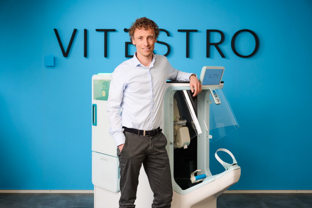
Vitestro has secured $70 million in oversubscribed Series B financing to accelerate development and commercialization of its Aletta Autonomous Robotic Phlebotomy Device (ARPD), a platform designed to automate routine blood collection in clinical settings.
In March 2025, The Dark Reportreported on Vitestro’s development of the ARPD—one of the last manual steps in the laboratory testing workflow. The company had recently received CE mark approval in Europe and reported clinical trial results showing a 95% first-stick success rate and strong patient acceptance, positioning the technology as a potential solution to phlebotomy staffing shortages and pre-analytical variability in clinical laboratories.
For clinical laboratory leaders facing persistent staffing shortages and rising specimen volumes, the investment highlights growing industry interest in automating one of healthcare’s most common clinical procedures.
“Closing our Series B financing reflects strong conviction in our mission to establish a new standard in autonomous robotic venous access and diagnostic blood collection,” said Toon Overbeeke, chief executive officer and co-founder of Vitestro. “Diagnostic blood collection remains the highest-volume invasive medical procedure globally, with billions of procedures performed annually.” (Photo credit: Vitestro)
Vitestro plans to use the capital to advance the next generation of its Aletta platform, conduct additional clinical studies, and scale manufacturing as the company prepares for broader commercial rollout in Europe and eventual entry into the United States through the FDA’s de novo regulatory pathway.
The Aletta system combines multimodal imaging, robotics, and artificial intelligence to autonomously identify veins, guide needle insertion, and collect blood samples with high precision. The platform is designed to perform routine diagnostic blood draws, potentially helping laboratories standardize collection quality while reducing human-dependent variability.
For laboratory executives, the technology could offer a new strategy to stabilize phlebotomy operations as workforce shortages persist. By automating routine blood draws, robotic platforms like Aletta may allow organizations to improve workflow predictability, support existing staff, and maintain patient throughput in high-volume outpatient settings.
A forthcoming issue of The Dark Report will feature interviews with Vitestro executives and provide deeper analysis of what autonomous robotic phlebotomy could mean for clinical laboratories, including its potential impact on staffing, workflow efficiency, and specimen quality.
Roche’s IVDR-certified Elecsys pTau181 blood test helps rule out Alzheimer’s early, supporting faster decisions in primary care.
Roche has received CE Mark approval for its Elecsys pTau181 test, developed in collaboration with Eli Lilly. The test measures phosphorylated Tau 181 (pTau181), a biomarker associated with amyloid pathology—one of the hallmarks of Alzheimer’s disease. Clinicians can use this test alongside other clinical data to help rule out Alzheimer’s as the cause of cognitive decline, potentially reducing the need for further diagnostic procedures in patients who test negative.
This is a minimally invasive blood test and was shown to be effective in clinical care settings such as primary care offices. The news is the latest in a series of breakthroughs in Alzheimer’s detection. This past June, The Dark Daily reported two of these stories:
This latest development from the Swiss-based company further shows how these developments are happening on a large and international scale. This also marks the second collaboration of Roche and American-based Eli Lilly on an Alzheimer’s biomarker test. As stated by Fierce Biotech, a 2023 breakthrough was a ‘side quest’ in Eli Lilly’s ongoing crusade against Alzheimer’s disease.
Removing Barriers to Care
As stated in the press release, up to 75% of individuals who live with symptoms of Alzheimer’s disease are never diagnosed. The ones who are diagnosed wait an average of approximately three years to seek testing after they experience an onset of symptoms.
“The burden of Alzheimer’s disease on society and healthcare systems is increasing as the world’s population ages,” said Matt Sause, CEO of Roche Diagnostics.
“With Elecsys pTau181, doctors can give patients and their caregivers the clarity they need when establishing the cause of cognitive decline. By enabling an earlier and less invasive diagnosis, this test has the potential to improve patient outcomes and decrease costs for healthcare systems worldwide,” Sause noted.
First of its Kind
According to Roche, this approval is the first of its kind for a blood test that rules out Alzheimer’s disease. This international study included 787 patients. The press release also stated, “The study showed the test was able to rule out Alzheimer’s disease with a high negative predictive value (NPV) of 93.8% based on a 22.5% prevalence of amyloid positivity according to positron emission tomography (PET) scans, with 83.6% sensitivity.”
This global study’s uniqueness came from the recruitment of a diverse patient pool that was specifically designed to reflect the patients who could benefit the most from this kind of testing.
Great Strides in Alzheimer’s Research
This news follows other key developments in detection and treatment of Alzheimer’s disease. Roche also reported success with a new Alzheimer’s drug. Trontinemab, an antibody drug, performed well in clinical trials with 91% of participants showing almost complete amyloid plaque removal according to Gene Online. Roche will now go onto two large scale clinical trials for 2025 to further test the effectiveness of the drug.
With these advances in testing and treatment of Alzheimer’s disease, pathology professionals would be wise to keep on the pulse of the news. About 75% of people living with Alzheimer’s disease globally remain undiagnosed according to the Gene Online reporting––in the coming years we could see that number dramatically go down.
New study shows how artificial neural networks are leading to improved microscopic capabilities and precision that could help researchers better understand causes of tumors
Pathologists, microbiologists, and clinical laboratory scientists engaged in research into cellular activity will be interested to learn that advances in artificial intelligence (AI) are leading to new opportunities for future research studies to observe and capture—in real time—biological processes in living cells/samples.
“EPFL biophysicists have now found a way to automate microscope control for imaging biological events in detail while limiting stress on the sample, all with the help of artificial neural networks. Their technique works for bacterial cell division, and for mitochondrial division,” according to Phys.org.
Even better, the EPFL scientists are making the control framework available as an open-source software plug-in for the microscope control program Micro-Manager. This enables clinical laboratories to integrate artificial intelligence into existing microscope control software, Phys.org noted.
“An intelligent microscope is kind of like a self-driving car. It needs to process certain types of information, subtle patterns that it then responds to by changing its behavior,” Suliana Manley, PhD, biophysicist and a professor in the Laboratory of Experimental Biophysics at the École Polytechnique Fédérale de Lausanne, told Phys.org. Clinical laboratories and anatomic pathology groups may want to explore the capabilities of this intelligent microscope technology. (Photo copyright: EPFL.)
EPFL Study Breakdown
Suliana Manley, PhD, biophysicist, professor, and head of the Laboratory of Experimental Biophysics at the EPFL, is the principal investigator of the “Intelligent microscope” study. She has spent her career focusing on the development of high-resolution optical instruments and the organization and dynamics of proteins.
Manley and her team learned how to detect mitochondrial divisions in bacteria such as Caulobacter Crescentus. These divisions can occur once every few minutes, but last only seconds. Their infrequency and ability to occur anywhere within the mitochondrial network makes them difficult to spot and photograph, Physics World noted.
To overcome this barrier, the EPFL team trained the neural network to look for mitochondrial constrictions—the shape change in mitochondria that leads to division—and combined that data with observations of the protein DRP1, the presence of which is required for spontaneous divisions to occur, Phys.org reported.
“A common goal of fluorescence microscopy is to collect data on specific biological events. Yet, the event-specific content that can be collected from a sample is limited, especially for rare or stochastic processes,” wrote the EPFL scientists in their Nature Methods paper. “We developed an event-driven acquisition framework, in which neural-network-based recognition of specific biological events triggers real-time control in an instant structured illumination microscope … we capture mitochondrial and bacterial divisions at imaging rates that match their dynamic timescales, while extending overall imaging durations. Because event-driven acquisition allows the microscope to respond specifically to complex biological events, it acquires data enriched in relevant content.”
Manley’s intelligent fluorescent microscope responds more rapidly than human control can achieve. When the microscope senses constrictions and DRP1 protein levels are high, it switches into high-speed image capture mode and gathers multiple images with minute detail. Conversely, when both constrictions and protein are low, the microscope goes into low-speed imaging mode, which preserves the integrity of the sample by avoiding excessive exposure to light, Phys.org noted.
Automating microscope control limits the stress placed on samples, increasing the likelihood of more accurate results. It also allows microscope control for imaging biological events in detail, as it eliminates human error that naturally occurs from trying to keep up with events in real time, Phys.org reported.
“Using a neural network, we can detect much more subtle events and use them to drive changes in acquisition speed,” Manley told Phys.org. “The potential of intelligent microscopy includes measuring what standard acquisitions would miss. We capture more events, measure smaller constrictions, and can follow each division in greater detail.”
What’s Next for EPFL?
Manley and her EPFL team plan to continue working with neural networks to detect different events and bring about different hardware responses.
“For example, we envision harnessing optogenetic perturbations to modulate transcription at key moments in cell differentiation,” she told Physics World. “We also think of using event detection as a means of data compression, selecting for storage or analysis the pieces of data that are most relevant to a given study.”
As research continues in bacterial cell division, there will be a point where the technology enables researchers to observe cell activity and what conditions cause abnormal (tumor) cells to be created. That would be the first step to then investigating ways to stop the cellular process that creates abnormal cells.
It should not surprise pathologists and clinical laboratory managers that researchers and technology developers are exploring ways to “turbocharge” classic light microscopy. Advances in image analysis, combined with machine learning algorithms, are making it possible to tease new insights from the images viewed with a standard microscope.
Should this AI-driven technology prove viable in clinical settings, it could contribute to easing the shortage of qualitied phlebotomists for medical laboratories worldwide
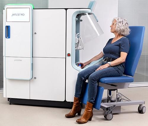
Could phlebotomists one day be out of a job? If European medical technology company Vitestro has its way, that could someday become a reality in European hospitals and in clinical laboratories worldwide. Headquartered in the Netherlands, the company has raised EUR 12.7 million ($14,057,947.50 US) in Series A financing to bring to market “the world’s first autonomous blood drawing device,” BioWorld Med Tech reported.
According to Vitestro’s website, the “device combines AI-based, ultrasound-guided 3D reconstruction with robotic needle insertion, ensuring accurate and secure blood collection. The procedure is performed fully automatically, from tourniquet to bandage application.”
This is another example of how artificial intelligence companies are finding opportunities in staffing shortages the healthcare industry is experiencing globally. In this case, the novel technology could help address the lack of qualified phlebotomists. And clinical laboratories around the world could become the proving grounds for new AI-driven devices that end up replacing human healthcare workers.
“This financing round marks a new phase of growth for Vitestro which brings the company closer to its mission of improving the venipuncture procedure for hundreds of millions of patients per year,” said Vitestro CEO and co-founder Toon Overbeeke (above), in a press release. “We look forward to growing the business and transforming patient care with Sonder Capital, leveraging their expertise in successfully commercializing medical robotic technologies.” If proven viable, clinical laboratories around the world suffering from shortages of phlebotomists could benefit from AI-driven autonomous blood draw stations. (Photo copyright: LinkedIn.)
Next Evolution for Clinical Laboratories
According to the Centers for Disease Control and Prevention (CDC), there are 14 billion clinical laboratory tests ordered annually in the US and 70% of medical decisions depend on laboratory results. One of the more common clinical laboratory procedures—venous blood draws—is pivotal in clinical diagnostics, but a worldwide shortage of skilled phlebotomists is having an impact on this critical testing method.
With the announcement of its completion of a EUR 12.7-million Series A financing round to bring the “world’s first” autonomous blood draw device to market, Vitestro seems poised to impact both the shortage and the job prospects of existing phlebotomists. This financing round was led by San Carlos, California-based Sonder Capital and included investors with experience in the clinical laboratory and medical technology industries.
“Automating this ubiquitous procedure is the next evolution for clinical laboratories, allowing them to improve quality of care for patients while building a more sustainable operation,” stated Andy McGibbon, Managing Partner at Sonder Capital in a March press release.
According to Investopedia, Series A financing refers to “an investment in a privately-held start-up company after it has shown progress in building its business model and demonstrates the potential to grow and generate revenue. It often refers to the first round of venture money a firm raises after seed and angel investors.”
Vitestro says it will utilize the capital from this financing round to accelerate production development, prepare market authorization in the European Union, and initiate production.
Vitestro’s autonomous blood drawing device prototype (above) has been tested on more than 1,000 volunteers and patients. Vitestro plans to continue its studies on the device this year and anticipates entering the European market with the device sometime in 2024. Development of this technology is something that phlebotomists and clinical laboratory managers will want to track. (Photo copyright: Vitestro.)
Coming to a Clinical Laboratory Near You
“Medical robotics will make optimal outcomes available to everyone. I strongly believe Vitestro will set the world standard in autonomous blood drawing,” said Fred Moll, MD, Managing Partner of Sonder Capital in the press release. Moll, who has been heralded as the “father of robotic surgery,” was also appointed as a non-executive board member of Vitestro. Moll co-founded Intuitive Surgical, Inc., Hansen Medical, Restoration Robotics, and Auris Health (acquired by Ethicon, a Johnson and Johnson company).
On April 12, Vitestro announced that leading Dutch clinical laboratory OLVG Lab will be the first healthcare provider to begin using their blood-drawing device. A number of hospitals, clinical laboratories, and blood drawing departments are preparing to use the device and OLVG Lab plans to have the system fully operational by late next year, according to a press release. OLVG lab provides laboratory services to hospitals, clinics, and care providers in the greater Amsterdam area.
“Robotization has become an important topic in diagnostics. Vitestro’s technology will improve the standardization and optimization of the sampling procedure. And it helps solve staff shortages in our blood drawing department,” said Anja Leyte, director of OLVG Lab, in the press release. “But more importantly, the patients are also very positive. Our staff are really enthusiastic as well and can’t wait to start using this breakthrough technology in our healthcare.”
Vitestro’s device is still in the testing phase but could prove to be very beneficial to clinical laboratories and help alleviate the shortage of trained phlebotomists. An automated blood draw machine might also improve the consistency of the blood draw experience for both patients and healthcare professionals.
Even as Balwani’s trial moves ahead, Hulu’s miniseries ‘The Dropout’ chronicles the pair’s romance and the company’s downfall while providing controversial subject matter for various media outlets
Unlike Theranos founder Elizabeth Holmes’ criminal trial for fraud which generated daily headlines across the nation, the related fraud trial of ex-Theranos COO Ramesh “Sunny” Balwani is not getting the same news coverage. Therefore, media have shifted their reporting to Balwani’s personal relationship with the Holmes, which is clearly having its moment in the media spotlight.
The release of the Hulu miniseries “The Dropout”—which chronicles Holmes’ failed attempt to revolutionize the clinical laboratory industry by developing a device capable of performing multiple clinical blood tests using a finger-stick of blood—created the initial media and TV-viewer buzz.
Now a diverse range of media, including Fortune, The New York Post, and The Guardian, are turning their attention to the former Theranos executives’ private relationship during the time when they were in charge at the failed medical laboratory company.
As “The Dropout” outlines, Holmes gained celebrity status after dropping out of Stanford University at age 19 and founding Theranos in 2003. Years later, when Theranos claimed its Edison blood-testing device could conduct hundreds of blood tests using a finger-prick of blood, the startup’s valuation soared to nearly $9 billion in 2014, making Holmes a billionaire based on her 50% stake in the company, Investopedia reported.
In “What Happened to Elizabeth Holmes and Sunny Balwani? Where the Shamed Theranos Execs are Today,” Fortune used the release of “The Dropout” to publish an update on Holmes and Balwani. The magazine notes Holmes’ family connections—she was a descendant of the founders of America’s first yeast company and the daughter of a former Enron executive and congressional aide—helped her early efforts at fundraising for Theranos.
Fortune also stated that Holmes’ “pedigreed background” enabled her to attract “luminaries” such as former Secretary of State Henry Kissinger and former CDC Director William Foege to the Theranos board and gained her access to high-profile investors.
In U.S. District Court Northern District of California, ex-Theranos president and COO Ramesh “Sunny” Balwani (above) faces charges for allegedly defrauding patients and investors about Theranos. His defense team has attempted to distance their client from the day-to-day decision-making in the clinical laboratory company, while prosecution witnesses are attempting to show Balwani not only invested money in the startup but orchestrated many of the company’s actions. Balwani has pleaded not guilty to all charges. (Photo copyright: David Paul Morris, Fortune.)
Theranos, Holmes Cloaked in Secrecy, according to Fortune
While Holmes sought the spotlight when promoting Theranos, Fortune maintains the company’s work culture and Holmes herself were clocked in secrecy. The article states Holmes hired bodyguards to serve as her chauffeurs, installed bulletproof glass in her office windows, and did not allow workers in separate departments to discuss projects with one another.
Balwani met Holmes in 2002 while both were studying in Beijing as part of a Mandarin language summer program. He was 37 and married at the time, while Holmes was an 18-year-old high school student. Balwani was attending an MBA program at the University of California, Berkeley, which he entered after selling his shares in software company Commerce One in 2000 for nearly $40 million.
The New York Post reported Balwani sold the upscale Silicon Valley home he previously shared with Holmes for $15.8 million this past January. The 6,800-square-foot, five-bedroom, seven-bathroom house in Atherton, Calif., is a one-acre property, which The Post states was purchased by the couple for $9 million in 2013. Balwani bought out Holmes’ 50% stake in 2018.
Aron Solomon, a Chief Legal Analyst for legal marketing firm Esquire Digital, is not surprised by the interest in all things Theranos-related.
“We are seeing a ton of interest following the Holmes trial, and I don’t think it’s going to go away,” he told The Guardian.
Potential Reason for Delay in Holmes’ Sentencing
Holmes was convicted in January on four counts of fraud, but she is not expected to be sentenced until September. Amanda Kramer, JD, a partner in the White Collar Defense and Investigations practice at Covington and Burling, LLP, and a former federal prosecutor, suggests that Holmes’ sentencing date may have been delayed until after Balwani’s trial due to the potential for new information to come to light.
“It’s not typical for a case to be sentenced eight months out, but this is not a typical case in many senses,” Kramer told NPR. “And some facts established in Balwani’s trial might prove to be relevant in Holmes’ sentencing.”
So, it appears clinical laboratory directors and pathologists may find more interesting insights about the problems at Theranos emerging from court testimony when it is time for Holmes to be sentenced and during the remaining days of Balwani’s trial. Stay tuned. Dark Daily will continue to bring you the relevant facts of the case.
By automating clinical chemistry and immunoassay testing, clinical laboratory leaders can improve throughput while reducing the stress on staff, laboratory expert says
The American Society for Clinical Pathology regularly conducts a vacancy survey of medical laboratories throughout the US. While the problem of lab department vacancy rates has been ongoing, the last survey reported showed increased rates for laboratory positions across all departments surveyed. Last year, burnout among healthcare workers reached a crisis level, reported Clinical Laboratory News.
As a result, staffing the clinical laboratory with qualified lab professionals resounds as a top concern—and at a time when expectations are perhaps the highest they have ever been for performance in healthcare operations, from general hospitals to the most complex integrated delivery networks.
Even in the midst of the clinical laboratory workforce shortage and chronic strain, laboratory leaders must still improve their labs’ processes and workflows; increase productivity; and expand routine and specialty testing to better serve patient populations.
Faced with unrelenting pressures to do more with less, lab directors are turning to automating certain departments of the laboratory as a way to:
Relieve the problems caused by an ongoing workforce shortage;
Improve workflows and processes through standardization;
Keep lab staff working on the most important tasks; and
Enhance the laboratory’s reach and grow the lab business in necessary ways.
How UMC Southern Nevada Prioritized STAT Runs, Consolidated Operations
One case in point highlights the University Medical Center (UMC) of Southern Nevada’s clinical laboratory. Located in Las Vegas, UMC is among the largest public hospitals in the United States. It is part of a recent master-planned Las Vegas Medical District (LVMD), and it is the only Level I trauma center in Nevada.
The laboratory needed to improve turnaround time and expand the test menu, among other goals, explained Scott Keigley, one of two General Laboratory Services Managers at UMC. While limited laboratory automation had already been applied broadly, the lab took its automation initiative one step further by connecting three high-volume automated clinical chemistry and immunoassay analyzers (CC/IA), an automated hematology line, and a coagulation analyzer.
The University Medical Center of Southern Nevada improved efficiency andstreamlined workflow by integrating a consolidated automated clinical chemistry and immunoassay analyzer (above) into the laboratory’s workflow. (Photo copyright: Siemens Healthineers)
An immediate benefit that UMC realized was consolidation of clinical lab operations. “Up until implementing our automated platform, we had a dedicated laboratory in our emergency room specifically to triage our emergency room tests,” Keigley explained. “You’re talking about not only a duplication of consumables, resources, and supplies, but also personnel.
“A big part of automating was showing our administration we were going to be able to eliminate that emergency room lab and still turn our results around as quickly and as efficiently without it,” Keigley added.
One of the ways that using an automated platform enabled consolidation of lab operations was by decreasing the turnaround times of STAT samples. “Our STAT turnaround times are way below many of the national thresholds or standards,” Keigley explained. “I’ll use troponin as an example. National threshold is 60 minutes from received to result, but we average about 30 minutes.
“Throughput definitely increased,” Keigley added, emphasizing that this increased throughput was actually accompanied by a reduced workload. “We’ve seen a reduction in the amount of hands-on time required to do the daily maintenance and quality controls. Once the daily maintenance and controls are completed, the chemistry department can usually be run by one person.”
Choosing a Consolidated Automated Chemistry and Immunoassay Platform
Described as flexible for adding components, modular, and scalable, a consolidated clinical chemistry and immunoassay analyzer (CC/IA) can run from 1 million to 3 million tests per year. Designed with innovative technological internal controls and sample handling—and other capabilities that include automated instrument calibration, maintenance, and quality control (QC) functions—the CC/IA platform also works as a standalone and is a first step toward implementing laboratory automation.
At UMC, multiple factors influenced the decision to add the platform, explained Keigley. “One reason was the increased productivity that it (the Atellica Solution) from Siemens Healthineers offers. This technology frees up our techs to do what we went to school to do. I can show anyone how to load samples on these analyzers in five minutes, but that’s not what it’s about.
“We were able to expand our test menu and our services. The platform allowed us to grow.” Keigley estimates that UMC’s test menu grew up to 20% after the change, both expanding the types of testing that could be offered and decreasing the number of send-outs. He estimates that the chemistry lab now processes about 2.6 million reportable results per year.
There were several (QC) features that Keigley believes UMC’s laboratory benefits from. The key QC features Keigley identified include onboard temperature-controlled storage, programmable run times, and barcode-labelled tube options from the control manufacturer that eliminate manual programming.
Operational Evaluation—Nexus Global Solutions, Inc. (Nexus), Plano, TX
While the primary driving factor in UMC’s decision to use the Atellica Solution platform was based on its individual laboratory’s needs, a recent study commissioned by Siemens Healthineers illustrated the benefits of this system.
An operational comparison report by Nexus found that there are multiple advantages associated with this integrated automation platform—as a standalone component—when compared to a similar offering.
Specifically, the Nexus report found:
Start-up and maintenance time was almost an hour and a half less;
Manual start-up time requirements were 28 minutes, compared to 46 minutes;
From 65% to 69% of samples had a faster turnaround time; and
A system footprint that used 20square feet less space and four fewer analyzers.
Clinical laboratory leaders can review the methodology and results of the Nexus Global report by clicking on this link: www.siemens-healthineers.com/operational.
This article was produced in partnership with Siemens Healthineers.