Artificial Intelligence in the Operating Room: Dutch Scientists Develop AI Application That Informs Surgical Decision Making during Cancer Surgery
Speedy DNA sequencing and on-the-spot digital imaging may change the future of anatomic pathology procedures during surgery
Researchers at the Center for Molecular Medicine (CMM) at UMC Utrecht, a leading international university medical center in the Netherlands, have paired artificial intelligence (AI) and machine learning with DNA sequencing to develop a diagnostic tool cancer surgeons can use during surgeries to determine in minutes—while the patient is still on the operating table—whether they have fully removed all the cancerous tissue.
The method, “involves a computer scanning segments of a tumor’s DNA and alighting on certain chemical modifications that can yield a detailed diagnosis of the type and even subtype of the brain tumor,” according to The New York Times, which added, “That diagnosis, generated during the early stages of an hours-long surgery, can help surgeons decide how aggressively to operate, … In the future, the method may also help steer doctors toward treatments tailored for a specific subtype of tumor.”
This technology has the potential to reduce the need for frozen sections, should additional development and studies confirm that it accurately and reliably shows surgeons that all cancerous cells were fully removed. Many anatomic pathologists would welcome such a development because of the time pressure and stress associated with this procedure. Pathologists know that the patient is still in surgery and the surgeons are waiting for the results of the frozen section. Most pathologists would consider fewer frozen sections—with better patient outcomes—to be an improvement in patient care.
The UMC Utrecht scientist published their findings in the journal Nature titled, “Ultra-Fast Deep-Learned CNS Tumor Classification during Surgery.”

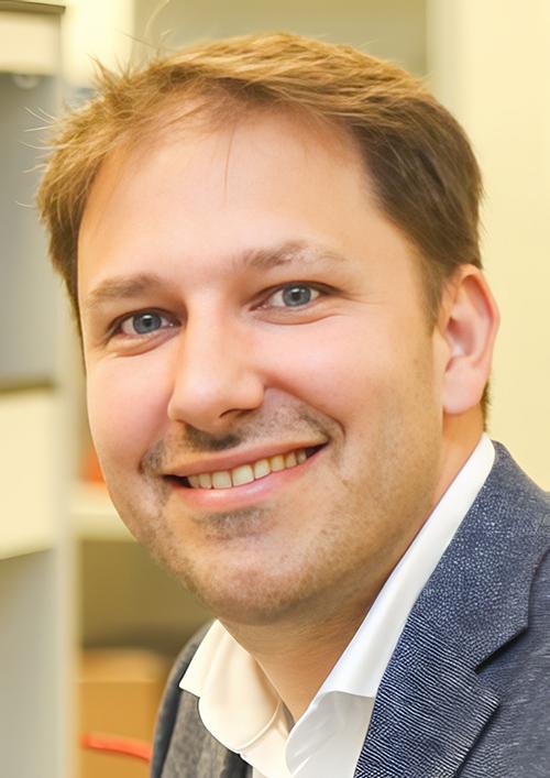
“It’s imperative that the tumor subtype is known at the time of surgery,” Jeroen de Ridder, PhD (above), associate professor in the Center for Molecular Medicine at UMC Utrecht and one of the study leaders, told The New York Times. “What we have now uniquely enabled is to allow this very fine-grained, robust, detailed diagnosis to be performed already during the surgery. It can figure out itself what it’s looking at and make a robust classification,” he added. How this discovery affects the role of anatomic pathologists and pathology laboratories during cancer surgeries remains to be seen. (Photo copyright: UMC Utrecht.)
Rapid DNA Sequencing Impacts Brain Tumor Surgeries
The UMC Utrecht scientists employed Oxford Nanopore’s “real-time DNA sequencing technology to address the challenges posed by central nervous system (CNS) tumors, one of the most lethal type of tumor, especially among children,” according to an Oxford Nanopore news release.
The researchers called their new machine learning AI application the “Sturgeon.”
According to The New York Times, “The new method uses a faster genetic sequencing technique and applies it only to a small slice of the cellular genome, allowing it to return results before a surgeon has started operating on the edges of a tumor.”
Jeroen de Ridder, PhD, an associate professor in the Center for Molecular Medicine at UMC Utrecht, told The New York Times that Sturgeon is “powerful enough to deliver a diagnosis with sparse genetic data, akin to someone recognizing an image based on only 1% of its pixels, and from an unknown portion of the image.” Ridder is also a principal investigator at the Oncode Institute, an independent research center in the Netherlands.
The researchers tested Sturgeon during 25 live brain surgeries and compared the results to an anatomic pathologist’s standard method of microscope tissue examination. “The new approach delivered 18 correct diagnoses and failed to reach the needed confidence threshold in the other seven cases. It turned around its diagnoses in less than 90 minutes, the study reported—short enough for it to inform decisions during an operation,” The New York Times reported.
But there were issues. Where the minute samples contain healthy brain tissue, identifying an adequate number of tumor markers could become problematic. Under those conditions, surgeons can ask an anatomic pathologist to “flag the [tissue samples] with the most tumor for sequencing, said PhD candidate Marc Pagès-Gallego, a bioinformatician at UMC Utrecht and a co-author of the study,” The New York Times noted.
“Implementation itself is less straightforward than often suggested,” Sebastian Brandner, MD, a professor of neuropathology at University College London, told The Times. “Sequencing and classifying tumor cells often still required significant expertise in bioinformatics as well as workers who are able to run, troubleshoot, and repair the technology,” he added.
“Brain tumors are also the most well-suited to being classified by the chemical modifications that the new method analyzes; not all cancers can be diagnosed that way,” The Times pointed out.
Thus, the research continues. The new method is being applied to other surgical samples as well. The study authors said other facilities are utilizing the method on their own surgical tissue samples, “suggesting that it can work in other people’s hands.” But more work is needed, The Times reported.
UMC Utrecht Researchers Receive Hanarth Grant
To expand their research into the Sturgeon’s capabilities, the UMC Utrecht research team recently received funds from the Hanarth Fonds, which was founded in 2018 to “promote and enhance the use of artificial intelligence and machine learning to improve the diagnosis, treatment, and outcome of patients with cancer,” according to the organization’s website.
The researchers will investigate ways the Sturgeon AI algorithm can be used to identify tumors of the central nervous system during surgery, a UMC Utrecht news release states. These type of tumors, according to the researchers, are difficult to examine without surgery.
“This poses a challenge for neurosurgeons. They have to operate on a tumor without knowing what type of tumor it is. As a result, there is a chance that the patient will need another operation,” said de Ridder in the news release.
The Sturgeon application solves this problem. It identifies the “exact type of tumor during surgery. This allows the appropriate surgical strategy to be applied immediately,” the news release notes.
The Hanarth funds will enable Jeroen and his team to develop a variant of the Sturgeon that uses “cerebrospinal fluid instead of (part of) the tumor. This will allow the type of tumor to be determined already before surgery. The main challenge is that cerebrospinal fluid contains a mixture of tumor and normal DNA. AI models will be trained to take this into account.”
The UMC Utrecht scientists’ breakthrough is another example of how organizations and research groups are working to shorten time to answer, compared to standard anatomic pathology methods. They are combining developing technologies in ways that achieve these goals.
—Kristin Althea O’Connor
Related Information:
Ultra-fast Deep-Learned CNS Tumor Classification during Surgery
New AI Tool Diagnoses Brain Tumors on the Operating Table
Pediatric Brain Tumor Types Revealed Mid-Surgery with Nanopore Sequencing and AI
AI Speeds Up Identification Brain Tumor Type
Four New Cancer Research Projects at UMC Utrecht Receive Hanarth Grants
Rapid Nanopore Sequencing, Machine Learning Enable Tumor Classification during Surgery


