Use of such precision diagnostics offer ‘early detection, localization, and the opportunity to monitor response to therapy,’ say the MIT scientists
Oncologists and medical laboratory scientists know that most clinical laboratory tests currently used to diagnose cancer are either based on medical imaging technologies—such as CT scans and mammography—or on molecular diagnostics that detect cancer molecules in the body’s urine or blood.
Now, in a study being conducted at the Massachusetts Institute of Technology (MIT), researchers have developed diagnostic nanoparticles that can not only detect cancer cells in bodily fluids but also image the cancer’s location. This is the latest example of how scientists are combining technologies in new ways in their efforts to develop more sensitive diagnostic tests that clinical laboratories and other providers can use to detect cancer and other health conditions.
Precision diagnostics such as molecular, imaging, and analytics technologies are key tools in the pursuit of precision medicine.
“Therapeutic outcomes in oncology may be aided by precision diagnostics that offer early detection, localization, and the opportunity to monitor response to therapy,” the authors wrote, adding, “Through tailored target specificities, this modular platform has the capacity to be engineered as a pan-cancer test that may guide treatment decisions for numerous tumor type.”
Development of Multimodal Diagnostics
The MIT scientists are developing a “multimodal” diagnostic that uses molecular screening combined with imaging techniques to locate where a cancer began in the body and any metastases that are present.
“In principle, this diagnostic could be used to detect cancer anywhere in the body, including tumors that have metastasized from their original locations,” an MIT new release noted.
“This is a really broad sensor intended to respond to both primary tumors and their metastases,” said biological engineer Sangeeta Bhatia, MD, PhD (above), in the news release. Bhatia is the John and Dorothy Wilson Professor of Health Sciences and Technology and Electrical Engineering and Computer Science at MIT and senior author of the study.
“It can trigger a urinary signal and also allow us to visualize where the tumors are,” she added. Bhatia previously worked on the development of cancer diagnostics that can produce synthetic biomarkers which are detectable in urine samples.
“The vision is that you could use this in a screening paradigm—alone or in conjunction with other tests—and we could collectively reach patients that do not have access to costly screening infrastructure today,” said Sangeeta Bhatia, MD, PhD (above), in the MIT news release. “Every year you could get a urine test as part of a general check-up. You would do an imaging study only if the urine test turns positive to then find out where the signal is coming from. We have a lot more work to do on the science to get there, but that’s where we would like to go in the long run.” (Photo copyright: NBC News.)
Precision Diagnostic Assists Assessment of Response to Cancer Therapy
For their research, the scientists added a radioactive tracer known as copper-64 to the nanoparticles. This enabled the particles to be used for positron emission tomography (PET) imaging. The particles were coated with a peptide that induced them to accumulate at tumor sites and insert themselves into cell membranes, producing a strong imaging signal for tumor detection.
The researchers tested their diagnostic nanoparticles in mouse models of metastatic colon cancer where tumor cells had traversed to the liver or the lungs. After treating the cancer cells with a chemotherapy regimen, the team successfully used both urine and imaging to determine how the tumors were responding to the treatment.
Bhatia is hopeful that this type of diagnostic could be utilized in assessing how patients are responding to treatment therapies and the monitoring of tumor recurrence or metastasis, especially for colon cancer.
What is unique about the approach used by Bhatia’s team is that one application of the copper-64 tracer can be used in vivo, in combination with imaging technology. The other application of the copper-64 tracer is in vitro in a urine specimen that can be tested by clinical laboratories.
“Those patients could be monitored with the urinary version of the test every six months, for instance. If the urine test is positive, they could follow up with a radioactive version of the same agent for an imaging study that could indicate where the disease had spread,” Bhatia said in the news release. “We also believe the regulatory path may be accelerated with both modes of testing leveraging a single formulation.”
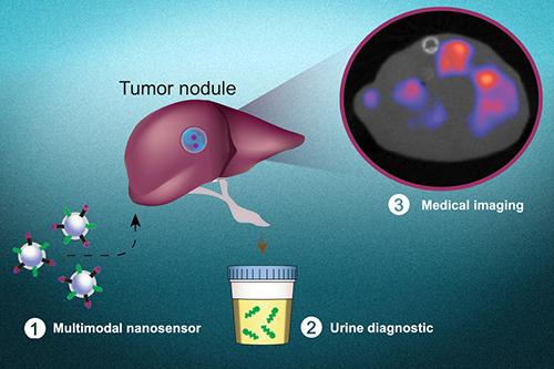
The graphic above, taken from the MIT news release, shows how “multimodal nanosensors (1) are engineered to target and respond to hallmarks in the tumor microenvironment. The nanosensors provide both a noninvasive urinary monitoring tool (2) and an on-demand medical imaging agent (3) to localize tumor metastasis and assess response to therapy,” the news release states. (Photo and caption copyright: Massachusetts Institute of Technology.)
Precision Medicine Cancer Screening Using Nano Technologies
Bhatia hopes that the nanoparticle technology may be used as a screening tool in the future to detect any type of cancer.
Her previous research with nanoparticle technology determined that a simple urine test could diagnose bacterial pneumonia and indicate if antibiotics could successfully treat that illness, the news release noted.
Nanoparticle-based technology might be adapted in the future to be part of a screening assay that determines if cancer cells are present in a patient. In such a scenario, clinical laboratories would be performing tests on urine samples while imaging techniques are simultaneously being used to diagnose and monitor cancers.
Surgical pathologists may also want to monitor the progress of this research, as it has the potential to be an effective tool for monitoring cancer patients following surgery, chemotherapy, or radiation therapy.
Studies presented at the Alzheimer’s Association International Conference point to the p-tau217 protein as an especially useful biomarker
Researchers disclosed a potentially useful biomarker for Alzheimer’s Disease at a major conference this summer. The good news for clinical laboratories is that the biomarker is found in blood. If further research confirms these early findings, medical laboratories could one day have a diagnostic test for this condition.
That possibility emerged from the Alzheimer’s Association International Conference (AAIC), which was held online July 27-31. Researchers presented findings from multiple studies that suggested blood/plasma levels of a protein known as phospho-tau217 (p-tau217) can indicate brain anomalies associated with Alzheimer’s.“Changes in brain proteins amyloid and tau, and their formation into clumps known as plaques and tangles, respectively, are defining physical features of Alzheimer’s disease in the brain,” states an AAIC press release. “Buildup of tau tangles is thought to correlate closely with cognitive decline. In these newly reported results, blood/plasma levels of p-tau217, one of the forms of tau found in tangles, also seem to correlate closely with buildup of amyloid.”
At present, “there is no single diagnostic test that can determine if a person has Alzheimer’s disease,” the association states on its website. Clinicians will typically review a patient’s medical history and conduct tests to evaluate memory and other everyday thinking skills. That may help determine that an individual has dementia, but not necessarily that Alzheimer’s is the cause.
“Currently, the brain changes that occur before Alzheimer’s dementia symptoms appear can only be reliably assessed by positron-emission tomography (PET) scans, and from measuring amyloid and tau proteins in [cerebrospinal] fluid (CSF),” the association states. “These methods are expensive and invasive. And, too often, they are unavailable because they are not covered by insurance or difficult to access, or both.”
In the AAIC press release, Alzheimer’s Association Chief Science Officer Maria C. Carrillo, PhD, said that a clinical laboratory blood test “would fill an urgent need for simple, inexpensive, non-invasive and easily available diagnostic tools for Alzheimer’s.
“New testing technologies could also support drug development in many ways,” she added. “For example, by helping identify the right people for clinical trials, and by tracking the impact of therapies being tested. The possibility of early detection and being able to intervene with a treatment before significant damage to the brain from Alzheimer’s disease would be game changing for individuals, families, and our healthcare system.”
However, she cautioned, “these are early results, and we do not yet know how long it will be until these tests are available for clinical use. They need to be tested in long-term, large-scale studies, such as Alzheimer’s clinical trials.”
The study, led by Oskar Hansson, MD, of Lund University in Sweden, included 1,402 participants. About half of these were enrolled in BioFINDER-2, an ongoing dementia study in Sweden. In this group, researchers were most interested in the test’s ability to distinguish Alzheimer’s from other neurodegenerative disorders that cause dementia.
Diagnostic accuracy was between 89% and 98%, the researchers reported, which was similar to the performance of PET imaging and CSF tests. P-tau217 was more accurate than magnetic resonance imaging (MRI) as well as other biomarkers, such as p-tau181.
“Today the majority of individuals with Alzheimer’s disease around the world do not get a timely diagnosis, which results in suboptimal symptomatic treatment and care,” Oskar Hansson, MD, said in an Eli Lilly news release. “With rising prevalence of Alzheimer’s disease, more patients will be evaluated in primary care and other clinics where CSF and PET biomarkers are not available. Blood-based biomarkers, like plasma p-tau217, together with digital tools for checking memory performance, such as smartphone-based apps, can considerably improve the diagnostic work-up of Alzheimer’s disease patients in such clinics.” (Photo copyright: Alzheimer’s Fund.)
Another cohort consisted of 81 participants in the Brain and Body Donation Program at Banner Sun Health Research Institute in Sun City, Ariz. In this program, elderly volunteers submit to periodic clinical assessments and agree to donate their organs and tissue for study after they die.
Here, the researchers’ primary goal was to determine the test’s ability to distinguish between individuals with and without Alzheimer’s. Researchers ran the p-tau217 test on plasma samples collected within 2.9 years of death and compared the results to postmortem examinations of the brain tissue. Accuracy was 89% in individuals with amyloid plaques and tangles, and 98% in individuals with plaques and more extensive tangles.
The third cohort consisted of 622 members of a large extended family in Colombia whose members share a genetic mutation that makes them susceptible to early-onset Alzheimer’s, The New York Times reported. Among the members, 365 were carriers of the mutation. In this group, levels of plasma p-tau217 increased by age, and “a significant difference from noncarriers was seen at age 24.9 years,” the researchers wrote in Jama Network. That’s about 20 years before the median age when mild cognitive impairment typically begins to appear in carriers.
Other Alzheimer Biomarker Studies Presented at AAIC
Suzanne Schindler, MD, PhD, a neurologist and instructor in the Department of Neurology at the Washington University School of Medicine (WUSM) in St. Louis, presented results of an Alzheimer’s Disease (AD) study that used mass spectrometry to analyze amyloid and p-tau variants in blood samples collected from participants. The researchers compared these with CSF and PET results and found that some of the of p-tau isoforms, especially p-tau217, had a strong concordance.
“These findings indicate that blood plasma Aβ and p-tau measures are highly precise biomarkers of brain amyloidosis, tauopathy, and can identify stages of clinical and preclinical AD,” stated an AAIC press release on the studies.
The WUSM researches launched the effort to develop and validate Alzheimer’s blood biomarkers called the Study to Evaluate Amyloid in Blood and Imaging Related to Dementia (SEABIRD) in April 2019. It runs through August 2023 and will seek to enroll more than 1,100 participants in the St. Louis area.
Another study presented at the conference compared the performance of p-tau217 and p-tau181 in distinguishing between Alzheimer’s and Frontotemporal Lobar Degeneration (FTLD), another condition that causes dementia. Study author Elisabeth Thijssen, MSc, of the UC San Francisco Memory and Aging Center reported that both biomarkers could be useful in differential diagnosis, but that p-tau217 was “potentially superior” for predicting a tau positive PET scan result.
For decades, physicians have wanted a diagnostic test for Alzheimer’s Disease that could identify this condition early in its development. This would allow the patient and the family to make important decisions before the onset of severe symptoms. Such a clinical laboratory test would be ordered frequently and thus would be a new source of revenue for medical laboratories.
Scientists worldwide engaged in research to develop a biomarker for dementia are predicting success, though some say additional research will be needed
Could a blood test for Alzheimer’s disease soon be on clinical laboratory test menus nationwide? Perhaps so. A recent Associated Press (AP) article that was picked up by NBC News and other healthcare publications reported that experimental test results presented during the Alzheimer’s Association International Conference (AAIC) in July suggest the Holy Grail of dementia tests—one where the specimen can be collected in a doctor’s office during a routine screening exam—may be close at hand.
The AP story noted that “half a dozen research groups gave new results on various experimental tests, including one that seems 88% accurate at indicating Alzheimer’s risk.” And Richard Hodes, MD, Director of the National Institute on Aging, told AP, “In the past year, we’ve seen a dramatic acceleration in progress [on Alzheimer’s tests]. This has happened at a pace that is far faster than any of us would have expected.”
This could be a boon for medical laboratories seeking way to contribute more value to patient care. Especially among Alzheimer’s patients, who account for as many as 70% of all dementia cases.
Plasma Biomarker for Predicting Alzheimer’s
One of the experimental blood tests presented at the AAIC involved a 2018 study into “the potential clinical utility of plasma biomarkers in predicting brain amyloid-β burden at an individual level. These plasma biomarkers also have cost-benefit and scalability advantages over current techniques, potentially enabling broader clinical access and efficient population screening,” the researchers stated an article they published in Nature.
AP also reported that Japanese scientists at the AAIC
presented results of a validation test conducted on 201 people who had either
Alzheimer’s, other types of dementia, or little or no symptoms. They found that
the test “correctly identified 92% of people who had Alzheimer’s and correctly
ruled out 85% who did not have it, for an overall accuracy of 88%.”
Akinori Nakamura, MD, PhD, of the National Center for
Geriatrics and Gerontology in Obu, Japan, was a member of the research team and
first author of the research paper. He told the AP that the test results “closely
matched those from the top tests used now—three types of brain scans and a
mental assessment exam.”
Eric McDade, DO (above), Associate Professor of Neurology at Washington University in St. Louis, told Neurology Today, “The results reported here provide a relatively high level of confidence given that this is a relatively well characterized population with an amyloid PET scan to provide confirmation of a significant level of amyloid plaque burden in the brain.” Could this level of physician confidence lead to a clinical laboratory test based on the plasma biomarker? (Photo copyright: Washington University.)
Koichi Tanaka is a Japanese engineer who won the Nobel prize winner for chemistry. He heads the Koichi Tanaka Research Lab at Shimadzu Corp. (OTCMKTS:SHMZF) in Kyoto, Japan, and was on the team that developed the Amyloid beta biomarker test that was presented at AAIC. He told Bloomberg, “Our finding overturned the common belief that it wouldn’t be possible to estimate amyloid accumulation in the brain from blood. We’re now being chased by others, and the competition is intensifying.”
But Tanaka cautions that the test needs further study before
it is ready for clinical use, and that for now “it belongs in the hands of drug
developers and research laboratories,” Bloomberg reported.
Other Studies into Developing an Alzheimer’s Biomarker
Alzheimer’s is usually diagnosed after symptoms appear, such
as memory loss. To arrive at their diagnoses, doctors often rely on medical
history, brain imaging (MRI, CT), PET, and measurement of amyloid in spinal
fluid.
An article published on Alzforum, a website and news service dedicated to the research and treatment for Alzheimer’s and other related disorders, noted a study by King’s College London researchers who, using mass spectrometry, “found a panel of biomarkers that predicted with almost 90% accuracy whether cognitively normal people had a positive amyloid scan.”
Nicholas Ashton, PhD, neuroscientist and Wallenberg Postdoctoral Fellow at University of Gothenburg in Sweden, and first author of the King’s College study, explained that “Amyloid-burden and neurofilament light polypeptide (NFL) peptides were important in predicting Alzheimer’s, but alone they weren’t as predictable as when we combined them with novel proteins related to amyloid PET.”
The researchers published their study earlier this year in Science Advances. “Using an unbiased mass spectrometry approach, we have found and replicated with high accuracy, specificity, and sensitivity a plasma protein classifier reflecting amyloid-beta burden in a cognitively unimpaired cohort,” the researchers wrote.
Meanwhile, researchers at Washington University School of Medicine St. Louis, along with the German Center for Neurodegenerative Diseases, a member of the Helmholtz Association, stated in a news release that a blood test they developed works by detecting leaks of NFL before the onset of symptoms. When the protein is found in cerebrospinal fluid, it could be a sign that Alzheimer’s may develop, as well as point to other neurodegenerative conditions such as multiple sclerosis, brain injury, or stroke, the researchers stated.
“This is something that would be easy to incorporate into a screening test in a neurology clinic,” Brian Gordon, PhD, Assistant Professor of Radiology at Washington University’s Mallinckrodt Institute of Radiology, and an author of the study, stated in the news release.
These parallel studies into screening for Alzheimer’s by
researchers worldwide are intriguing. The favorable results suggest that
someday there may be a screen for Alzheimer’s using a clinical laboratory blood
test.
With Alzheimer’s affecting nearly six million Americans of all ages, such an assay would enable clinical laboratories to help many people.
Both pathologists and clinical laboratory managers are likely to be intrigued with how swiftly mobile computing technology can adapted for use with healthcare images. Earning the honors as the first mobile app to be cleared by the FDA for use with radiology images is the Mobile MIM software, developed by MIM Software, Inc. of Cleveland, Ohio.