Rice University Researchers Develop ‘Molecular Jackhammer’ That Kills Cancer Cells
Research could lead to similar treatments for other diseases, as well as creating a demand for a new line of oncology tests for clinical labs and pathology groups
Cancer treatment has come a long way in the past decades, and it seems poised to take another leap forward thanks to research being conducted at Rice University in Houston. Molecular scientists there have developed what they call a “molecular jackhammer” that uses special molecules and near-infrared light to attack and kill cancer cells.
The technique has been effective in research settings. Should it be cleared for use in patient care, it could change the way doctors treat cancer patients while giving clinical laboratories a new diagnostic tool that could guide treatment decisions.
The researchers “found that the atoms of a small dye molecule used for medical imaging can vibrate in unison—forming what is known as a plasmon [a quantum of plasma oscillation]—when stimulated by near-infrared light, causing the cell membrane of cancerous cells to rupture,” a Rice University news release noted.
The small dye molecule is called aminocyanine, a type of fluorescent synthetic dye that is already in use in medical imaging.
“These molecules are simple dyes that people have been using for a long time,” said physical chemistry scientist Ciceron Ayala-Orozco, PhD, the researcher who led the study, in the news release. “They’re biocompatible, stable in water, and very good at attaching themselves to the fatty outer lining of cells. But even though they were being used for imaging, people did not know how to activate these as plasmons.”
The Rice University scientists published their findings in the journal Nature Chemistry titled, “Molecular Jackhammers Eradicate Cancer Cells by Vibronic-Driven Action.”
“The method had a 99% efficiency against lab cultures of human melanoma cells, and half of the mice with melanoma tumors became cancer-free after treatment,” according to the Rice University news release.
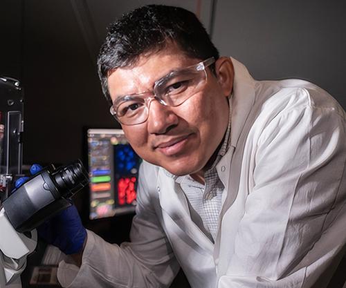
“I spent approximately four years working with these ideas on using molecular forces and what is called blue-light activated molecular motors,” Ciceron Ayala-Orozco, PhD (above), told Oncology Times. “At some point, I connected the dots that what I wanted to do is use a simple molecule, not necessarily a motor, that absorbs NIR light in similar ways as plasmonic nanoparticles do and go deeper into the tissue. When activated, we found that the molecules vibrate even faster than our minds can imagine and serve as a force to break the cancer cells apart.” Once approved for use treating cancer patients, clinical laboratories working with oncologists may play a key role in diagnosing candidates for the new treatment. (Photo copyright: Rice University.)
How the Technique Works
Nuclei of the aminocyanine molecules oscillate in sync when exposed to near-infrared radiation and pummel the surface of the cancer cell. These blows are so powerful they rupture the cell’s membrane sufficiently enough to destroy it.
“The speed of this type of therapy can completely kill the cancer much faster than, say, photodynamic therapy,” Ayala-Orozco noted. “The mechanical action through the molecular jackhammer is immediate, within a few minutes.”
One advantage to near-infrared light is that it can infiltrate deeper into the body than visible light and access organs and bones without damaging tissue.
“Near-infrared light can go as deep as 10 centimeters (four inches) into the human body as opposed to only half a centimeter (0.2 inches), the depth of penetration for visible light, which we used to activate the nanodrills,” said James Tour, PhD, T. T. and W. F. Chao Professor of Chemistry, Professor of Materials Science and NanoEngineering at Rice University, in the news release. “It is a huge advance.”
The molecular plasmons identified by the team had a near-symmetrical structure. The plasmons have an arm on one side that does not contribute to the motion, but rather anchors the molecule to the lipid bilayer of the cell membrane. The scientists had to prove that the motion could not be categorized as a form of either photodynamic or photothermal therapy.
“What needs to be highlighted is that we’ve discovered another explanation for how these molecules can work,” Ayala-Orozco said in the Rice news release. “This is the first time a molecular plasmon is utilized in this way to excite the whole molecule and to actually produce mechanical action used to achieve a particular goal—in this case, tearing apart cancer cells’ membrane.
“This study is about a different way to treat cancer using mechanical forces at the molecular scale,” he added.
New Ways to Treat Cancer
The likelihood of cancer cells developing a resistance to these molecular jackhammers is extremely low, which renders them a safer and more cost effective method for inducing cancer cell death.
“The whole difference about this is because it’s a mechanical action, it’s not relying on some chemical effect,” Tour told KOMO News. “It’s highly unlikely that the cell will be able to battle against this. Once it’s cell-associated, the cell is toast once it gets hit by light. Only if a cell could prevent a scalpel from being able to cut it in half, could it prevent this.
“It will kill all sorts of cell types. With our other mechanical action molecules, we’ve demonstrated that they kill bacteria; we’ve demonstrated that they kill fungi. If a person has lost the ability to move a limb, if you can stimulate the muscle with light, that would be quite advantageous. Cancer is just the beginning,” he added.
“From the medical point of view, when this technique is available, it will be beneficial and less expensive than methods such as photothermal therapy, photodynamics, radio-radiation, and chemotherapy,” said Jorge Seminario, PhD, Professor in the Artie McFerrin Department of Chemical Engineering at Texas A&M University in a news release.
Researchers from Texas A&M University and the University of Texas-MD Anderson Cancer Center participated in the study.
“This is one of the very few theoretical-experimental approaches of this nature. Usually, research in the fields related to medicine does not use first principles quantum-chemistry techniques like those used in the present work, despite the strong benefit of knowing what the electrons and nuclei of all atoms are doing in molecules or materials of interest,” Seminario noted.
“It’s really a tremendous advance. What this is going to do is open up a whole new mode of treatment for medicine,” Tour said. “It’s just like when radiation came in [and] when immunotherapy came in. This is a whole new modality. And when a new modality comes in, so much begins to open up.
“Hopefully, this is going to change medicine in a big way,” he added.
More research and clinical studies are needed before this new technology is ready for patient care. Clinical laboratories and anatomic pathology groups will likely be involved identifying patients who would be good candidates for the new treatment. These molecular jackhammers could be a useful tool in the future fight against cancer, which is ranked second (after heart disease) as the most common cause of death in the US.
—JP Schlingman
Related Information:
New Molecular Jackhammer Technique Achieves 99% Cancer Treatment Success in Labs
Scientists Destroy 99% of Cancer Cells in the Lab Using Vibrating Molecules
Molecular Jackhammers Drill Pathway to Killing Cancer Cells
Molecular Jackhammers Eradicate Cancer Cells by Vibronic-driven Action
Molecular Jackhammers’ “Good Vibrations” Eradicate Cancer Cells
Molecular Jackhammers’ Non-Invasive Approach to Destroy Cancer Cells