With further research, clinical laboratories may soon be performing macrobiotic testing to measure certain bacterial levels in patients’ gut bacteria
New insights from the University of Chicago (UChicago) into how human microbiota (aka, gut bacteria) play a role in food allergies has the potential to change the way a number of gastrointestinal health conditions are diagnosed and treated. This would give microbiologists and clinical laboratories a greater role in helping physicians diagnose, treat, and monitor patients with these health issues.
Past research has shown that certain gut bacteria can prevent antigens that trigger allergic reactions from entering the bloodstream. For example, Clostridium bacteria in the stomach produce a short-chain fatty acid known as butyrate, a metabolite that promotes the growth of healthy bacteria in the gut. This helps keep the microbiome in balance.
One way butyrate is created in the gut is through the fermentation of fiber. However, a lack of fiber in the diet can deplete the production of butyrate and cause the microbiome to be out of balance. When this happens, a state known as dysbiosis occurs that disrupts the microbiome and can lead to food allergies.
Without butyrate, the gut lining can become permeable and allow food to leak out of the gastrointestinal tract and into the body’s circulatory system. This reaction can trigger a potentially fatal anaphylactic response in the form of a food allergy. Thus, eating enough fiber is critical to the production of butyrate and to maintaining a balanced microbiome.
But today’s western diet can be dangerously low in soluble fiber. Therefore, the scientists at the University of Chicago have developed “a special type of polymeric molecule to deliver a crucial metabolite produced by these bacteria directly to the gut, where it helps restore the intestinal lining and allows the beneficial bacteria to flourish. … these polymers, called micelles, can be designed to release a payload of butyrate, a molecule that is known to help prevent food allergies, directly in the small and large intestines,” according to a UChicago news release.
This will be of interest to microbiologists, in particular. It’s another example of researchers connecting a specific species of bacteria in the human microbiome to a specific benefit.
“It’s very unlikely that butyrate is the only relevant metabolite, but the beauty of this platform is that we can make polymers with other microbial metabolites that could be administered in conjunction with butyrate or other therapies,” said Cathryn Nagler, PhD (above), Bunning Family Professor in the Biological Sciences Division and Pritzker School of Molecular Engineering at UChicago and a senior author of the study. “So, the potential for the polymer platform is pretty much wide open.” As further research validates these findings, clinical labs are likely to be doing microbiomic testing to monitor these therapies. (Photo copyright: University of Chicago.)
Restoring Butyrate in the Gut
One way to treat this anomaly has been through a microbiota transplant—also called a fecal biota transplant—where the administration of a solution of fecal matter is transplanted from a donor into the intestinal tract of the recipient. This transplant alters the recipient’s gut microbial composition to a healthier state, but it has had mixed results.
So, the UChicago researchers went in another direction (literally). They created an oral solution of butyrate and administered it to mice in the lab. The purpose of the solution was to thwart an allergic reaction when the mice were exposed to peanuts.
But there was a problem with their oral solution. It was repulsive.
“Butyrate has a very bad smell, like dog poop and rancid butter, and it also tastes bad, so people wouldn’t want to swallow it,” Shijie Cao, PhD, Postdoctoral Scientist at the Pritzker School of Molecular Engineering at UChicago and one of the researchers who worked on the project, told Medical News Today.
The researchers developed a new configuration of polymers that masked the butyrate. They then delivered these polymer micelles directly into the digestive systems of mice that lacked healthy gut bacteria or a proper gut linings.
The treatment restored the microbiome by increasing the production of peptides that obliterate harmful bacteria. This allowed more of the beneficial butyrate-producing bacteria to emerge, which protected the mice from an anaphylactic reaction to peanuts and even reduced the symptom severity in an ulcerative colitis model.
“We were delighted to see that our drug both replenished the levels of butyrate present in the gut and helped the population of butyrate-producing bacteria to expand,” said Cathryn Nagler, PhD, Bunning Family Professor in the Biological Sciences Division and Pritzker School of Molecular Engineering at the University of Chicago and a senior author of the study, in the press release. “That will likely have implications not only for food allergy and inflammatory bowel disease (IBD), but also for the whole set of non-communicable chronic diseases that have been rising over the last 30 years, in response to lifestyle changes and overuse of antibiotics in our society.”
Future Benefits of UChicago Treatment
According to data from the Asthma and Allergy Foundation of America, about 20 million Americans suffered from food allergies in 2021. This includes approximately 16 million (6.2%) of adults and four million (5.8%) of children. The most common allergens for adults are shellfish, peanuts, and tree nuts, while the most common allergens for children are milk, eggs, and peanuts.
The best way to prevent an allergic reaction to a trigger food is strict avoidance. But this can be difficult to ensure outside of the home. Therefore, scientists are searching for ways to prevent food allergies from happening in the first place. The micelle technology could be adapted to deliver other metabolites and molecules which may make it a potential platform for treating allergies as well as other inflammatory gastrointestinal diseases.
“It’s a very flexible chemistry that allows us to target different parts of the gut,” said Jeffrey Hubbell, PhD, Eugene Bell Professor in Tissue Engineering and Vice Dean and Executive Officer at UChicago’s Pritzker School of Molecular Engineering and one of the project’s principal investigators, in the UChicago news release. “And because we’re delivering a metabolite like butyrate, it’s antigen-agnostic. It’s one agent for many different allergic indications, such as peanut or milk allergies. Once we begin working on clinical trials, that will be a huge benefit.”
Nagler and Hubbell have co-founded a company called ClostraBio to further the development of butyrate micelles into a commercially available treatment for peanut and other food allergies. They hope to begin clinical trials within the next 18 months and expand the technology to other applications as well.
Further research and clinical trials are needed to prove the validity of using polymer micelles in the treatment of diseases. But it is possible that clinical laboratories will be performing microbiomic testing in the future to help alleviate allergic reactions to food and other substances.
Wearable microneedle sensors that track multiple biomarkers in interstitial fluid are finding their way into chronic disease monitoring and sample collecting for clinical laboratory testing
Wearable devices that replace finger sticks and blood draws for monitoring biomarkers of chronic diseases such as diabetes are the holy grail of non-invasive (or at least minimally invasive) technologies that collect specimens for clinical laboratory testing.
Now, in their quest for alternatives to invasive phlebotomy blood draws, engineers at University of California San Diego’s (UCSD) Center for Wearable Sensors have added their own wearable device to the mix. The scientists developed a “lab-on-the-skin” multi-tasking microneedle sensor that monitors multiple biomarkers simultaneously, according to a UCSD news release.
“This is like a complete lab on the skin,” said Joseph Wang, PhD (above), Distinguished Professor of Nanoengineering at UC San Diego and Director of UCSD’s Center of Wearable Sensors, in a news release. “It is capable of continuously measuring multiple biomarkers at the same time, allowing users to monitor their health and wellness as they perform their daily activities.” UC San Diego’s microneedle patch for monitoring biomarkers of disease certainly would be popular with patients who must regularly undergo painful blood draws for clinical laboratory testing. (Photo copyright: UC San Diego.)
Advantage of Monitoring Multiple Biomarkers in Real Time
While current glucose monitors on the market only measure glucose, the UCSD wearable device also monitors alcohol and lactate, providing other additional information to diabetics when engaged in activities that affect those biomarkers.
For example, UCSD’s microneedle sensor allows diabetics to monitor their glucose level when drinking alcohol, which can lower glucose levels. Additionally, monitoring lactate while exercising also could be beneficial since physical activity influences the body’s ability to regulate glucose.
“With our wearable, people can see the interplay between their glucose spikes or dips with their diet, exercise, and drinking of alcoholic beverages. That could add to their quality of life as well,” said Farshad Tehrani, a nanoengineering PhD graduate researcher in Wang’s lab at UCSD and one of the co-first authors of the study, in the news release.
UC San Diego’s wearable microneedle patch (above) is about the size of a stack of six quarters and simultaneously monitors glucose, alcohol, and lactate levels continuously. It affixes to the skin through a patch of microneedles each about one-fifth the width of a human hair. The microneedles barely penetrate the surface of the skin to sample biomolecules in the interstitial fluid and are not painful. The quarter-sized patch is worn on the upper arm and transmits its data to a smartphone app. The microneedle patch is disposable, and the reusable electronic case is rechargeable using an off-the-shelf wireless charging pad. (Photo copyright: Laboratory for Nanobioelectronics/UC San Diego.)
Other Microneedle Wearable Monitoring Patches
The quest for a painless alternative to in-patient blood draws for many clinical laboratory tests has been ongoing worldwide for years.
In “Researchers Develop ‘Smart’ Microneedle Adhesive Bandage System for Monitoring Sodium, Glucose, pH, and More,” Dark Daily reported on a proof-of-concept study conducted by scientists from Israel and China who developed a “smart” microneedle adhesive bandage that measures and monitors in real time three critical biomarkers that currently require invasive blood draws for medical laboratory tests commonly performed on patients in hospitals.
While further research and validation of studies are needed before UC San Diego’s wearable microneedle sensor patch can be deployed to monitor chronic diseases, it is in good company. Diabetics and other suffers of similar chronic diseases can look forward to a future where they can monitor their health conditions in real time without the need for invasive blood draws and clinical laboratory testing.
Columbia University’s MediSCAPE enables surgeons to examine tissue structures in vivo and a large-scale clinical trial is planned for later this year
Scientists at Columbia University in New York City have developed a high-speed 3D microscope for diagnosis of cancers and other diseases that they say could eventually replace traditional biopsy and histology “with real-time imaging within the living body.”
The technology is designed to enable in situ tissue analysis. Known as MediSCAPE, the microscope is “capable of capturing images of tissue structures that could guide surgeons to navigate tumors and their boundaries without needing to remove tissues and wait for pathology results,” according to a Columbia University news story.
“The way that biopsy samples are processed hasn’t changed in 100 years, they are cut out, fixed, embedded, sliced, stained with dyes, positioned on a glass slide, and viewed by a pathologist using a simple microscope. This is why it can take days to hear news back about your diagnosis after a biopsy,” said Hillman in the Columbia news story.
“Our 3D microscope overcomes many of the limitations of prior approaches to enable visualization of cellular structures in tissues in the living body. It could give a doctor real-time feedback about what type of tissue they are looking at without the long wait,” she added in I News.
Hillman’s team previously used the technology—originally dubbed SCAPE for “Swept Confocally Aligned Planar Excitation” microscopy—to capture 3D images of neurological activity in living samples of worms, fish, and flies. In their recent study, the researchers tested the technology with human kidney tissue, a human volunteer’s tongue, and a mouse with pancreatic cancer.
“This was something I didn’t expect—that I could actually look at structures in 3D from different angles,” said nephropathologist and study co-author Shana M. Coley, MD, PhD (above), Director, Transplant Translational Research and Multiplex Imaging Center at Arkana Laboratories, in the Columbia news story. At the time of the Columbia study, Coley was an assistant professor at Columbia University and a renal pathologist at the Columbia University Medical Center. “We found many examples where we would not have been able to identify a structure from a 2D section on a histology slide, but in 3D we could clearly see its shape. In renal pathology in particular, where we routinely work with very limited amounts of tissue, the more information we can derive from the sample, the better for delivering more effective patient care,” she added. (Photo copyright: Arkana Laboratories.)
How MediSCAPE Works
Unlike traditional 3D microscopes that use a laser to scan tiny spots of a tissue sample and then assemble those points into a 3D image, the MediSCAPE 3D microscope “illuminates the tissue with a sheet of light—a plane formed by a laser beam that is focused in a special way,” I News reported.
The MediSCAPE microscope thus captures 2D slices which are rapidly stacked into 3D images at a rate of more than 10 volumes per second, according to I News.
“One of the first tissues we looked at was fresh mouse kidney, and we were stunned to see gorgeous structures that looked a lot like what you get with standard histology,” said optical systems engineer and the study’s lead author, Kripa Patel, PhD, in the Columbia news story. “Most importantly, we didn’t add any dyes to the mouse—everything we saw was natural fluorescence in the tissue that is usually too weak to see.
“Our microscope is so efficient that we could see these weak signals well,” she continued, “even though we were also imaging whole 3D volumes at speeds fast enough to rove around in real time, scanning different areas of the tissue as if we were holding a flashlight.”
A big advantage of the technology, Hillman noted, is the ability to scan living tissue in the body.
“Understanding whether tissues are staying healthy and getting good blood supply during surgical procedures is really important,” she said in the Columbia news story. “We also realized that if we don’t have to remove (and kill) tissues to look at them, we can find many more uses for MediSCAPE, even to answer simple questions such as ‘what tissue is this?’ or to navigate around precious nerves. Both of these applications are really important for robotic and laparoscopic surgeries, where surgeons are more limited in their ability to identify and interact with tissues directly.”
Clinical Trials and FDA Clearance
Early versions of the SCAPE microscopes were too large for practical use by surgeons, so Columbia post-doctoral research scientist Wenxuan Liang, PhD, co-author of the study, helped the team develop a smaller version that would fit into an operating room.
Later this year, the researchers plan to launch a large-scale clinical trial, I News reported. The Columbia scientists hope to get clearance from the US Food and Drug Administration (FDA) to develop a commercialized version of the microscope.
“They will initially seek permission to use it for tumor screening and guidance during operations—a lower and easier class of approval—but ultimately, they hope to be allowed to use it for diagnosis,” Liang wrote.
Charles Evans, PhD, research information manager at Cancer Research UK, told I News, “Using surgical biopsies to confirm a cancer diagnosis can be time-consuming and distressing for patients. And ensuring all the cancerous tissue is removed during surgery can be very challenging unaided.”
He added, “more work will be needed to apply this technique in a device that’s practical for clinicians and to demonstrate whether it can bring benefits for people with cancer, but we look forward to seeing the next steps.”
Will the Light Microscope be Replaced?
In recent years, research teams at various institutions have been developing technologies designed to enhance or even replace the traditional light microscope used daily by anatomic pathologists across the globe.
And digital scanning algorithms for creating whole-slide images (WSIs) that can be analyzed by pathologists on computer screens are gaining in popularity as well.
Such developments may spark a revolution in surgical pathology and could signal the beginning of the end of the light microscope era.
Surgical pathologists should expect to see a steady flow of technologically advanced systems for tissue analysis to be submitted to the FDA for pre-market review and clearance for use in clinical settings. The light microscope may not disappear overnight, but there are a growing number of companies actively developing different technologies they believe can diagnose either or both tissue and digital images of pathology slides with accuracy comparable to a pathologist.
Skin patch technologies could enable clinical laboratories to monitor patients’ vitals and report to medical professionals in real time
Pathologists and clinical laboratory leaders have read many Dark Daily ebriefings on the development of skin patches over the years that do everything from monitoring fatigue in the military to being a complete lab-on-skin technology. Now, researchers at the University of California San Diego (UCSD) have developed a wearable patch that can monitor cardiovascular signals and other various biochemical levels in the body simultaneously.
The researchers believe there is enormous potential for such a patch in helping patients monitor conditions such as hypertension or diabetes. They also foresee a scenario where the patch could be used in settings where vitals must be constantly monitored. They hope to develop future versions of the patch that can detect more biomarkers within the body.
“This type of wearable would be very helpful for people with underlying medical conditions to monitor their own health on a regular basis,” Lu Yin, a PhD student and co-first author of the study, told New Atlas. “It would also serve as a great tool for remote patient monitoring, especially during the COVID-19 pandemic when people are minimizing in-person visits to the clinic,” she added.
Combining Precision Medicine with Telehealth and the Internet of Things
About the size of a postage stamp and consisting of stretchy polymers that conform to the skin, the UCSD patch monitors blood pressure and contains sensors that measure different biochemical levels in the body, such as:
The sensors are carefully arranged on the patch to eliminate interference between the signals, noted a UCSD press release.
In their published research, the UCSD researchers wrote of their new skin patch monitoring device, “Intertwined with concepts of telehealth, the internet of medical things, and precision medicine, wearable sensors offer features to actively and remotely monitor physiological parameters. Wearable sensors can generate data continuously without causing any discomfort or interruptions to daily activity, thus enhancing the self-monitoring compliance of the wearer, and improving the quality of patient care.” (Photo copyright: University of California San Diego.)
“Each sensor provides a separate picture of a physical or chemical change. Integrating them all in one wearable patch allows us to stitch those different pictures together to get a more comprehensive overview of what’s going on in our bodies,” said Sheng Xu, PhD, Principle Investigator, Xu Research Group at UCSD, Assistant Professor in the Department of NanoEngineering Department, and a co-first author of the study, in the press release.
The UCSD researchers developed their skin patch to monitor specific biomarkers that can affect blood pressure.
“Let’s say you are monitoring your blood pressure and you see spikes during the day and think that something is wrong,” co-first author Juliane Sempionatto, PhD, a postdoctoral researcher at California Institute of Technology (Caltech) and co-first author of the study, told New Atlas. “But a biomarker reading could tell you if those spikes were due to an intake of alcohol or caffeine. This combination of sensors can give you that type of information,” she added.
The blood pressure sensor sits near the center of the patch and consists of a set of small transducers welded to the patch via a conductive link. Voltage applied to the transducers send ultrasound waves through the body which bounce off arteries and create echoes that are detected by the sensor and converted into an accurate blood pressure reading.
The chemical sensor releases the drug pilocarpine into the skin to induce sweat and then measures the chemicals contained in the sweat to provide readings of certain biochemical levels.
The glucose sensor located in the patch emits a mild electrical current to the body that stimulates the release of interstitial fluid and then reads the glucose level in that fluid.
“The novelty here is that we take completely different sensors and merge them together on a single small platform as small as a stamp,” Joseph Wang, D.Sc, SAIC Endowed Chair, Distinguished Professor of NanoEngineering, Director of the Center for Wearable Sensors at UCSD, and co-author of the study told New Atlas. “We can collect so much information with this one wearable and do so in a non-invasive way, without causing discomfort or interruptions to daily activity.” (Photo copyright: University of Southern California San Diego.)
Skin Patch Measurements Closely Match Those of Traditional Devices
Test subjects wore the patch on their neck while performing various combinations of the following tasks:
exercising on a stationary bicycle,
eating a high-sugar meal,
drinking an alcoholic beverage, and
drinking a caffeinated beverage.
The results of the measurements taken from the patch closely matched measurements collected by traditional monitoring devices such as a:
For now, the patch must be connected to an external power source which transmits the reading to a counter-top machine, but the researchers hope to create a wireless version in the future.
“There are opportunities to monitor other biomarkers associated with various diseases,” Sempionatto said in the UCSD press release. “We are looking to add more clinical value to this device.”
Other Similar Skin Patch Monitoring Technologies
Though an important breakthrough, the UCSD’s device is not the first skin patch monitor to be developed.
Multiple research and clinical studies are underway that hope to prove the accuracy and safety of wearable devices at detecting and monitoring certain health conditions. It’s a worthy goal.
Skin patches, such as the one created at UCSD, could enable clinical laboratories to provide value-added service to medical professionals and patients alike. Medical labs could potentially monitor skin patch readings in real-time and notify physicians and patients of changes in biomarkers that require attention.
Further, as this technology is developed, it will likely find a ready market with the latest generation of consumers who are more willing than previous generations to buy their own diagnostic tests for home use. These “next-generation” healthcare consumers have demonstrated their willingness to use Apple watches, Fitbits, and similar wearable devices to monitor their condition during exercise and other health metrics.
Pathologists and clinical laboratory managers should not overlook the potential for robust consumer demand to accelerate development and market adoption of such skin patches.
Painless technology could one day replace some phlebotomy blood draws as the go-to specimen-collection method for clinical laboratory testing and health monitoring
Clinical laboratories have long sought a non-invasive way to do useful medical laboratory testing without the need for either a venipuncture or a needle stick. Now engineers at the McKelvey School of Engineering at Washington University in St. Louis in Missouri have developed a disposable microneedle patch that one day could be a painless alternative to some blood draws for diagnostics tests and health monitoring.
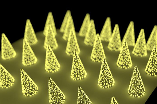
The technology uses an easy-to-administer low-cost patch that can be applied to the skin like an adhesive bandage. The patch is virtually painless because the microneedles are too small to reach nerve receptors. Another unique aspect to this innovative approach to collecting a specimen for diagnostic testing is that the Washington University in St. Louis (WashU) research team designed the microneedle patch to include plasmonic-fluor. These are ultrabright gold nanolabels that light up target protein biomarkers and can make the biomarkers up to 1,400 times brighter at low concentrations, compared to traditional fluorescent labels.
The patch, states a WashU news release, “… can be applied to the skin, capture a biomarker of interest and, thanks to its unprecedented sensitivity, allow clinicians to detect its presence.”
The technology is low cost, easy for clinicians or patients themselves to use, and could eliminate the need for a trip to patient service center where a phlebotomist would draw blood for clinical laboratory testing, the news release states.
“We have created a platform technology that anyone can use. And they can use it to find their own biomarker of interest,” study leader Srikanth Singamaneni, PhD (above), Lilyan and E. Lisle Hughes Professor in the Department of Mechanical Engineering and Materials Sciences at Washington University in St. Louis, said in the WashU news release. Singamaneni and his colleagues are developing a new specimen collection method that might someday be widely used by clinical laboratories. (Photo copyright: Washington University in St. Louis.)
“We used the microneedle patch in mice for minimally invasive evaluation of the efficiency of a cocaine vaccine, for longitudinal monitoring of the levels of inflammatory biomarkers, and for efficient sampling of the calvarial periosteum [a skull membrane]—a challenging site for biomarker detection—and the quantification of its levels of the matricellular protein periostin, which cannot be accurately inferred from blood or other systemic biofluids,” the researchers wrote. “Microneedle patches for the minimally invasive collection and analysis of biomarkers in interstitial fluid might facilitate point-of-care diagnostics and longitudinal monitoring.”
Mark Prausnitz, PhD, Regents’ Professor, J. Erskine Love Jr. Chair in Chemical and Biomolecular Engineering, and Director of the Center for Drug Design, Development, and Delivery at Georgia Tech, told WIRED, “Blood is a tiny fraction of the fluid in our body. Other fluids should have something useful—it’s just hard to get those fluids.”
“Previously, concentrations of a biomarker had to be on the order of a few micrograms per milliliter of fluid,” said Zheyu (Ryan) Wang, a PhD candidate in Srikanth Singamaneni’s lab at McKelvey School of Engineering and a lead author of the paper, in the WashU news release. By using plasmonic-fluor, researchers were able to detect biomarkers on the order of picograms per milliliter—one millionth of the concentration.
“That’s orders of magnitude more sensitive,” Wang said.
Unlike blood, dermal interstitial fluid often does not contain high enough concentrations of biomarkers to be easily detectable. To overcome this hurdle, the Washington University in St. Louis research team developed a microneedle patch with plasmonic-fluor—ultrabright gold nanolabels (above)—which lit up target protein biomarkers, making them roughly 1,400 times brighter at low concentrations than when using traditional fluorescent labels commonly used in many medical laboratory tests. (Photo copyright: Washington University in St. Louis.)
Can Microneedles Be Used as a Diagnostic Tool?
As reported in WIRED, the polystyrene patch developed by Srikanth Singamaneni’s lab at McKelvey School of Engineering removes interstitial fluid from the skin and turns the needles into “biomarker traps” by coating them with antibodies known to bind to specific proteins, such as Interleukin 6 (IL-6). Once the microneedles are mixed with plasmonic-fluor, the patch will glow if the IL-6 biomarkers are present.
The development of such a highly sensitive biomarker-detection method means skin becomes a potential pathway for using microneedles to diagnose conditions, such as myocardial infarction or to measure COVID-19 antibodies in vaccinated persons.
“Now we can actually use this tool to understand what’s going on with interstitial fluid, and how we’re going to be able to use it to answer healthcare-related or medical problems,” Maral Mousavi, PhD, Assistant Professor of Biomedical Engineering, Viterbi School of Engineering at the University of Southern California, told WIRED. “I think it has the potential to be that kind of a game changer.”
Because the WashU study is a proof-of-concept in mice, it may be many years before this technology finds its way to clinical application. Many skin biomarkers will need to be verified for direct links to disease before microneedle patches will be of practical use to clinicians for diagnostics. However, microneedle patch technology has already proven viable for the collection of blood.
In 2017, Massachusetts-based Seventh Sense Biosystems (7SBio) received 510(k) clearance for a new microneedle blood collection device. Called TAP, the device is placed on the upper arm and blood collection starts with a press of a button. The process takes two to three minutes.
Initially, the FDA clearance permitted only healthcare workers to use the device “to collect capillary blood for hemoglobin A1c (HbA1c) testing, which is routinely used to monitor blood sugar levels in diabetic or pre-diabetic patients,” a Flagship Pioneering news release noted.
Then, in 2019, the FDA extended its authorization “to include blood collection by laypersons. Regulators are also allowing the device to be used ‘at-home’ for wellness testing,” a 7SBio news release stated. This opened the door for a microneedle device to be used for home care blood collection.
“No one likes getting blood drawn, but blood is the single-most important source of medical information in healthcare today, with about 90% of all diagnostic information coming from blood and its components,” Howard Weisman, former CEO of 7SBio and current CEO of PaxMedica, a clinical-stage biopharmaceutical company, said in the Flagship Pioneering news release. “TAP has the potential to transform blood collection from an inconvenient, stressful, and painful experience to one people can do themselves anywhere, making health monitoring much easier for both healthcare professionals and patients.”
As microneedle technology continues to evolve, clinical laboratories should expect patches to be used in a growing number of drug delivery systems and diagnostic tests. But further research will be needed to determine whether interstitial fluid can provide an alternate pathway for diagnosing disease.