


Researchers at Washington University in St. Louis Use Microneedle Patch with Fluorescent Nanolabels to Detect Biomarkers in Skin’s Interstitial Fluid
Painless technology could one day replace some phlebotomy blood draws as the go-to specimen-collection method for clinical laboratory testing and health monitoring
Clinical laboratories have long sought a non-invasive way to do useful medical laboratory testing without the need for either a venipuncture or a needle stick. Now engineers at the McKelvey School of Engineering at Washington University in St. Louis in Missouri have developed a disposable microneedle patch that one day could be a painless alternative to some blood draws for diagnostics tests and health monitoring.
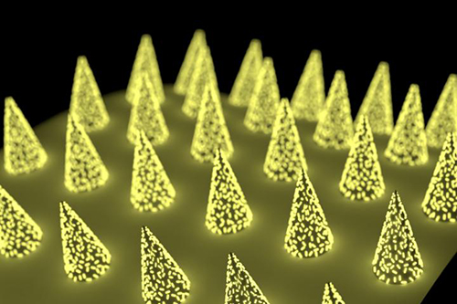
The technology uses an easy-to-administer low-cost patch that can be applied to the skin like an adhesive bandage. The patch is virtually painless because the microneedles are too small to reach nerve receptors. Another unique aspect to this innovative approach to collecting a specimen for diagnostic testing is that the Washington University in St. Louis (WashU) research team designed the microneedle patch to include plasmonic-fluor. These are ultrabright gold nanolabels that light up target protein biomarkers and can make the biomarkers up to 1,400 times brighter at low concentrations, compared to traditional fluorescent labels.
The patch, states a WashU news release, “… can be applied to the skin, capture a biomarker of interest and, thanks to its unprecedented sensitivity, allow clinicians to detect its presence.”
The technology is low cost, easy for clinicians or patients themselves to use, and could eliminate the need for a trip to patient service center where a phlebotomist would draw blood for clinical laboratory testing, the news release states.

The WashU researchers published their study, titled, “Microneedle Patch for the Ultrasensitive Quantification of Protein Biomarkers in Interstitial Fluid,” in the journal Nature Biomedical Engineering.
Minimally Invasive Specimen Collection
“We used the microneedle patch in mice for minimally invasive evaluation of the efficiency of a cocaine vaccine, for longitudinal monitoring of the levels of inflammatory biomarkers, and for efficient sampling of the calvarial periosteum [a skull membrane]—a challenging site for biomarker detection—and the quantification of its levels of the matricellular protein periostin, which cannot be accurately inferred from blood or other systemic biofluids,” the researchers wrote. “Microneedle patches for the minimally invasive collection and analysis of biomarkers in interstitial fluid might facilitate point-of-care diagnostics and longitudinal monitoring.”
Mark Prausnitz, PhD, Regents’ Professor, J. Erskine Love Jr. Chair in Chemical and Biomolecular Engineering, and Director of the Center for Drug Design, Development, and Delivery at Georgia Tech, told WIRED, “Blood is a tiny fraction of the fluid in our body. Other fluids should have something useful—it’s just hard to get those fluids.”
“Previously, concentrations of a biomarker had to be on the order of a few micrograms per milliliter of fluid,” said Zheyu (Ryan) Wang, a PhD candidate in Srikanth Singamaneni’s lab at McKelvey School of Engineering and a lead author of the paper, in the WashU news release. By using plasmonic-fluor, researchers were able to detect biomarkers on the order of picograms per milliliter—one millionth of the concentration.
“That’s orders of magnitude more sensitive,” Wang said.
Can Microneedles Be Used as a Diagnostic Tool?
As reported in WIRED, the polystyrene patch developed by Srikanth Singamaneni’s lab at McKelvey School of Engineering removes interstitial fluid from the skin and turns the needles into “biomarker traps” by coating them with antibodies known to bind to specific proteins, such as Interleukin 6 (IL-6). Once the microneedles are mixed with plasmonic-fluor, the patch will glow if the IL-6 biomarkers are present.
The development of such a highly sensitive biomarker-detection method means skin becomes a potential pathway for using microneedles to diagnose conditions, such as myocardial infarction or to measure COVID-19 antibodies in vaccinated persons.
“Now we can actually use this tool to understand what’s going on with interstitial fluid, and how we’re going to be able to use it to answer healthcare-related or medical problems,” Maral Mousavi, PhD, Assistant Professor of Biomedical Engineering, Viterbi School of Engineering at the University of Southern California, told WIRED. “I think it has the potential to be that kind of a game changer.”
Because the WashU study is a proof-of-concept in mice, it may be many years before this technology finds its way to clinical application. Many skin biomarkers will need to be verified for direct links to disease before microneedle patches will be of practical use to clinicians for diagnostics. However, microneedle patch technology has already proven viable for the collection of blood.
In 2017, Massachusetts-based Seventh Sense Biosystems (7SBio) received 510(k) clearance for a new microneedle blood collection device. Called TAP, the device is placed on the upper arm and blood collection starts with a press of a button. The process takes two to three minutes.
Initially, the FDA clearance permitted only healthcare workers to use the device “to collect capillary blood for hemoglobin A1c (HbA1c) testing, which is routinely used to monitor blood sugar levels in diabetic or pre-diabetic patients,” a Flagship Pioneering news release noted.
Then, in 2019, the FDA extended its authorization “to include blood collection by laypersons. Regulators are also allowing the device to be used ‘at-home’ for wellness testing,” a 7SBio news release stated. This opened the door for a microneedle device to be used for home care blood collection.
“No one likes getting blood drawn, but blood is the single-most important source of medical information in healthcare today, with about 90% of all diagnostic information coming from blood and its components,” Howard Weisman, former CEO of 7SBio and current CEO of PaxMedica, a clinical-stage biopharmaceutical company, said in the Flagship Pioneering news release. “TAP has the potential to transform blood collection from an inconvenient, stressful, and painful experience to one people can do themselves anywhere, making health monitoring much easier for both healthcare professionals and patients.”
As microneedle technology continues to evolve, clinical laboratories should expect patches to be used in a growing number of drug delivery systems and diagnostic tests. But further research will be needed to determine whether interstitial fluid can provide an alternate pathway for diagnosing disease.
—Andrea Downing Peck
Related Information:
Microneedle Patch for the Ultrasensitive Quantification of Protein Biomarkers in Interstitial Fluid
Forget Blood—Your Skin Might Know If You’re Sick
First-Ever Device for Fast and Virtually Painless Blood Draw Receives FDA Clearance
Microneedle Patch with Plasmonic Fluor, Ultrabright Gold Nanolables (IMAGE)
Microneedle Patch Could Replace Standard Tuberculosis Skin Test
Seventh Sense Biosystems Unlocks Market for Consumer Blood Collection Through Layperson Clearance