Genetic data captured by this new technology could lead to a new understanding of how different types of cells exchange information and would be a boon to anatomic pathology research worldwide
What if it were possible to map the interior of cells and view their genetic sequences using chemicals instead of light? Might that spark an entirely new way of studying human physiology? That’s what researchers at the Massachusetts Institute of Technology (MIT) believe. They have developed a new approach to visualizing cells and tissues that could enable the development of entirely new anatomic pathology tests that target a broad range of cancers and diseases.
Scientists at MIT’s Broad Institute and McGovern Institute for Brain Research developed this new technique, which they call DNA Microscopy. They published their findings in Cell, titled, “DNA Microscopy: Optics-free Spatio-genetic Imaging by a Stand-Alone Chemical Reaction.”
Joshua Weinstein, PhD, a postdoctoral associate at the Broad Institute and first author of the study, said in a news release that DNA microscopy “is an entirely new way of visualizing cells that captures both spatial and genetic information simultaneously from a single specimen. It will allow us to see how genetically unique cells—those comprising the immune system, cancer, or the gut for instance—interact with one another and give rise to complex multicellular life.”
The news release goes on to state that the new technology “shows
how biomolecules such as DNA and RNA are organized in cells and tissues,
revealing spatial and molecular information that is not easily accessible
through other microscopy methods. DNA microscopy also does not require
specialized equipment, enabling large numbers of samples to be processed
simultaneously.”
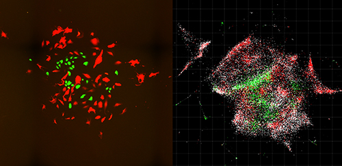
The images above, taken from the MIT study, compares optical imaging of a cell population (left) with an inferred visualization of the same cell population based on the information provided by DNA microscopy (right). Scale bar = 100 μm (100 micrometers). This technology has the potential to be useful for anatomic pathologists at some future date. (Photo and caption copyrights: Joshua Weinstein, PhD, et al/Cell.)
New Way to Visualize Cells
The MIT researchers saw an opportunity for DNA microscopy to
find genomic-level cell information. They claim that DNA microscopy images
cells from the inside and enables the capture of more data than with
traditional light microscopy. Their new technique is a chemical-encoded
approach to mapping cells that derives critical genetic insights from the
organization of the DNA and RNA in cells and tissue.
And that type of genetic information could lead to new precision medicine treatments for chronic disease. New Atlas notes that “ Speeding the development of immunotherapy treatments by identifying the immune cells best suited to target a particular cancer cell is but one of the many potential application for DNA microscopy.”
In their published study, the scientists note that “Despite enormous progress in molecular profiling of cellular constituents, spatially mapping [cells] remains a disjointed and specialized machinery-intensive process, relying on either light microscopy or direct physical registration. Here, we demonstrate DNA microscopy, a distinct imaging modality for scalable, optics-free mapping of relative biomolecule positions.”
How DNA Microscopy Works
The New York Times (NYT) notes that the advantage of DNA microscopy is “that it combines spatial details with scientists’ growing interest in—and ability to measure—precise genomic sequences, much as Google Street View integrates restaurant names and reviews into outlines of city blocks.”
And Singularity Hub notes that “ DNA microscopy, uses only a pipette and some liquid reagents. Rather than monitoring photons, here the team relies on ‘bar codes’ that chemically tag onto biomolecules. Like cell phone towers, the tags amplify, broadcasting their signals outward. An algorithm can then piece together the captured location data and transform those GPS-like digits into rainbow-colored photos. The results are absolutely breathtaking. Cells shine like stars in a nebula, each pseudo-colored according to their genomic profiles.”
“We’ve used DNA in a way that’s mathematically similar to photons in light microscopy,” Weinstein said in the Broad Institute news release. “This allows us to visualize biology as cells see it and not as the human eye does.”
In their study, researchers used DNA microscopy to tag RNA
molecules and map locations of individual human cancer cells. Their method is
“surprisingly simple” New Atlas reported. Here’s how it’s done,
according to the MIT news release:
Small synthetic DNA tags (dubbed “barcodes” by the MIT team) are added to biological samples;
The “tags” latch onto molecules of genetic material in the cells;
The tags are then replicated through a chemical reaction;
The tags combine and create more unique DNA labels;
The scientists use a DNA sequencer to decode and reconstruct the biomolecules;
A computer algorithm decodes the data and converts it to images displaying the biomolecules’ positions within the cells.
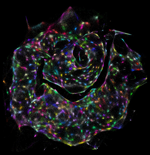
The visualization above was created from data gathered by DNA microscopy, which peers inside individual cells. It demonstrates how DNA microscopy enables scientists to identify different cells (colored dots) within a sample—with no prior knowledge of what the sample looks like. (Photo and caption copyright: Joshua Weinstein, PhD, et al./Cell.)
“The first time I saw a DNA microscopy image, it blew me away,” said Aviv Regev, PhD, a biologist at the Broad Institute, a Howard Hughes Medical Institute (HHMI) Investigator, and co-author of the MIT study, in an HHMI news release. “It’s an entirely new category of microscopy. It’s not just a technique; it’s a way of doing things that we haven’t ever considered doing before.”
Precision Medicine Potential
“Every cell has a unique make-up of DNA letters or genotype. By capturing information directly from the molecules being studied, DNA microscopy opens up a new way of connecting genotype to phenotype,” said Feng Zhang, PhD, MIT Neuroscience Professor,
Core Institute Member of the Broad Institute, and
Investigator at the McGovern Institute for Brain Research at MIT, in the HHMI
news release.
In other words, DNA microscopy could someday have applications in precision medicine. The MIT researchers, according to Stat, plan to expand the technology further to include immune cells that target cancer.
The Broad Institute has applied for a patent on DNA
microscopy. Clinical laboratory and anatomic pathology group leaders seeking
novel resources for diagnosis and treatment of cancer may want to follow the MIT
scientists’ progress.
MUSE microscope speeds up some anatomic pathology laboratory processes and removes exposure to toxic fixative chemicals
Because they handle tissue specimens, histotechnologists, anatomic pathologists, and hospital nurses are exposed to deadly chemicals such as formaldehyde, formalin, Xylene, and Toluene. The risks associated with these chemicals has been covered regularly by Dark Daily as recently as 2018 and as far back as 2011. (See, “Europe Implements New Anatomic Pathology Guidelines to Reduce Nurse Exposure to Formaldehyde and Other Toxic Histology Chemicals,” January 3, 2018; and, “Health of Pathology Laboratory Technicians at Risk from Common Solvents like Xylene and Toluene,” July 5, 2011.)
Now, scientists at the University of California at Davis (UC Davis) have developed a microscope that uses ultraviolet light (UV) to illuminate tissue samples. The UV microscope removes the need for traditional histology processes involved with preparation of tissue to produce conventional slides and makes it possible for anatomic pathologists to evaluate tissues without formalin fixation, according to a UC Davis news release.
“Here, we introduce a simple, non-destructive slide-free technique that, within minutes, provides high-resolution diagnostic histological images resembling those obtained from conventional hematoxylin and eosin histology,” the researchers wrote in their paper, published in Nature Biomedical Engineering.
High-resolution Biopsy Images in Minutes
The UV microscope relies on technology that UC Davis researchers dubbed MUSE, which stands for Microscopy with Ultraviolet Surface Excitation. According to the researchers, MUSE produces high-resolution images of biopsies and other fresh tissue samples that are ready for a pathologist’s review within minutes.
“MUSE eliminates any need for conventional tissue processing with formalin fixation, paraffin embedding, or thin-sectioning. It doesn’t require lasers, confocal, multiphoton, or optical coherence tomography instrumentation. And the simple technology makes it well-suited for deployment wherever biopsies are obtained and evaluated,” stated Richard Levenson, MD, MUSE Microscopy CEO, Professor, and Vice Chair for Strategic Technologies in the Department of Pathology and Laboratory Medicine at UC Davis, in the news release.
Ultraviolet microscopy is distinguished by its ability to magnify samples and enable views with greater resolution. This is due to the shorter wavelength of ultraviolet light, which improves image resolution beyond the diffraction limit of optical microscopes using normal white light, according to News Medical.
The unique ultraviolet light microscope tool may soon enable clinical laboratories and anatomic pathology groups to accurately report on biopsies to physicians and patients faster, for less money, and without exposure to deadly chemicals. This would be timely considering the pressure on the pathology industry to switch to value-based reimbursement from fee-for-service billing, and to embrace personalized medicine.
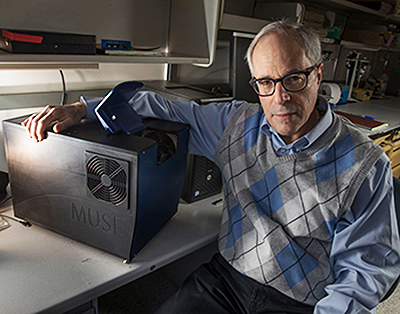
“It has become increasingly important to submit relevant portion of often tiny tissue samples for DNA and other molecular functional tests,” notes Richard Levenson, MD, MUSE Microscopy CEO, Professor, and Vice Chair for Strategic Technologies in the Department of Pathology and Laboratory Medicine at UC Davis, shown above with MUSE. “Making sure that the submitted material actually contains tumor in sufficient quantity is not always easy and sometimes just preparing conventional microscope slices can consume most of or even all of small specimens. MUSE is important because it quickly provides images from fresh tissue without exhausting the sample.” (Photo and caption copyright: UC Davis.)
Traditional Microscopy is Time-Consuming, Hazardous, Expensive
Light microscopy, a time-honored technology, has been available to pathologists for more than 200 years. It is the cornerstone for cancer diagnostics and pathology, the UC Davis researchers acknowledged. But it requires time-consuming and expensive processes, which are especially glaring in a resource-challenged healthcare industry, they pointed out.
“Histological examination of tissues is central to the diagnosis and management of neoplasms and many other diseases. However, commonly used bright-field microscopy requires prior preparation of micrometer-thick tissue sections mounted on glass slides—a process that can require hours or days, contributes to cost, and delays access to critical information,” they wrote in their paper.
“MUSE promises to improve the speed and efficiency of patient care in both state-of-the art and low-resource settings, and to provide opportunities for rapid histology in research,” they continued.
No Histology Slide Preparation Needed
MUSE developers also called attention to the use of hazardous chemicals, such as formalin, in lab processes, which has been linked to cancers including myeloid leukemia, nasopharyngeal cancer, and sinonasal cancer, according to a National Academy of Sciences report. Still, more than 300 million slides are prepared in the US each year at a cost of several billion dollars to the healthcare industry, according to the MUSE Website.
MUSE, however, penetrates tissue samples by using ultraviolet light at short wavelengths—below the 300-nanometer range. The MUSE ultraviolet microscope can reach several microns-deep into tissues.
That’s enough, the researchers claim, to be comparable with the thickness of tissue slices anatomic pathologists use with traditional microscope slides. However, MUSE requires no conventional tissue processing associated with histology slides.
How Does it Work?
MUSE is comprised of an optical system with UV light-emitting diodes (LEDs), a UV compatible stage, and a conventional microscope. That’s according to Photonics Online, which described the process:
“UV light at 280 nanometer spectral range illuminates about one square millimeter of specimen;
“Surface is limited to a few nanometers deep to make high-contrast images possible;
“Excitation light, at sub-300 nanometer spectral region, elicits bright emission from tissue specimens;
“Specimens, which were stained with conventional florescent dyes, emit photons;
“Photons are captured using glass-based microscope optics;
“Images are comparable to the hematoxylin and eosin versions histologists and pathologists are accustomed to.”
The result, according the MUSE website, “is stunning detailed images conveying a degree of resolution, structure, and depth unachievable until now by any single technology.”
Other Alternative Histology Processes Under the Microscope
MUSE is not the only approach being studied that could create cellular images without sectioning tissue samples. Anatomic and histopathology laboratory leaders looking to differentiate their labs should keep watch on the development of MUSE and other alternatives to current histology methods, especially once these new devices become green-lighted by the Food and Drug Administration (FDA) for use in patient care.