Oct 31, 2018 | Laboratory Hiring & Human Resources, Laboratory Management and Operations, Laboratory News, Laboratory Operations, Laboratory Pathology, Laboratory Testing
Only 3% of histopathology departments that responded to the Royal College of Pathologists’ workforce census reported enough staff to meet clinical demand
There is a chronic shortage of histopathologists in the United Kingdom (UK) and it is being blamed for cancer treatment waiting times that now reach the worst-ever levels, as National Health Service (NHS) training initiatives and other steps fail to keep pace with growing demand for diagnostic services.
For US anatomic pathologists and clinical laboratory managers, headlines from the UK reveal the impact a shortage of trained histopathologists (UK’s version of anatomic pathologists) and support technical staff can have on patient care when longer wait times for pathology support and diagnosis become the norm.
Royal College of Pathologists Report
The extent of the UK-wide histopathology staff shortages was highlighted in a recently released 2017 workforce census by the Royal College of Pathologists (RCPath). Of the 103 histopathology departments that responded to a survey, only 3% said they had enough staff to meet the current clinical demand! And 45% of departments had to outsource work, while half of the departments were forced to use more expensive temporary workers.
“The cost of staff shortages across histopathology departments is high for both patients and for our health services,” Jo Martin, PhD, President of the Royal College of Pathologists, noted in a statement announcing the survey results. “For patients, it means worrying delays in diagnosis and treatment. For NHS hospitals, it means spending more resources on [temporary] doctors to fill staffing gaps, or outsourcing services. We estimate this cost [to be] £27 million ($35.2 million) each year across the UK health service—money that could be better invested in staff and new diagnostic equipment.”

Royal College of Pathologists President Jo Martin, PhD, is calling on the National Health Service to take additional steps to increase the number of pathologists working in the United Kingdom, including more funded training places. That’s following the release of a Royal College of Pathologists workforce survey, which reported only 3% of histopathology departments in the UK have enough staff to meet clinical demand. (Photo copyright: Twitter.)
According to iNews, NHS England recorded its worst cancer treatment waiting times on record in July 2018, with more than 3,000 people waiting longer than two months for cancer treatment to begin. NHS’ target is for 85% of patients to begin cancer treatment within 62 days of being referred by their general practitioner.
Anatomic pathologists in the United States should consider how the UK’s average delay in starting cancer treatment affects patients in that country. It is a metric that patients in the US would not tolerate. However, in the UK’s single payer system, the government’s National Health Service sets the budgets for training new physicians, including histopathologists. For many years, the pathology profession in the UK has regularly advocated for increasing the number of histopathologists trained each year by the medical schools in that country.
In July, the number of patients waiting for treatment longer than 60 days fell to 78.2%, the 31st month in a row the target was breached, iNews reported.
“We know that histopathology consultant shortages contribute to at least part of that problem,” Martin told iNews.
The RCPath report highlights the intense pressures on histopathologists—most of whom working in understaffed laboratories—face from increased workloads, as new NHS cancer screening initiatives, an aging population, and a shift toward precision medicine result in a rising number of cases being referred to labs.
“Demand for pathology services has grown significantly in recent years and continues to grow,” Martin noted in the RCPath statement. “The pathology workforce has not increased in line with this demand. If this trend continues unchecked, clinical services could be in jeopardy. Making sure pathology services can cope with current and future demand is essential if we are to ensure early diagnosis and improve outcomes for patients.”
Lack/Loss of Trained Histopathologists an Ongoing Problem
This is not the first time the alarm has been sounded in the UK over the lack of investment in trained pathologists along with a growing shortage of trained professional staff. In 2017, Dark Daily reported on calls by pathologists and other physicians for the UK government to increase funding for trained medical laboratory professionals to avert a predicted critical shortage in laboratory staffing within the next decade. (See “Pathologists and Physicians in United Kingdom Comment on How Shortage of Medical Laboratory Professionals Could Soon Delay Essential Diagnostic, Therapeutic Testing,” February 6, 2017.)
In its most recent workforce report, The Royal College of Pathologists is reiterating its call for:
- Increased funding for training;
- Better information technology (IT) for day-to-day work;
- Capital investment to implement digital pathology more widely; and,
- Development of advanced clinical practitioner apprenticeships to help more biomedical scientists become independent practitioners who would work alongside medically-qualified histopathologists.
Long-term, the organization is calling for additional training slots for pathologists in universities as well as other changes to draw more scientists into the field.
Aging Pathology Staff Means Shortages in US a Possibility
The NHS has stopped short of declaring the pathologist shortage a crisis. Instead, a Department of Health and Social Care spokesperson in an interview with the BBC highlighted recent initiatives taken in response to the shortage. “There are hundreds more pathologists in the NHS compared to 2010 and hospitals have reduced spending on temporary agency staff by over half a billion pounds in the last year,” the spokesperson noted. “We are listening to staff, encouraging more flexible working, and have increased medical training places for home-grown doctors by 25%, to ensure the NHS has the workforce it needs for the future.”
Despite those steps, the NHS may have more work to do. According to the Royal College of Pathologists workplace survey, a quarter of all histopathologists in the UK are 55 or older, adding an approaching retirement crisis to the existing shortage.
US anatomic pathology groups and clinical laboratories should monitor these events with a keen eye. The American pathology industry is challenged by many of the same trends, such as an aging workforce and lackluster replacement initiatives. Time will tell if the US learns from the UK’s experience.
—Andrea Downing Peck
Related Information:
College Report Finds Severe Staff Shortages Across Services Vital to Cancer Diagnosis
Pathologist Shortage ‘Delaying Cancer Diagnosis’
Cancer Treatment Waiting Times Worsening Amid Shortage of Pathology Staff
Histopathology Staff Shortages ‘Affecting Cancer Diagnoses’
Cancer Waiting Times at Their Worst Ever Level
Meeting Pathology Demand
Pathology Staff Shortages Causing Delays to Cancer Diagnosis, Says Report
Pathologists and Physicians in United Kingdom Comment on How Shortage of Medical Laboratory Professionals Could Soon Delay Essential Diagnostic, Therapeutic Testing
Jul 18, 2018 | Instruments & Equipment, Laboratory Instruments & Laboratory Equipment, Laboratory News, Laboratory Operations, Laboratory Testing, Management & Operations
MUSE microscope speeds up some anatomic pathology laboratory processes and removes exposure to toxic fixative chemicals
Because they handle tissue specimens, histotechnologists, anatomic pathologists, and hospital nurses are exposed to deadly chemicals such as formaldehyde, formalin, Xylene, and Toluene. The risks associated with these chemicals has been covered regularly by Dark Daily as recently as 2018 and as far back as 2011. (See, “Europe Implements New Anatomic Pathology Guidelines to Reduce Nurse Exposure to Formaldehyde and Other Toxic Histology Chemicals,” January 3, 2018; and, “Health of Pathology Laboratory Technicians at Risk from Common Solvents like Xylene and Toluene,” July 5, 2011.)
Now, scientists at the University of California at Davis (UC Davis) have developed a microscope that uses ultraviolet light (UV) to illuminate tissue samples. The UV microscope removes the need for traditional histology processes involved with preparation of tissue to produce conventional slides and makes it possible for anatomic pathologists to evaluate tissues without formalin fixation, according to a UC Davis news release.
“Here, we introduce a simple, non-destructive slide-free technique that, within minutes, provides high-resolution diagnostic histological images resembling those obtained from conventional hematoxylin and eosin histology,” the researchers wrote in their paper, published in Nature Biomedical Engineering.
High-resolution Biopsy Images in Minutes
The UV microscope relies on technology that UC Davis researchers dubbed MUSE, which stands for Microscopy with Ultraviolet Surface Excitation. According to the researchers, MUSE produces high-resolution images of biopsies and other fresh tissue samples that are ready for a pathologist’s review within minutes.
“MUSE eliminates any need for conventional tissue processing with formalin fixation, paraffin embedding, or thin-sectioning. It doesn’t require lasers, confocal, multiphoton, or optical coherence tomography instrumentation. And the simple technology makes it well-suited for deployment wherever biopsies are obtained and evaluated,” stated Richard Levenson, MD, MUSE Microscopy CEO, Professor, and Vice Chair for Strategic Technologies in the Department of Pathology and Laboratory Medicine at UC Davis, in the news release.
Ultraviolet microscopy is distinguished by its ability to magnify samples and enable views with greater resolution. This is due to the shorter wavelength of ultraviolet light, which improves image resolution beyond the diffraction limit of optical microscopes using normal white light, according to News Medical.
The unique ultraviolet light microscope tool may soon enable clinical laboratories and anatomic pathology groups to accurately report on biopsies to physicians and patients faster, for less money, and without exposure to deadly chemicals. This would be timely considering the pressure on the pathology industry to switch to value-based reimbursement from fee-for-service billing, and to embrace personalized medicine.
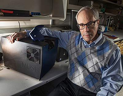
“It has become increasingly important to submit relevant portion of often tiny tissue samples for DNA and other molecular functional tests,” notes Richard Levenson, MD, MUSE Microscopy CEO, Professor, and Vice Chair for Strategic Technologies in the Department of Pathology and Laboratory Medicine at UC Davis, shown above with MUSE. “Making sure that the submitted material actually contains tumor in sufficient quantity is not always easy and sometimes just preparing conventional microscope slices can consume most of or even all of small specimens. MUSE is important because it quickly provides images from fresh tissue without exhausting the sample.” (Photo and caption copyright: UC Davis.)
MUSE is being commercialized and investors sought by MUSE Microscopy, Inc.
Traditional Microscopy is Time-Consuming, Hazardous, Expensive
Light microscopy, a time-honored technology, has been available to pathologists for more than 200 years. It is the cornerstone for cancer diagnostics and pathology, the UC Davis researchers acknowledged. But it requires time-consuming and expensive processes, which are especially glaring in a resource-challenged healthcare industry, they pointed out.
“Histological examination of tissues is central to the diagnosis and management of neoplasms and many other diseases. However, commonly used bright-field microscopy requires prior preparation of micrometer-thick tissue sections mounted on glass slides—a process that can require hours or days, contributes to cost, and delays access to critical information,” they wrote in their paper.
“MUSE promises to improve the speed and efficiency of patient care in both state-of-the art and low-resource settings, and to provide opportunities for rapid histology in research,” they continued.
No Histology Slide Preparation Needed
MUSE developers also called attention to the use of hazardous chemicals, such as formalin, in lab processes, which has been linked to cancers including myeloid leukemia, nasopharyngeal cancer, and sinonasal cancer, according to a National Academy of Sciences report. Still, more than 300 million slides are prepared in the US each year at a cost of several billion dollars to the healthcare industry, according to the MUSE Website.
MUSE, however, penetrates tissue samples by using ultraviolet light at short wavelengths—below the 300-nanometer range. The MUSE ultraviolet microscope can reach several microns-deep into tissues.
That’s enough, the researchers claim, to be comparable with the thickness of tissue slices anatomic pathologists use with traditional microscope slides. However, MUSE requires no conventional tissue processing associated with histology slides.
How Does it Work?
MUSE is comprised of an optical system with UV light-emitting diodes (LEDs), a UV compatible stage, and a conventional microscope. That’s according to Photonics Online, which described the process:
- “UV light at 280 nanometer spectral range illuminates about one square millimeter of specimen;
- “Surface is limited to a few nanometers deep to make high-contrast images possible;
- “Excitation light, at sub-300 nanometer spectral region, elicits bright emission from tissue specimens;
- “Specimens, which were stained with conventional florescent dyes, emit photons;
- “Photons are captured using glass-based microscope optics;
- “A Python programing language solution, with a graphics unit, converts MUSE images in real-time;
- “Images are comparable to the hematoxylin and eosin versions histologists and pathologists are accustomed to.”
The result, according the MUSE website, “is stunning detailed images conveying a degree of resolution, structure, and depth unachievable until now by any single technology.”
Other Alternative Histology Processes Under the Microscope
MUSE is not the only approach being studied that could create cellular images without sectioning tissue samples. Anatomic and histopathology laboratory leaders looking to differentiate their labs should keep watch on the development of MUSE and other alternatives to current histology methods, especially once these new devices become green-lighted by the Food and Drug Administration (FDA) for use in patient care.
—Donna Marie Pocius
Related Information:
Microscope That Uses Ultraviolet Instead of Visible Light Emerging as Powerful Diagnostic Tool
Microscope with Ultraviolet Surface Excitation for Rapid Slide-Free Histology
Ultraviolet Microscope to Dramatically Speed-up Lab Tests
What is Ultraviolet Microscopy?
Europe Implements New Anatomic Pathology Guidelines to Reduce Nurse Exposure to Formaldehyde and Other Toxic Histology Chemicals
National Academy of Sciences Confirms That Formaldehyde Can Cause Cancer in a Finding That Has Implications for Anatomic Pathology and Histology Laboratories
Health of Pathology Laboratory Technicians at Risk from Common Solvents like Xylene and Toluene
Jan 16, 2017 | Laboratory Hiring & Human Resources, Laboratory Management and Operations, Laboratory News, Laboratory Operations, Laboratory Pathology, Laboratory Testing
15th Annual Frontiers in Laboratory Medicine (FiLM) takes place in Birmingham, England, on January 31–February 1, 2017, and features pathology experts from UK, France, Sweden, The Netherlands, Serbia, Canada, and the USA
Recent innovations in medical laboratory management and operations in Europe and the United Kingdom (UK) will be the subject of a major conference that takes place on January 31 through February 1, 2017, in Birmingham, England. It is the 15th annual Frontiers in Laboratory Medicine (FiLM).
“Medical laboratories throughout Europe are confronted with multiple challenges,” stated Robert Michel, Editor-in-Chief of The Dark Report and one of the conference organizers. “Funding for lab tests is shrinking, demand for lab tests is soaring, and many national health systems are taking forceful actions to consolidate labs into regional networks. All of these topics will be discussed at FiLM.” (more…)
Dec 5, 2016 | Laboratory Management and Operations, Laboratory News, Laboratory Operations
Last week involved a full slate of pathology meetings and medical laboratory site visits on both islands of New Zealand during Dark Daily’s visit to this Pacific nation
DATELINE: CHRISTCHURCH, NEW ZEALAND—There’s a good case to be made that the health system in this South Pacific nation is farther down the path of medical laboratory regionalization and consolidation than most other developed nations.
That’s one insight to be gleaned from a week’s worth of meetings with pathologists, clinical laboratory professionals, and health system administrators in the cities of Auckland on the North Island and Christchurch on the east coast of New Zealand’s South Island. Your Dark Daily Editor, Robert Michel, had the opportunity to speak at several conferences and workshops, along with visits to medical laboratories.
A note of explanation about nomenclature will be helpful to Dark Daily’s international readers. In Australia and New Zealand, “pathology laboratory” is the common term for the medical laboratories that typically test blood, urine, saliva, and similar specimens. (In the United States and Canada, “clinical laboratory” is used interchangeably with medical laboratory.) “Histopathology” (or anatomic pathology) is the common term for labs that handle tissue specimens in New Zealand and Australia. (In North America, anatomic pathology, or surgical pathology laboratory is used more frequently than histopathology.) (more…)
Nov 27, 2015 | Compliance, Legal, and Malpractice, Laboratory Management and Operations, Laboratory News, Laboratory Operations, Laboratory Pathology, Management & Operations
Newspapers in this populous nation regularly publish stories about clinical laboratories that produce inaccurate lab test results that cause patient harm
In the healthcare systems of many countries across the globe, one common issue involves the accuracy and reliability of medical laboratory test results produced by clinical pathology laboratories. In most of these countries, a lack of effective government regulation is a primary reason why shady lab operators continue to provide questionable lab test results to patients.
In India, professionally-trained pathologists and laboratory scientists have repeatedly called for tougher government regulation of medical laboratories, along with more diligent inspections and enforcement actions by the authorities. Newspapers in India regularly publish stories of the medical lab enforcement actions that do happen. (more…)


