The deal will enable Crosscope’s digital pathology platform to layer around Clarapath’s histology automation hardware, a combination that could improve quality and efficiencies in diagnostic services for future customers, according to a Clarapath press release.
Clarapath’s goal with its products is to automate certain manual processes in histology laboratories, while at the same time reducing variability in how specimens are processed and produced into glass slides. In an exclusive interview with Dark Daily, Eric Feinstein, CEO and President at Clarapath said he believes the resulting data about these activities can drive further changes.
“A histotechnologist turns a microtome wheel and makes decisions about a piece of tissue in real time,” noted Feinstein, who will speak at the Executive War College on Diagnostics, Clinical Laboratory, and Pathology Management on April 25-26 in New Orleans. “All of that real-time data isn’t captured. Imagine if we could take all of that data from thousands of histotechnologists who are cutting every day and aggregate it. Then you could start drawing definitive conclusions about best practices.”
“Clarapath’s foundation is about creating consistency and standardizing steps in histology—and uncovering the data that you need in order to accomplish those goals as a whole system,” Eric Feinstein (above), CEO and President at Clarapath told Dark Daily. “A histology lab’s workflow—from when the tissue comes in to when the glass slide is produced—should all be connected.” Many processes in histology and anatomic pathology continue to be manual. Automated solutions can contribute to improved productivity and reducing variability in how individual specimens are processed. (Photo copyright: Clarapath.)
Details Behind Clarapath’s Deal to Acquire Crosscope
As part of its acquisition, Clarapath of Hawthorne, New York, has retained all of Crosscope’s employees, who are located in Mountain View, California, and Bombay, India. Financial terms of the deal were not disclosed.
Clarapath’s flagship histology automation product is SectionStar, a tissue sectioning and transfer system designed to automate inefficient and manual activities in slide processing. The device offers faster and more efficient sample processing while reducing human involvement. Clarapath expects SectionStar be on the market in 2023. The company is currently taking pre-orders.
Meanwhile, Crosscope developed Crosscope Dx, a turnkey digital pathology solution that provides workflow tools and slide management as well as AI and machine learning to assist pathologists with their medical decision-making and diagnoses.
Adoption of Digital Pathology and Automation Can Be Challenging
Digital pathology has experienced growing popularity in the post-COVID-19 pandemic world. This is not only because remote pathology case reviews have become increasingly acceptable to physicians but also because of the ongoing shortages in clinical laboratory staffing.
“A pain point today for clinicians and laboratories is labor. That’s across the board,” Feinstein said. “We can help solve that with SectionStar.”
Feinstein does not believe adoption of digital pathology and histology automation is proceeding slowly, but he does acknowledge barriers to healthcare organizations installing the technologies.
“There are lots of little things that—from a workflow perspective—people have outsized expectations about,” he explained. “Clinicians and administrators are not used to innovating in a product sense. They may be innovating on how they deliver care or treatment pathways, but they’re not used to developing an engineering product and going through alpha and beta stages. That makes adopting new technology challenging.”
Medical laboratory managers and pathologists interested in pursuing histology automation and digital pathology should first determine what processes are sub-optimal or would benefit from the standardization hardware and software can offer. Being able to articulate those gains can help build the case for a return on investment to decision-makers.
Another resource to consider: Feinstein will speak about innovations for remote histology laboratory workers at the upcoming Executive War College for Clinical Laboratory, Diagnostics, and Pathology Management on April 25-26 in New Orleans. His session is titled, “Re-engineering the Classic Histology Laboratory: Enabling the Remote Histotechnologist with New Tools That Improve Productivity, Automate Processes, and Protect Quality.”
Google designed the suite to ease radiologists’ workload and enable easy and secure sharing of critical medical imaging; technology may eventually be adapted to pathologists’ workflow
Clinical laboratory and pathology group leaders know that Google is doing extensive research and development in the field of cancer diagnostics. For several years, the Silicon Valley giant has been focused on digital imaging and the use of artificial intelligence (AI) algorithms and machine learning to detect cancer.
Now, Google Cloud has announced it is launching a new medical imaging suite for radiologists that is aimed at making healthcare data for the diagnosis and care of cancer patients more accessible. The new suite “promises to make medical imaging data more interoperable and useful by leveraging artificial intelligence,” according to MedCity News.
In a press release, medical technology company Hologic, and healthcare provider Hackensack Meridian Health in New Jersey, announced they were the first customers to use Google Cloud’s new suite of medical imaging products.
“Hackensack Meridian Health has begun using it to detect metastasis in prostate cancer patients earlier, and Hologic is using it to strengthen its diagnostic platform that screens women for cervical cancer,” MedCity News reported.
“Google pioneered the use of AI and computer vision in Google Photos, Google Image Search, and Google Lens, and now we’re making our imaging expertise, tools, and technologies available for healthcare and life sciences enterprises,” said Alissa Hsu Lynch (above), Global Lead of Google Cloud’s MedTech Strategy and Solutions, in a press release. “Our Medical Imaging Suite shows what’s possible when tech and healthcare companies come together.” Clinical laboratory companies may find Google’s Medical Imaging Suite worth investigating. (Photo copyright: Influencive.)
.
Easing the Burden on Radiologists
Clinical laboratory leaders and pathologists know that laboratory data drives most healthcare decision-making. And medical images make up 90% of all healthcare data, noted an article in Proceedings of the IEEE (Institute of Electrical and Electronics Engineers).
More importantly, medical images are growing in size and complexity. So, radiologists and medical researchers need a way to quickly interpret them and keep up with the increased workload, Google Cloud noted.
“The size and complexity of these images is huge, and, often, images stay sitting in data siloes across an organization,” said Alissa Hsu Lynch, Global Lead, MedTech Strategy and Solutions at Google, told MedCity News. “In order to make imaging data useful for AI, we have to address interoperability and standardization. This suite is designed to help healthcare organizations accelerate the development of AI so that they can enable faster, more accurate diagnosis and ease the burden for radiologists,” she added.
According to the press release, Google Cloud’s Medical Imaging Suite features include:
Imaging Storage: Easy and secure data exchange using the international DICOM (digital imaging and communications in medicine) standard for imaging. A fully managed, highly scalable, enterprise-grade development environment that includes automated DICOM de-identification. Seamless cloud data management via a cloud-native enterprise imaging PACS (picture archiving and communication system) in clinical use by radiologists.
Imaging Lab: AI-assisted annotation tools that help automate the highly manual and repetitive task of labeling medical images, and Google Cloud native integration with any DICOMweb viewer.
Imaging Datasets and Dashboards: Ability to view and search petabytes of imaging data to perform advanced analytics and create training datasets with zero operational overhead.
Imaging AI Pipelines: Accelerated development of AI pipelines to build scalable machine learning models, with 80% fewer lines of code required for custom modeling.
Imaging Deployment: Flexible options for cloud, on-prem (on-premises software), or edge deployment to allow organizations to meet diverse sovereignty, data security, and privacy requirements—while providing centralized management and policy enforcement with Google Distributed Cloud.
First Customers Deploy Suite
Hackensack Meridian Health hopes Google’s imaging suite will, eventually, enable the healthcare provider to predict factors affecting variance in prostate cancer outcomes.
“We are working toward building AI capabilities that will support image-based clinical diagnosis across a range of imaging and be an integral part of our clinical workflow,” said Sameer Sethi, Senior Vice President and Chief Data and Analytics Officer at Hackensack, in a news release.
The New Jersey healthcare network said in a statement that its work with Google Cloud includes use of AI and machine learning to enable notification of newborn congenital disorders and to predict sepsis risk in real-time.
Hologic, a medical technology company focused on women’s health, said its collaboration integrates Google Cloud AI with the company’s Genius Digital Diagnostics System.
“By complementing our expertise in diagnostics and AI with Google Cloud’s expertise in AI, we’re evolving our market-leading technologies to improve laboratory performance, healthcare provider decision making, and patient care,” said Michael Quick, Vice President of Research and Development and Innovation at Hologic, in the press release.
Hologic says its Genius Digital Diagnostics System combines AI with volumetric medical imaging to find pre-cancerous lesions and cancer cells. From a Pap test digital image, the system narrows “tens of thousands of cells down to an AI-generated gallery of the most diagnostically relevant,” according to the company website.
Hologic plans to work with Google Cloud on storage and “to improve diagnostic accuracy for those cancer images,” Hsu Lynch told MedCity News.
Medical image storage and sharing technologies like Google Cloud’s Medical Imaging Suite provide an opportunity for radiologists, researchers, and others to share critical image studies with anatomic pathologists and physicians providing care to cancer patients.
One key observation is that the primary function of this service that Google has begun to deploy is to aid in radiology workflow and productivity, and to improve the accuracy of cancer diagnoses by radiologists. Meanwhile, Google continues to employ pathologists within its medical imaging research and development teams.
Assuming that the first radiologists find the Google suite of tools effective in support of patient care, it may not be too long before Google moves to introduce an imaging suite of tools designed to aid the workflow of surgical pathologists as well.
Acceptance of digital pathology and whole-slide imaging is now almost universal among academic health center pathology departments and the nation’s largest pathology companies
Across the United States, many private practice anatomic pathology groups now recognize that digital pathology is the path forward for the entire profession. During the past decade, most academic pathology departments and large pathology lab companies have incorporated digital pathology (DP) and whole-slide imaging (WSI) into many of their labs’ daily activities.
However, in community hospital-based anatomic pathology groups, there have been barriers to even the partial adoption of digital pathology. The two biggest barriers are well-known and discussed frequently at conferences and in the literature.
Some Pathologists Reluctant to Give Up Light Microscopes
One recognized barrier to wider adoption of DP is the reluctance of many long-serving pathologists to give up their familiar light microscopes and glass slides so they can make the transition to reading pathology images on a computer screen. These pathologists remain loyal to the tools and workflows that have served them well throughout their careers.
They generally oppose their group’s move to digital pathology when the subject is discussed in partner meetings and strategic retreats. Since many pathology groups require 100% of partners or shareholders to approve major business decisions, even one recalcitrant and stubborn pathologist-partner can block the motion to adopt digital pathology that is supported by most partners.
The second barrier is the fully-loaded cost to acquire, validate, implement, and use a digital pathology system with whole-slide imaging. A full-featured scanner can cost $250,000 or more and acquiring all the software, systems, and tools needed by a group to fully incorporate digital pathology into daily workflow can easily total $500,000 to $1,000,000.
This substantial commitment of a pathology group’s capital can trigger the same intense debates as the original question of whether the pathologists in the group should adopt DP and WSI. And, not surprisingly, in most pathology groups the same dynamics come into play when votes are tallied on the motion for the pathology group to commit the funds necessary to acquire a digital pathology system, the scanners, and associated tools.
Just one or two partner holdouts can block the decision to spend the money, despite that most of the pathologist partners are ready to make the commitment.
More Community Pathology Groups Considering Digital Pathology
Yet, the momentum in favor of adopting DP and WSI continues to build. “Those pathology labs that are early adopters report multiple clinical and financial benefits. These can include generating positive financial outcomes—including the ability to attract new clients, increasing case referrals, and generating new sources of revenue to the group. In turn, the increased revenue can allow the group to increase pathologist compensation,” said Robert L. Michel, Editor-in-Chief of Dark Daily and its sister publication The Dark Report.
Every day, more anatomic pathologists in the United States use a digital pathology system with a workstation (like above) to view whole-slide images and manage their daily caseload. Most academic center pathology departments use digital pathology, as do many of the nation’s largest pathology lab companies. (Photo copyright: WizardHealth.)
“We are in a time when health insurers are hammering away at the reimbursement paid to anatomic pathologists,” Michel continued. “Year after year, payers cut reimbursement for technical component and professional component services. They exclude many pathology groups from payer networks. That is why more community pathology groups are recognizing several important benefits with the use of DP and WSI that can increase a pathology group’s revenue and boost its pathologist compensation.
Community Pathology Groups Can Use Digital Pathology to Add Value
Equally important, there are specific ways that digital pathology and whole-slide imaging increase the value of the clinical services pathologists deliver to their client physicians. These dual benefits of DP are often overlooked—or not discussed—when community pathology groups conduct their annual retreats and debate the key points of when to adopt—and how to fund—a digital pathology system for their group. These benefits range from giving physicians a faster diagnostic answer on their cancer cases to helping the group’s subspecialist pathologists get more case referrals from physicians in other states.
“It’s important for all surgical pathologists to recognize several realities in today’s pathology marketplace,” Michel noted. “First, almost every sector in healthcare is digitizing itself. Reinforcing this trend is the federal government’s mandates for interoperability across EHRs, HISs, and LISs. Any private pathology group practice that lags in its adoption of digital capabilities and digital images will find itself falling farther and farther behind as physicians switch their case referrals to other pathology labs that have converted to digital pathology and whole-slide images.
“Second, pathology groups that adopt DP and WSI put themselves in a position to build market share in their service region, while at the same time increasing case referrals for their in-house subspecialist pathologists from throughout the United States,” Michel continued. “Also, when the histology is done locally, the local pathology group can deliver faster diagnostic answers and provide digital images as appropriate to referring physicians and hospitals in that region without the need to transport glass slides by couriers.
“Third—and this is an often-overlooked benefit of digital pathology—the local pathology group with DP and WSI can recruit today’s graduating pathology residents and fellows who have trained on DP and WSI. These new pathologists typically limit their job search to pathology groups that have gone digital,” Michel noted. “Millennial pathologists trained with digital images in their residency program. They are eager to work with the automated image analysis algorithms now coming to market.”
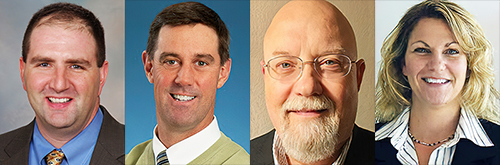
On Thursday, May 27, at 1:00 PM EDT, Keith Kaplan, MD, Chief Medical Officer of Corista (left), Andrew Evans, MD, Medical Director of Laboratory Medicine at Mackenzie Health (center left), William DeSalvo, President of Collaborative Advantage Consulting and Manager of Histology Operations at Sonora Quest Laboratories in Tempe, Ariz. (center right), and Lisa-Jean Clifford, COO and Chief Strategy Officer at Gestalt Diagnostics (right) will present “Adopting Digital Pathology on a Budget: Getting Started, Knowing What’s Feasible, and Funding Your DP from Overlooked Sources.” Anatomic pathologists, clinical laboratory directors, laboratory managers, clinical pathologists, and laboratory technicians will gain a critical understanding of which components a fully integrated digital pathology system requires, the differences between your lab’s existing LIS and a digital pathology system, budget-minded approaches to buying the components of a digital pathology system and implementing them in a stepwise fashion, and much more! (Photo copyright: Dark Daily.)
Recognizing the significant capital investment needed to acquire and deploy digital pathology and WSI, one goal of the webinar’s panel of experts is to identify ways that pathology groups can go digital on a budget. “We will do our best to identify different ways that pathology groups with limited financial resources can get into digital pathology,” said Keith Kaplan, MD, Chief Medical Officer at Corista in Concord, Mass., who will chair the upcoming webinar. “This may be the first public presentation where there is candid information about different financial strategies that your pathology group can utilize to acquire the scanners, the DP systems, and the associated tools needed for a full conversion to daily digital pathology.”
Don’t overlook how your participation in this webinar can be the foundation for helping your pathology group practice develop a timely, cost-effective path forward to introduce digital pathology and whole-slide imaging. Use of DP and WSI can become an important factor in helping your group offset payer prices cuts, develop new clients and sources of revenue, and increase pathologist compensation.
Click HERE to register today (or copy and paste this URL into your browser: https://www.darkdaily.com/webinar/adopting-digital-pathology-on-a-budget/). Make sure to have your pathology practice administrator and your histology manager join you for this important webinar.