Though smartphone apps are technically not clinical laboratory tools, anatomic pathologists and medical laboratory scientists (MLSs) may be interested to learn how health information technology (HIT), machine learning, and smartphone apps are being used to assess different aspects of individuals’ health, independent of trained healthcare professionals.
The issue that the Cedars Sinai researchers were investigating is the accuracy of patient self-reporting. Because poop can be more complicated than meets the eye, when asked to describe their bowel movements patients often find it difficult to be specific. Thus, use of a smartphone app that enables patients to accurately assess their stools in cases where watching the function of their digestive tract is relevant to their diagnoses and treatment would be a boon to precision medicine treatments of gastroenterology diseases.
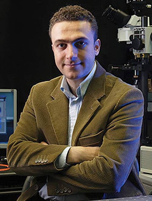
“This app takes out the guesswork by using AI—not patient input—to process the images (of bowel movements) taken by the smartphone,” said gastroenterologist Mark Pimentel, MD (above), Executive Director of Cedars-Sinai’s Medically Associated Science and Technology (MAST) program and principal investigator of the study, in a news release. “The mobile app produced more accurate and complete descriptions of constipation, diarrhea, and normal stools than a patient could, and was comparable to specimen evaluations by well-trained gastroenterologists in the study.” (Photo copyright: Cedars-Sinai.)
Pros and Cons of Bristol Stool Scale
In their paper, the scientists discussed the Bristol Stool Scale (BSS), a traditional diagnostic tool for identifying stool forms into seven categories. The seven types of stool are:
Type 1: Separate hard lumps, like nuts (difficult to pass).
Type 2: Sausage-shaped, but lumpy.
Type 3: Like a sausage, but with cracks on its surface.
Type 4: Like a sausage or snake, smooth and soft (average stool).
Type 5: Soft blobs with clear cut edges.
Type 6: Fluffy pieces with ragged edges, a mushy stool (diarrhea).
Type 7: Watery, no solid pieces, entirely liquid (diarrhea).
But even with the BSS, things can get murky for patients. Inaccurate self-reporting of stool forms by people with IBS and diarrhea can make proper diagnoses difficult.
“The problem is that whenever you have a patient reporting an outcome measure, it becomes subjective rather than objective. This can impact the placebo effect,” gastroenterologist Mark Pimentel, MD, Executive Director of Cedars-Sinai’s Medically Associated Science and Technology (MAST) program and principal investigator of the study, told Healio.
Thus, according to the researchers, AI algorithms can help with diagnosis by systematically doing the assessments for the patients, News Medical reported.
30,000 Stool Images Train New App
To conduct their study, the Cedars-Sinai researchers tested an AI smartphone app developed by Dieta Health. According to Health IT Analytics, employing AI trained on 30,000 annotated stool images, the app characterizes digital images of bowel movements using five parameters:
BSS,
Consistency,
Edge fuzziness,
Fragmentation, and
Volume.
“The app used AI to train the software to detect the consistency of the stool in the toilet based on the five parameters of stool form, We then compared that with doctors who know what they are looking at,” Pimentel told Healio.
AI Assessments Comparable to Doctors, Better than Patients
According to Health IT Analytics, the researchers found that:
AI assessed the stool comparable to gastroenterologists’ assessments on BSS, consistency, fragmentation, and edge fuzziness scores.
AI and gastroenterologists had moderate-to-good agreement on volume.
AI outperformed study participant self-reports based on the BSS with 95% accuracy, compared to patients’ 89% accuracy.
Additionally, the AI outperformed humans in specificity and sensitivity as well:
Specificity (ability to correctly report a negative result) was 27% higher.
Sensitivity (ability to correctly report a positive result) was 23% higher.
“A novel smartphone application can determine BSS and other visual stool characteristics with high accuracy compared with the two expert gastroenterologists. Moreover, trained AI was superior to subject self-reporting of BSS. AI assessments could provide more objective outcome measures for stool characterization in gastroenterology,” the Cedars-Sinai researchers wrote in their paper.
“In addition to improving a physician’s ability to assess their patients’ digestive health, this app could be advantageous for clinical trials by reducing the variability of stool outcome measures,” said gastroenterologist Ali Rezaie, MD, study co-author and Medical Director of Cedars-Sinai’s GI Motility Program in the news release.
The researchers plan to seek FDA review of the mobile app.
Opportunity for Clinical Laboratories
Anatomic pathologists and clinical laboratory leaders may want to reach out to referring gastroenterologists to find out how they can help to better serve gastro patients. As the Cedars-Sinai study suggests, AI smartphone apps can perform BSS assessments as good as or better than humans and may be useful tools in the pursuit of precision medicine treatments for patient suffering from painful gastrointestinal disorders.
“The SDPR will consolidate geographically fragmented EMR, PAS, and LIMS systems to create a detailed lifelong patient record and deliver cost savings,” NSW Health said in a news release.
NSW Health is the largest public health system in Australia with more than 220 public hospitals, 16 Local Health Districts, and three Specialty Networks. NSW Health Pathology operates more than 60 pathology laboratories (clinical laboratories in the US) and has 150 patient service centers.
“While this initiative will provide untold benefits to all the patients of NSW, we are excited about its potential for improving the health outcomes of our regional patients,” said Andrew Montague (above), former Chief Executive, Central Coast Local Health District in a press release. “By enabling greater collaboration across all local health districts and specialty health networks, the Single Digital Patient Record will provide clinicians with even better tools to keep the patient at the center of everything we do.” This project is more market evidence of the trend to bring clinical laboratory test results from multiple lab sites into a single data repository. (Photo copyright: Coast Community News.)
Cloud-based Realtime Access to Patient Records
Australia has a population of about 26 million and New South Wales, a state on the east coast, is home to more than eight million people. Though the scale of healthcare in Australia is much smaller than in the US, this is still a major project to pull patient data together from all the NSW hospitals, physicians’ offices, and other healthcare providers such as clinical laboratories and pathology practices.
With the change, NSW clinicians will benefit from a cloud–based system offering up real-time access to patients’ medical records, NSW Health Pathology Chief Executive Tracey McCosker told ITnews.
“Patients and our busy staff will benefit from clinical insights gained from the capture of important new data. Our work in pathology is vital to the diagnostic process and developing a statewide laboratory information management system will ensure we provide the best possible services,” McCosker told ITnews.
The KLAS Research report, “US Hospital Market Share 2022,” states that Epic, located in Verona, Wisconsin, has the largest US electronic health record (EHR) market share, Healthgrades noted. According to KLAS:
NSW Health’s decision to engage Epic came after a process involving 350 clinicians, scientists, and technical experts, Zoran Bolevich, MD, Chief Executive of eHealth NSW and NSW Health’s Chief Information Officer, told ITnews.
NSW Health’s Goal for Statewide Digital Patient Record
It was in December 2020 when NSW Health announced its plan to create the SDPR.
“Our vision is to be able to provide a single, holistic, statewide view of every patient—and for that information to be readily accessible to anyone involved in the patient’s care,” Bolevich said in the news release.
The SDPR, according to NSW Health, will address the following:
Challenges:
Current systems not connected statewide.
Inaccessible patient data.
Duplicative data collection.
Gaps in decision-making.
Goals:
Improve health outcomes.
Create patient centricity.
Leverage insights.
NSW’s government has already invested more than $106 million in the SDPR, Healthcare IT News reported.
Other Large EHR Rollouts
NSW Health is not the only large organization to take on such an ambitious project of creating a large-scale digital patient record. And not always to a successful conclusion.
The US Department of Veterans Affairs (VA)—also intent on EHR modernization—recently announced it is suspending roll-out of the Oracle Cerner EHR at VA centers until June 2023 to address technical issues affecting appointments, referrals, and test results.
Four VA centers in Washington, Oregon, and Ohio already went live with the system in 2022.
“We are delaying all future deployments of the new EHR while we fully assess performance and address every concern. Veterans and clinicians deserve a seamless, modernized health record system, and we will not rest until they get it,” said Deputy Secretary of Veterans Affairs Donald Remy, JD, in a news release.
For its part, Oracle Cerner wrote federal lawmakers noting the importance of continuing the project, which will move the VA away from its former VistA health information system.
“Modernization requires change and some short-term pain for the long-term benefits of a modern technology infrastructure,” noted Oracle Cerner Executive Vice President Ken Glueck in the letter, Becker’s Health IT reported. “A modernization project of this scale and scope necessarily involves time to untangle the decades of customized processes established in support of VistA, which inevitably involves challenges.”
NSW Health’s goal is to build a single repository of health information—including lab test results from multiple clinical laboratory sites. When finished NSW Health expects that sharing patient data will contribute to producing better healthcare outcomes.
However, the VA’s experience—and several other similar attempts at large-scale electronic patient record installations—suggest the work ahead will not be easy. But for NSW Health, it may be worth the effort.
Platform could be next breakthrough in quest for painless technology to replace in-patient phlebotomy blood draws for many clinical laboratory tests
In a proof-of-concept study, scientists from Israel and China have developed a “smart” microneedle adhesive bandage that measures and monitors in real time three critical biomarkers that currently require invasive blood draws for medical laboratory tests commonly performed on patients in hospitals.
According to a Technion news release, the microneedles are short, thin, and relatively painless because they only extend through the outer layer of skin to reach the interstitial fluid underneath. The needle system attaches to the patient’s skin using an adhesive patch and transfers data wirelessly to both doctor and patient in real time through cloud and Internet of Things (IoT) technologies.
Such a novel technology that allows inpatients to be monitored for key biomarkers without the need for a phlebotomist to collect blood for testing will be attractive and would likely improve the patient’s experience.
It also could reduce the volume of specimen required, potentially eliminating the invasive specimen collection procedure altogether.
“To adapt the technology to daily life, we have developed a unique [adhesive bandage] made of a flexible and soft polymer that stretches and contracts along with the skin and therefore does not interfere with any action whatsoever,” said Hossam Haick, PhD (above), in a Technion news release. “Since it is important for us that the system is available to everyone, we made sure to use relatively inexpensive materials, so the final product will not be expensive. The technology we have developed represents a leap forward in diagnosing diseases and continuous physiological monitoring at home and in the clinic.” Such a real-time monitoring device could eliminate clinical laboratory testing for certain biomarkers that currently require invasive blood draws. (Photo copyright: Technion-Israel Institute of Technology.)
Leap Forward in Diagnostic Testing and Disease Monitoring
As pathologists and medical laboratory scientists are aware, sodium is a prominent prognostic biomarker for assessing certain blood conditions such as dysnatremia, the presence of too much or too little sodium. It’s an essential element found in blood cells and blood fluid that plays a vital role in transmitting signals to the nervous system, as well as in other biological functions.
Led by Hossam Haick, PhD, head of the LNDB (Laboratory for Nanomaterials-based Devices) group and Dean of Certification Studies at Technion, the team of scientists tested their device’s effectiveness at monitoring patients’ blood for both hypernatremia (high concentration of sodium in the blood) as well as hyponatremia (low concentration of sodium in the blood).
Both conditions can affect neurological function and lead to loss of consciousness and coma. Thus, early monitoring is critical.
“As of now, detection and monitoring of sodium levels in the human body is carried out by means of laborious and bulky laboratory equipment, or by offline analysis of various bodily fluids,” the study’s authors explained in the news release. Use of the smart microneedle patch, they added, allows the patient to continue about their day as normal, as well as gives their doctor time to attend to more patients.
The “innovative stretchable, skin-conformal and fast-response microneedle extended-gate FET (field-effect transistor) biosensor [integrated with] a wireless-data transmitter and the Internet-of-Things cloud for real-time monitoring and long-term analysis [could] eventually help [bring] unlimited possibilities for efficient medical care and accurate clinical decision-making,” noted the study’s authors in Advanced Materials.
More research will be needed to determine whether this latest medical technology breakthrough will lead to a viable minimally invasive method for measuring, diagnosing, and monitoring medical conditions, but Technion’s platform appears to be another step toward a long-sought alternative to painful blood draws.
Further, pathologists and clinical laboratory managers should expect more products to hit the market that are designed to collect a lab specimen without the need for a trained phlebotomist. Companies developing these products recognize that recruiting and retaining trained phlebotomist is an ongoing concern for medical labs. Thus, to have a method of collecting a lab specimen that is simple and can be done by anyone—including patients themselves—would be an important benefit.
The technology is similar to the concept of a liquid biopsy, which uses blood specimens to identify cancer by capturing tumor cells circulating in the blood.
According to the American Cancer Society, lung cancer is responsible for approximately 25% of cancer deaths in the US and is the leading cause of cancer deaths in both men and women. The ACS estimates there will be about 236,740 new cases of lung cancer diagnosed in the US this year, and about 130,180 deaths due to the disease.
Early-stage lung cancer is typically asymptomatic which leads to later stage diagnoses and lowers survival rates, largely due to a lack of early disease detection tools. The current method used to detect early lung cancer lesions is low-dose spiral CT imaging, which is costly and can be risky due to the radiation hazards of repeated screenings, the news release noted.
MGH’s newly developed diagnostic tool detects lung cancer from alterations in blood metabolites and may lead to clinical laboratory tests that could dramatically improve survival rates of the deadly disease, the MGH scientist noted in a news release.
“Our study demonstrates the potential for developing a sensitive screening tool for the early detection of lung cancer,” said Leo Cheng, PhD (above), in the news release. Cheng is Associate Professor of Radiology at Harvard Medical School and Associate Biophysicist in Radiology at Massachusetts General Hospital. “The predictive model we constructed can identify which people may be harboring lung cancer. Individuals with suspicious findings would then be referred for further evaluation by imaging tests, such as low-dose CT, for a definitive diagnosis,” he added. Oncologists may soon have a clinical laboratory test for screening patients with early-stage lung cancer. (Photo copyright: OCSMRM.)
Detecting Lung Cancer in Blood Metabolomic Profiles
The MGH scientists created their lung-cancer predictive model based on magnetic resonance spectroscopy which can detect the presence of lung cancer from alterations in blood metabolites.
The researchers screened tens of thousands of stored blood specimens and found 25 patients who had been diagnosed with non-small-cell lung carcinoma (NSCLC), and who had blood specimens collected both at the time of their diagnosis and at least six months prior to the diagnosis. They then matched these individuals with 25 healthy controls.
The scientists first trained their statistical model to recognize lung cancer by measuring metabolomic profiles in the blood samples obtained from the patients when they were first diagnosed with lung cancer. They then compared those samples to those of the healthy controls and validated their model by comparing the samples that had been obtained from the same patients prior to the lung cancer diagnosis.
The predictive model yielded values between the healthy controls and the patients at the time of their diagnoses.
“This was very encouraging, because screening for early disease should detect changes in blood metabolomic profiles that are intermediate between healthy and disease states,” Cheng noted.
The MGH scientists then tested their model with a different group of 54 patients who had been diagnosed with NSCLC using blood samples collected before their diagnosis. The second test confirmed the accuracy of their model.
Predicting Five-Year Survival Rates for Lung Cancer Patients
Values derived from the MGH predictive model measured from blood samples obtained prior to a lung cancer diagnosis also could enable oncologists to predict five-year survival rates for patients. This discovery could prove to be useful in determining clinical strategies and personalized treatment decisions.
The researchers plan to analyze the metabolomic profiles of the clinical characteristics of lung cancer to understand the entire metabolic spectrum of the disease. They hope to create similar models for other illnesses and have already created a model that can distinguish aggressive prostate cancer by measuring the metabolomics profiles of more than 400 patients with that disease.
In addition, they are working on a similar model to screen for Alzheimer’s disease using blood samples and cerebrospinal fluid.
More research and clinical studies are needed to validate the utilization of blood metabolomics models as early screening tools in clinical practice. However, this technology might provide pathologists and clinical laboratories with diagnostic tests for the screening of early-stage lung cancer that could save thousands of lives each year.
Though the new technology could speed diagnoses of cancers and other skin diseases, it would also greatly reduce dermatopathology biopsy referrals and revenue
What effect would elimination of tissue biopsies have on dermatopathology and clinical laboratory revenue? Quite a lot. Dermatologists alone account for a significant portion of skin biopsies sent to dermatopathologists. Thus, any new technology that can “eliminate the need for invasive skin biopsies” would greatly reduce the number of histopathological referrals and reduce revenue to those practices.
“What if we could entirely bypass the biopsy process and perform histology-quality staining without taking tissue and processing tissue in a noninvasive way? Can we create images that diagnosticians can benefit from?” asked Aydogan Ozcan, PhD (above), Chancellor’s Professor of Electrical and Computer Engineering at UCLA’s Samueli School of Engineering, one of the scientists who developed UCLA’s new virtual histology method, during an interview with Medical Device + Diagnostic Industry (MD+DI). (Photo copyright: Nature.)
Could Skin Biopsies be Eliminated?
The UCLA researchers believe their innovative deep learning-enabled imaging framework could possibly circumvent the need for skin biopsies to diagnose skin conditions.
“Here, we present a deep learning-based framework that uses a convolutional neural network to rapidly transform in vivo RCM images of unstained skin into virtually-stained hematoxylin and eosin-like images with microscopic resolution, enabling visualization of the epidermis, dermal-epidermal junction, and superficial dermis layers.
“This application of deep learning-based virtual staining to noninvasive imaging technologies may permit more rapid diagnoses of malignant skin neoplasms and reduce invasive skin biopsies,” the researchers added in their published study.
According to the published study, the UCLA team trained their neural network under an adversarial machine learning scheme to transform grayscale RCM images into virtually stained 3D microscopic images of normal skin, basal cell carcinoma, and pigmented melanocytic nevi. The new images displayed similar morphological features to those shown with the widely used hematoxylin and eosin (H&E) staining method.
“In our studies, the virtually stained images showed similar color contrast and spatial features found in traditionally stained microscopic images of biopsied tissue,” Ozcan told Photonics Media. “This approach may allow diagnosticians to see the overall histological features of intact skin without invasive skin biopsies or the time-consuming work of chemical processing and labeling of tissue.”
The framework covers different skin layers, including the epidermis, dermal-epidermis, and superficial dermis layers. It images deeper into tissue without being invasive and can be quickly performed.
“The virtual stain technology can be streamlined to be almost semi real time,” Ozcan told Medical Device + Diagnostic Industry (MD+DI). “You can have the virtual staining ready when the patient is wrapping up. Basically, it can be within a couple of minutes after you’re done with the entire imaging.”
Currently, medical professionals rely on invasive skin biopsies and histopathological evaluations to diagnose skin diseases and cancers. These diagnostic techniques can result in unnecessary biopsies, scarring, multiple patient visits and increased medical costs for patients, insurers, and the healthcare system.
Improving Time to Diagnosis through Digital Pathology
Another advantage of this virtual technology, the UCLA researchers claim, is that it can provide better images than traditional staining methods, which could improve the ability to diagnose pathological skin conditions and help alleviate human error.
“The majority of the time, small laboratories have a lot of problems with consistency because they don’t use the best equipment to cut, process, and stain tissue,” dermatopathologist Philip Scumpia, MD, PhD, Assistant Professor of Dermatology and Dermatopathology at UCLA Health and one of the authors of the research paper, told MD+DI.
“What ends up happening is we get tissue on a histology slide that’s basically unevenly stained, unevenly put on the microscope, and it gets distorted,” he added, noting that this makes it very hard to make a diagnosis.
Scumpia also added that this new technology would allow digital images to be sent directly to the pathologist, which could reduce processing and laboratory times.
“With electronic medical records now and the ability to do digital photography and digital mole mapping, where you can obtain a whole-body imaging of patients, you could imagine you can also use one of these reflectance confocal devices. And you can take that image from there, add it to the EMR with the virtual histology stain, which will make the images more useful,” Scumpia said. “So now, you can track lesions as they develop.
“What’s really exciting too, is that there’s the potential to combine it with other artificial intelligence, other machine learning techniques that can give more information,” Scumpia added. “Using the reflectance confocal microscope, a clinician who might not be as familiar in dermatopathology could take images and send [them] to a practitioner who could give a more expert diagnosis.”
Faster Diagnoses but Reduced Revenue for Dermatopathologists, Clinical Labs
Ozcan noted that there’s still a lot of work to be done in the clinical assessment, validation, and blind testing of their AI-based staining method. But he hopes the technology can be propelled into a useful tool for clinicians.
“I think this is a proof-of-concept work, and we’re very excited to make it move forward with further advances in technology, in the ways that we acquire 3D information [and] train our neural networks for better and faster virtual staining output,” he told MD+DI.
Though this new technology may reduce the need for invasive biopsies and expedite the diagnosis of skin conditions and cancers—thus improving patient outcomes—what affect might it have on dermatopathology practices?
More research and clinical studies are needed before this new technology becomes part of the diagnosis and treatment processes for skin conditions. Nevertheless, should virtual histology become popular and viable, it could greatly impact the amount of skin biopsy referrals to pathologists, dermatopathologists, and clinical laboratories, thus diminishing a great portion of their revenue.
MIT’s deep learning artificial intelligence algorithm demonstrates how similar new technologies and smartphones can be combined to give dermatologists and dermatopathologists valuable new ways to diagnose skin cancer from digital images
According to an MIT press release, “The paper describes the development of an SPL [Suspicious Pigmented Lesion] analysis system using DCNNs [Deep Convolutional Neural Networks] to more quickly and efficiently identify skin lesions that require more investigation, screenings that can be done during routine primary care visits, or even by the patients themselves. The system utilized DCNNs to optimize the identification and classification of SPLs in wide-field images.”
The MIT scientists believe their AI analysis system could aid dermatologists, dermatopathologists, and clinical laboratories detect melanoma, a deadly form of skin cancer, in its early stages using smartphones at the point-of-care.
“Our research suggests that systems leveraging computer vision and deep neural networks, quantifying such common signs, can achieve comparable accuracy to expert dermatologists,” said Luis Soenksen, PhD (above), Venture Builder in Artificial Intelligence and Healthcare at MIT and first author of the study in an MIT press release. “We hope our research revitalizes the desire to deliver more efficient dermatological screenings in primary care settings to drive adequate referrals.” The MIT study demonstrates that dermatologists, dermatopathologists, and clinical laboratories can benefit from using common technologies like smartphones in the diagnosis of disease. (Photo copyright: Wyss Institute Harvard University.)
Improving Melanoma Treatment and Patient Outcomes
Melanoma develops when pigment-producing cells called melanocytes start to grow out of control. The cancer has traditionally been diagnosed through visual inspection of SPLs by physicians in medical settings. Early-stage identification of SPLs can drastically improve the prognosis for patients and significantly reduce treatment costs. It is common to biopsy many lesions to ensure that every case of melanoma can be diagnosed as early as possible, thus contributing to better patient outcomes.
“Early detection of SPLs can save lives. However, the current capacity of medical systems to provide comprehensive skin screenings at scale are still lacking,” said Luis Soenksen, PhD, Venture Builder in Artificial Intelligence and Healthcare at MIT and first author of the study in the MIT press release.
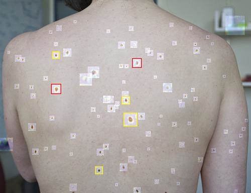
The researchers trained their AI system by using 20,388 wide-field images from 133 patients at the Gregorio Marañón General University Hospital in Madrid, as well as publicly available images. The collected photographs were taken with a variety of ordinary smartphone cameras that are easily obtainable by consumers.
They taught the deep learning algorithm to examine various features of skin lesions such as size, circularity, and intensity. Dermatologists working with the researchers also visually classified the lesions for comparison.
When the algorithm is “shown” a wide-field image like that above taken with a smartphone, it uses deep convolutional neural networks to analyze individual pigmented lesions and screen for early-stage melanoma. The algorithm then marks suspicious images as either yellow (meaning further inspection should be considered) or red (indicating that further inspection and/or referral to a dermatologist is required). Using this tool, dermatopathologists may be able to diagnose skin cancer and excise it in-office long before it becomes deadly. (Photo copyright: MIT.)
“Our system achieved more than 90.3% sensitivity (95% confidence interval, 90 to 90.6) and 89.9% specificity (89.6 to 90.2%) in distinguishing SPLs from nonsuspicious lesions, skin, and complex backgrounds, avoiding the need for cumbersome individual lesion imaging,” the MIT researchers noted in their Science Translational Medicine paper.
In addition, the algorithm agreed with the consensus of experienced dermatologists 88% of the time and concurred with the opinions of individual dermatologists 86% of the time, Medgadget reported.
Modern Imaging Technologies Will Advance Diagnosis of Disease
According to the American Cancer Society, about 106,110 new cases of melanoma will be diagnosed in the United States in 2021. Approximately 7,180 people are expected to die of the disease this year. Melanoma is less common than other types of skin cancer but more dangerous as it’s more likely to spread to other parts of the body if not detected and treated early.
More research is needed to substantiate the effectiveness and accuracy of this new tool before it could be used in clinical settings. However, the early research looks promising and smartphone camera technology is constantly improving. Higher resolutions would further advance development of this type of diagnostic tool.
In addition, MIT’s algorithm enables in situ examination and possible diagnosis of cancer. Therefore, a smartphone so equipped could enable a dermatologist to diagnose and excise cancerous tissue in a single visit, without the need for biopsies to be sent to a dermatopathologist.
Currently, dermatologists refer a lot of skin biopsies to dermapathologists and anatomic pathology laboratories. An accurate diagnostic tool that uses modern smartphones to characterize suspicious skin lesions could become quite popular with dermatologists and affect the flow of referrals to medical laboratories.