


The device automates the process used by pathology labs to process biopsy specimens and could be applied to automate other scientific processes
Hoping to speed up the processing of human biopsies to reduce the time required to diagnose cancers, two undergraduate engineering students at the University of Washington (UW) have developed a cheap, miniaturized device that could one day be used in anatomic pathology laboratories.
The protype is a low-cost, credit-card-sized device that automates the processing of human tissue biopsies using fluid transport. The device could help pathologists diagnose pancreatic cancer earlier and faster, to hopefully treat patients before it progresses to a deadly stage, according to a UW press release.
Unlike cancers that can be diagnosed early with fine needle biopsy, such as breast cancer, pancreatic cancer is one of the deadliest forms of cancers. It kills 94% of victims within five years because diagnosis usually is too late to effectively treat the disease.
Microfluidic Lab Device is Simple to Manufacture and Use
The two innovators who developed this device are Ronnie Das, Ph.D., a UW postdoctoral researcher in bioengineering, and Chris Burfeind, a UW undergraduate student in mechanical engineering. They say their microfluidic device would be simple to manufacture and use.
Das and Burfeing built a mold using a petri dish. They then inserted Teflon tubes into the petri dish and poured gooey silicon material into the mold. After the material hardened, they removed the disposable container and were left with an intact, small, transparent instrument with seamless curved and straight channels.
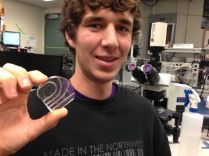
Pictured above is Chris Burfeind, a University of Washington undergraduate student in mechanical engineering. He is holding the microfluidic device which he built in a petri dish using silicon and Teflon tubes. Designed to be simple to manufacture and use, this transparent, flexible, credit-card-sized microfluidic device processes biopsy specimens using the same steps as a pathology lab. (Photo copyright University of Washington)
Traditionally, anatomic pathology labs cut biopsy specimens into thin slices. These are then put onto slides, stained and optically analyzed in 2-D for abnormalities.
UW Technology Harnesses Microfluidics to Process Biopsy Specimens
This flexible device allows a piece of tissue to pass through the channels and undergo a series of steps that replicate what happens in a pathology lab. Tissue taken from the biopsy with a syringe needle can be deposited directly into the UW device to begin processing, without the need to be handled by histotechnologists. Using microfluidics, the tissue is propelled down a channel to be stained and washed. It then moves along the channel and out of the device.
In this video, a piece of tissue first is deposited into the microfluidic device by a needle that is used clinically by pathologists to take biopsy samples from patients. The tissue then is propelled down a channel, where both a stain and a wash process the tissue, simulating steps in a pathology lab. Finally, the tissue is moved along the channel and out of the device.
A More Robust, Next-Gen Device to Offer 3-D Imaging of Specimens
The instrument developers worked with Melissa Upton, M.D., a UW pathologist. They plan to combine all of the steps into a more robust device that is capable of 3-D imaging. This version will be built and optimized for use in a medical laboratory, noted the UW press release. Future versions of the device could include layers of channels that would allow more analyses of a single piece of tissue, without adding more bulk to the device.
With this technology, whole tissue biopsies processed and analyzed for 3-D imaging would offer a more complete picture of the cellular makeup of a tumor, compared to the current methods of processing tissue, observed Das. “As soon as you cut a piece of tissue, you lose information about it. If you can keep the original tissue biopsy intact, you can see the whole story of abnormal cell growth,” he explained. “You can also see connections, cell morphology and structure as it looks in the body.”
In this video, tissue biopsies first are seen moving through the transparent channels of a microfluidic device. In the second cut, an optical clearing fluid illuminates the original channels. Moving whole tissue samples through such a device has never been done before.
“This new process is expected to help the pathologist make a more rapid diagnosis and be able to determine more accurately how invasive the cancer has become, leading to improved prognosis,” said Eric Seibel, Ph.D., a UW Research Professor of Mechanical Engineering and Director of the department’s Human Photonics Laboratory.
The UW device could potentially be used by pathologists to diagnose cancers in a matter of minutes, noted Das. He also believes it could be packaged as an over-the-counter diagnostic test kit to process biopsies in remote regions of the world. The final process tissues would then be shipped to a pathologist to determine if cancer is present.
Device Concept Could Automate Other Scientific Processes
The researchers noted that this is the first time specimens larger than a single-cell organism have successfully moved through a microfluidic device. This breakthrough presents opportunities for automating scientific processes traditionally done by humans across the sciences.
This research was funded by the National Science Foundation Bioengineering division and U.S. Department of Education Graduate Assistance in Areas of National Need program.
One observation about innovative tissue processing device developed by Das and Burfeind is that they are combining a variety of technologies to create a tissue-processing capability with the potential to shorten the time required to deliver the anatomic pathologist the sample he or she requires to make an accurate diagnosis. This is another example of how researchers are following unorthodox paths to deliver useful advances to the fields of anatomic pathology and clinical laboratory testing.
—By Patricia Kirk
Related Information:
Credit card-sized device could analyze biopsy, help diagnose pancreatic cancer in minutes
Credit card-sized device could analyze biopsy, help diagnose pancreatic cancer in minutes
Credit card-sized device could analyze biopsy, help diagnose pancreatic cancer in minutes

Exciting breakthrough and we hope to see new development in other cancer diagnoses in women- ovarian cancer etc