Researchers say they can see, identify, and count blood cells in vivo, with a system that could eventually move some routine high-volume tests out of centralized medical labs
It would be disruptive to many medical laboratories if routine hematology testing—particularly the traditional complete blood count (CBC)—were to move out of the central clinical laboratory and become a real-time, non-invasive point-of-care test (POCT) that provides the same information that is similar to the traditional complete blood count (CBC).
Israeli researchers developed a microscope with cellular resolution that uses a rainbow of light to image blood cells in vivo as they flow through a microvessel. Experts familiar with the research project say the technology has the potential to find a ready role in clinical diagnostics.
“We have invented a new optical microscope that can see individual blood cells as they flow inside our body,” declared Lior Golan, a graduate student in the Department of Biomedical Engineering at the Israel Institute of Technology (aslo called: Technion). He was quoted in a press release from The Optical Society (OSA).
Titled “Noninvasive imaging of flowing blood cells using label-free spectrally encoded flow cytometry,” the paper was published in the OSA journal, Biomedical Optics Express.
Device Promises Rapid Diagnostic Tests without Need of a Needle Stick
Pathologists and medical laboratory managers may be surprised to learn of how close the researchers are to developing a practical system that can be used in clinical settings. The authors of the paper provided the following conclusion: “In summary, an optical system that allows noninvasive imaging of blood cells in vivo has been demonstrated. With no fluorescence labeling, we have shown high resolution imaging of red and white blood cells, derived important parameters which are useful for clinical diagnosis, and observed unique dynamics of cellular flow. By providing direct information on cells’ morphology and flow, SEFC offers a new set of tools which could be utilized for a wide range of screening and diagnostics applications, and would open new possibilities in the clinical research and practice.”

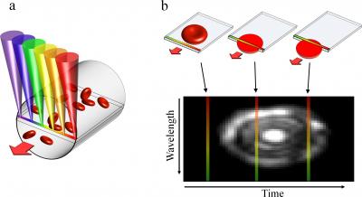
Using a technology called spectrally encoded confocal microscopy (SECM), a research team at the Israeli Institute of Technology has demonstrated the ability to identify red and blood cells in vivo as they flow through a microvessel. In (a) above, a single line within a blood vessel is imaged with multiple colors of light that encode lateral positions. Next, in (b) above, a single cell crossing the spectral line produces a two-dimensional image with one axis encoded by wavelength and the other by time. The image below shows Individual red blood cells (RBCs) flowing in a small-diameter capillary. (Images copyright by BioMedical Optics Express.)
The new microscope developed by Golan and his colleagues offers a number of advantages over current technology used by clinical laboratories:
- It is non-invasive;
- It does away with the need to collect and handle specimens;
- It shortens the often-long wait times for results;
- Its portability would make it feasible for doctors serving rural populations with limited access to medical laboratories to screen large numbers of people for common blood disorders; and,
- This device might be able to help detect warning signs, such as high white blood cell count, before a patient develops severe medical problems.
The device works by shining light through the lower lip. This site is rich in blood vessels, has no pigment to block light, and does not lose blood flow in trauma. The researchers were able to image blood flowing through a microvessel. They successfully measured the average diameter of the red and white blood cells and calculated the percentage volume of the various cell types, often a key diagnostic measurement.
The approach uses a technique called spectrally encoded confocal microscopy (SECM). SECM creates images by splitting a light beam into its constituent colors, which spread out in a line. In scanning moving blood cells, a probe is pressed against the skin. The line of light is then directed across a blood vessel near the surface of the skin. The blood cells scatter light as they cross the line. This information is then collected and analyzed.
“An important feature of the technique is its reliance on reflected light from the flowing cells to form their images, thus avoiding the use of fluorescent dyes that could be toxic,” Golan observed. “Since the blood cells are in constant motion, their appearance is distinctively different from the static tissue surrounding them.”
Researchers Are Working to Widen View and Increase Depth
The microscope’s narrow field of view was an early challenge that the researchers had to overcome. It made it difficult to locate suitable capillary vessels to image. The team solved the problem by adding a green LED and camera. This provided a wider view in which the blood vessels appeared dark because hemoglobin absorbs green light.
“Unfortunately, the green channel does not help in finding the depth of the blood vessel,” Golan observed. “Adjusting the imaging depth of the probe for imaging a small capillary is still a challenge we will address in future research.”
The researchers are working on a second-generation prototype that may expand the possibilities for imaging sites beyond the lip. They are also working to miniaturize the system for easier use and portability. “We hope to have a thumb-size prototype within the next year,” Golan stated.
Most people do not like to be stuck with a needle and for some patients the phlebotomy experience can be traumatic. This is the reason why many different research projects are investigating technologies that can diagnose without the need to use a needle or other invasive method to collect a specimen.
Technion’s new non-invasive device is one more example of the rapid evolution of potentially disruptive diagnostic technology. A microscope that can make non-invasive, in vivo tests feasible is especially important for pathologists and clinical laboratory directors. It is further indication of the continuing effort by technology companies to develop new diagnostic assays that have the potential to shift of high-volume routine tests out of the core laboratory and to point-of-care settings.
—Pamela Scherer McLeod
Related Information:
Spectrally encoded confocal microscopy
THE DARK REPORT: New Report: POC Market Will Grow 30% by 2013


Daily Dark Report:
This is a very interesting concept: However I do not think that Medical Laboratories have to worry about losing their “routine hematology” testing to this new Microscope and testing technique anytime soon.
It would be interesting to know what the projected cost of this Microscope and the projected cost per test might be.
How about the precision, accuracy and reproductibility of performing cell counts using this Microscope?
Last but certainly not least: What about the possibile interference issues? Lips like any other body part are subject to human individuality including shape, size and even color.
Sincerly,
Helen C. Ogden-Grable, MT(ASCP)PBT
Naples, Florida