Pathologists can Read Blood Samples Remotely and Render Diagnoses
Imagine using a miniaturized light field microscope attached to a mobile phone to support healthcare in remote and developing areas. One pioneering effort in this area won a 2010 Nokia Health Award and demonstrates to pathologists and clinical laboratory managers how innovative new technologies can be used to transform the way medical laboratory testing is performed.
A development team from the University of California at Berkeley (UCB) and the University of California at San Francisco (UCSF) decided to tackle the challenge of diagnosing disease in the rural areas of developing countries. The challenge is to provide advanced medical services to indigenous populations in remote regions where there is a lack of common medical services and equipment.

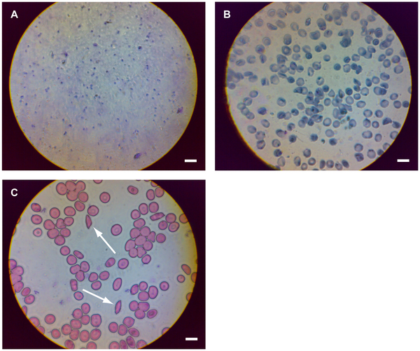
Mobile phone microscopy images of diseased blood smears. (Sourced from the PLoS ONE research article “Mobile Phone Based Clinical Microscopy for Global Health Application.”)
Their goal was to create a telemedicine solution to enable physicians to remotely examine patients. For physical injuries this can be quite effective. However, when it comes to diagnosing most diseases, it is necessary for tissue and blood samples to be taken and then examined by a pathologist as a necessary step for the attending physician to render a diagnosis.
The UCB/UCSF team recognized how remote microscopy or telemicroscopy—which involves attaching a special microscopic lens apparatus to a common cell or Smartphone—might be an effective solution. The combo-device enables caregivers to photograph or video record minute details, such as moles, skin abrasions, and even blood cells, which then can be transmitted to the cell phone of a consulting physician or pathologist for interpretation.
Minaturized Light Microscope Can Aid Pathologists
In 2009, the research group at UCB/UCSF developed a portable method for performing light microscopy from remote locations using cell phones. What sets their invention apart from existing light microscopy products is that their solution is small, cheap, robust, and portable.
“Unfortunately, much of the power of light microscopy, especially fluorescence imaging and the opportunity for remote consultation and electronic record keeping, remains inaccessible in rural and developing areas due to prohibitive equipment and training costs,” wrote the scientists in a research article title “Mobile Phone Based Clinical Microscopy for Global Health Applications” published in 2009 in the peer-reviewed online journal PLoS ONE. “This is especially problematic since the diagnosis, screening, and monitoring of treatment for many diseases and infections endemic to such areas—e.g., tuberculosis (TB), malaria, and sickle cell disease—depend on light microscopy as a screening tool or a definitive diagnostic test.
“Toward this end, we have built a mobile phone-mounted light microscope and demonstrated its potential for clinical use by imaging P. falciparum-infected and sickle red blood cells in brightfield and M. tuberculosis-infected sputum samples in fluorescence with LED excitation,” the group continued. “In all cases resolution exceeded that necessary to detect blood cell and micro-organism morphology, and with the tuberculosis samples, we took further advantage of the digitized images to demonstrate automated bacillus counting via image analysis software.
“We expect such a telemedicine system for global healthcare via mobile phone—offering inexpensive brightfield and fluorescence microscopy integrated with automated image analysis—to provide an important tool for disease diagnosis and screening, particularly in the developing world and rural areas where laboratory facilities are scarce but mobile phone infrastructure is extensive,” the group added.
Why Cell Phones Are Seen as a Pathology Testing Solution
Remote areas of the world typically lack many basic services—quality healthcare being among them. However, it is increasingly common for these same remote areas to have very good cell phone coverage. This allows telemedicine practitioners to use these communications networks to feed information about the patient’s specimens to pathologists located in cities or other regions.
In 2010, Professor Daniel A. Fletcher, Professor, Department of Bioengineering Faculty Scientist, Lawrence Berkeley National Laboratory Deputy Division Director, Physical Biosciences Division, LBL, and a member of the original research team that developed the telemicroscopy technology, won the prestigious 2010 Nokia Health Award for development of the CellScope, a small lens that “turns the camera of a standard cell phone into a diagnostic-quality microscope with a magnification of 5x-50x,” according to the Berkeley-based CellScope website.

CellScope combines a cell phone and a microscope and provides mobile microscopy for disease diagnosis and monitoring, thereby linking patients with high quality physicians no matter where they are in the world. (Sourced from the CellScope website. Photo by David Breslauer.)
“Cell phone microscopy will enable visualization of samples, followed by capture, organization, and transmission of images critical for diagnosis. Our preliminary work has demonstrated the technical feasibility of this concept, with parts added to a cell phone allowing us to capture and transmit images of blood samples anywhere in the world,” continued the site’s description of the technology.
Pathologists Can Leverage Cell Phone Networks in Support of Physicians
“Microscope-enabled mobile phones have the potential to significantly contribute to the technology available for global healthcare,” wrote the researchers, “particularly in the developing world and rural areas where mobile phone infrastructure is already ubiquitous but trained medical personnel, clinical laboratory facilities, and clinical expertise are scarce. By using existing communication infrastructure and expanding the capability of existing mobile phone technology, mobile phone microscopy systems could enable greater access to high-quality healthcare by allowing rapid, on or off-site microscopic evaluation of patient samples.
“Not only could such a mobile phone microscopy system help alleviate the problems of inadequate access to clinical microscopy in developing and rural areas,” they continued, “but it would provide those areas remote access to digital record keeping, automated sample analysis, expert diagnosticians, and epidemiological monitoring—the latter enhanced by the ease of location-tagging patient data by cellular triangulation or GPS location data. Combining the mobile phone microscopy system with automated sample preparation systems could address challenges associated with use by minimally-trained health workers and the time involved in imaging multiple fields of view.”
Of course, pathologists and clinical lab managers intuitively understand that the same technology that enables miniaturized light microscopes that can transmit data by cell phones can also be applied to reducing the size, the complexity, and the expense of light microscope products now typically used in clinical and pathology laboratories.
Further, should the various efforts to create viable miniaturized light microscopy/cell phone solutions finally make inroads into rural, remote locations, it will become possible for pathologists in more developed regions and cities to serve patients and referring doctors in these more remote regions. It represents one more trend that will contribute to the further globalization of clinical laboratory testing and laboratory medicine services.
Related Information:
Mobile Phone-based Clinical Microscopy for Global Health Applications
Portable, Low-Cost Imaging for Monitoring and Disease Diagnosis


please let me know more about portable,low cost for monitoring and disease diagnosis
regards
mukesh singh and associates