Study suggests AI-enabled technology can help clinical laboratories and hospital blood banks save thousands of dollars annually on expensive blood products
Artificial intelligence may prove to be a useful tool in helping hospitals better manage utilization of blood products. That’s one conclusion from a newly-published study done at New York’s Icahn School of Medicine at Mount Sinai. If so, this is a technology improvement that would be welcomed by blood bankers and clinical laboratory managers who must manage the cost and utilization of blood products.
There’s no way around it—blood is expensive. A typical 400- to 600-bed hospital likely budgets upwards of one million dollars annually just for blood products. Almost universally, in hospitals the medical laboratory manages the blood bank. This is where medical technologists trained in blood banking test patients and test blood to ensure whole blood units, or other blood products such as platelets, match and will not trigger a negative reaction when administered to the patient.
When left unmanaged, the cost and utilization of blood bank
products can put the budgets of hospital medical laboratories in the red. Hospitals
also invest a great deal of money training surgeons to accurately assess the
procedure and order the correct amount of blood components prior to surgery.
Therefore, new artificial intelligence (AI) technology that helps pinpoint patients’ blood loss during childbirth will be of interest to blood bankers and hospital laboratory administrators.
Can AI Help Clinical Labs Improve Utilization of Blood Products
in Hospitals?
Physicians at the Icahn School of Medicine at Mount Sinai recently investigated whether “Quantifying blood loss” would improve the use of blood during human childbirth. They published the results of their study in the International Journal of Obstetric Anesthesia.
Their research into 7,618 deliveries (vaginal and cesarean) involved “An observational study comparing blood loss, management, and outcomes between two historical cohorts (August 2016 to January 2017 and August 2017 to January 2018) at an academic tertiary care center. Patients in the intervention group (second period) had blood loss quantified compared with visual estimation for controls,” the research paper notes.
The researchers concluded that “Quantifying blood loss may
result in increased vigilance for vaginal and cesarean delivery. We identified
an association between quantifying blood loss and improved identification of
postpartum hemorrhage, patient management steps, and cost savings.”
The researchers, according to a press release, employed the Triton AI-enabled platform from Gauss Surgical, a silicon valley-based health technology company, to “monitor blood loss in all deliveries (vaginal and cesarean, n=3807) at Mount Sinai Hospital from August 2017 through January 2018 to support the institution’s stage-based hemorrhage protocol.”
The researchers found that use of a monitoring system was
associated with earlier postpartum hemorrhage
intervention and annual cost savings of $172,614 in lab costs and $36,614 in blood
bank costs.
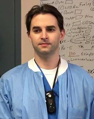
Daniel Katz, MD (above), Director of Obstetric Anesthesia Research and Associate Professor Anesthesiology, Perioperative and Pain Medicine, at Mount Sinai Hospital in New York, said in the news release, “This study demonstrates that efficiently obtaining accurate, real-time blood loss information is critical to the successful implementation of a stage-based hemorrhage protocol.” Katz was the study’s lead author. (Photo copyright: Mount Sinai Hospital.)
Measuring Blood Loss: The Eye versus AI
Gauss has secured Food and Drug Administration (FDA) clearance for Triton and more than 50 US hospitals are using it. Triton provides, in real-time, images of blood-saturated surgical sponges and canisters and uses computer vision and machine learning to pinpoint blood loss, reported MD+DI.
Traditionally, physicians visually estimate blood loss
during procedures. When they are off in their estimates of postpartum
hemorrhage, harmful postpartum health complications and deaths can occur, the
Mount Sinai researchers explained in their paper.
And although other vital signs—heart rate, rhythm, blood
pressure, oxygen level, etc.— are monitored with equipment in the surgical
suite, blood usage is not.
“Blood loss in surgery has been an enigma for decades since the dawn of medicine,” Siddarth Satish, Founder and Chief Executive Officer of Gauss, told MD+DI. “We monitor many other vital signs in surgery, but ultimately there hasn’t been any direct indicator of a patient’s hemoglobin loss.”
Bleeding Better Recognized, Less Blood Transfusions
After the Mount Sinai researchers used the Triton system to
monitor blood loss during 3,807 vaginal and cesarean deliveries from August
2017 to January 2018 at Mount Sinai Hospital, they compared their findings to
3,811 deliveries from August 2016 to January 2017, during which doctors relied
solely on visual estimation of blood loss.
The study found the following, according to the news
release:
Improved hemorrhage recognition in vaginal deliveries of 2.2% and cesarean sections of 12.6% compared to .5% and 6.4%, respectively;
Less blood transfusions needed (vaginal patients): 47% with Triton compared to 71%;
Reduced blood transfusion dose (cesarean section): 1.90 units with Triton compared to 2.52 units;
Cost savings: $209,228 a year (the total of aforementioned lab and blood bank costs).
“What we like about [Gauss] is that it somewhat embodies precision medicine in the sense that you’re using more precise tools of measurement in their first use case,” Garrett Vygantas, MD, MBA, Managing Director for OSF Ventures, the financing arm of OSF Healthcare, who also serves on Gauss Surgical’s board, told MD+DI.
Possible New Resource for Hospital Medical Laboratories
So, will AI quickly become an omnipresent overseer in surgical suites? Hardly. However, AI is in the early stages of finding places in healthcare where it can be useful. “A lot of people are predicting that AI will play a huge role in healthcare … I think it’ll be ever-present. There will be a little bit of AI in everything you’re doing, but I think the actual practice of medicine in its truest form is going to carry forward,” Satish told Fierce Healthcare.
Hospital medical laboratories and blood blanks looking for
new tools to manage blood use may want to look into AI-enabled systems like
Triton. Saving money is not the only benefit. Less transfused blood is better
for patient care as well.
Use of synthetic genetics to replicate an infectious disease agent is a scientific accomplishment that many microbiologists and clinical laboratory managers expected would happen
Microbiologists and infectious disease doctors are quite familiar with Escherichia coli (E. coli). The bacterium has caused much human sickness and even death around the globe, and its antibiotic resistant strains are becoming increasingly difficult to eradicate.
Now, scientists in England have created a synthetic “recoded” version of E. coli bacteria that is being used in a positive way—to fight disease. Their discovery is being heralded as an important breakthrough in the quest to custom-alter DNA to create synthetic forms of life that one day could be designed to fight specific infections, create new drugs, or produce tools to diagnose or treat disease.
Scientists worldwide working in the field of synthetic genomics are looking for ways to modify genomes in order to produce new weapons against infection and disease. This research could eventually produce methods for doctors—after diagnosing a patient’s specific strain of bacteria—to then use custom-altered DNA as an effective weapon against that patient’s specific bacterial infection.
This latest milestone is the result of a five-year quest by researchers at the Medical Research Council Laboratory of Molecular Biology (MRC-LMB) in Cambridge, England, to create a man-made version of the intestinal bacteria by redesigning its four-million-base-pair genetic code.
The MRC-LMB lab’s success marks the first time a living
organism has been created with a compressed genetic code.
The researchers published their findings in the journal Nature.
“This is a landmark in the emerging field of synthetic
genomics and finally applies the technology to the laboratory’s workhorse
bacterium,” they wrote. “Synthetic genomics offers a new way of life, while at
the same time moving synthetic biology towards a future in which genomes can be
written to design.”
All known forms of life on Earth contain 64 codons—a specific sequence of three consecutive nucleotides that corresponds with a specific amino acid or stop signal during protein synthesis. Jason Chin, PhD, Program Lead at MRC-LMB, said biologists long have questioned why there are 20 amino acids encoded by 64 codons.
“Is there any function to having more than one codon to encode each amino acid?” Chin asked during an interview with the Cambridge Independent. “What would happen if you made an organism that used a reduced set of codons?”
The MRC-LMB research team took an important step toward
answering that question. Their synthetic E. coli strain, dubbed Syn61,
was recoded through “genome-wide substitution of target codons by defined
synonyms.” To do so, researchers mastered a new piece-by-piece technique that
enabled them to recode 18,214 codons to create an organism with a 61-codon
genome that functions without a previously essential transfer RNA.
“Our synthetic genome implements a defined recoding and refactoring scheme–with simple corrections at just seven positions–to replace every known occurrence of two sense codons and a stop codon in the genome,” lead author Julius Fredens, PhD, a post-doctoral research associate at MRC, and colleagues, wrote in their paper.
Science Alert reports that the laboratory-created version of E. coli (above) “isn’t quite a dead ringer for its ancestor. The cells are a touch longer, and they reproduce 1.6 times slower. But the edited E. coli seems healthy and produces the same range and quantity of proteins as the non-edited versions.” (Photo copyright: Jason Chin/STAT.)
Joshua Atkinson, PhD, a postdoctoral research associate at Rice University in Houston, labeled the breakthrough a “tour de force” in the field of synthetic genomics. “This achievement sets a new world record in synthetic genomics by yielding a genome that is four times larger than the pioneering synthesis of the one-million-base-pair Mycoplasma mycoides genome,” he stated in Synthetic Biology.
“Synthetic genomics is enabling the simplification of
recoded organisms; the previous study minimized the total number of genes and
this new study simplified the way those genes are encoded.”
Manmade Bacteria That are Immune to Infections
Researchers from the J.
Craig Venter Institute in Rockville, Maryland, created the first synthetic
genome in 2010. According to an article in Nature,
the Venter Institute successfully synthesized the Mycoplasma mycoides genome
and used it “reboot” a cell from a different species of bacterium.
The MRC-LMB team’s success may prove more significant.
“This new synthetic E. coli should not be able to decode DNA from any other organism and therefore it should not be possible to infect it with a virus,” the MRC-LMB stated in a news release heralding the lab’s breakthrough. “With E. coli already being an important workhorse of biotechnology and biological research, this study is the first time any commonly used model organism has had its genome designed and fully synthesized and this synthetic version could become an important resource for future development of new types of molecules.”
Because the MRC-LMB team was able to remove transfer RNA and
release factors that decode three codons from the E. coli bacteria,
their achievement may be the springboard to designing manmade bacteria that are
immune to infections or could be turned into new drugs.
“This may enable these codons to be cleanly reassigned and
facilitate the incorporation of multiple non-canonical amino acids. This
greatly expands the scope of using non-canonical amino acids as unique tools
for biological research,” the MRC-LMB news release added.
Though synthetic genomics impact on clinical laboratory diagnostics is yet to be known, medical laboratory leaders should be mindful of the potential for rapid innovation in this field as proof-of-concept laboratory innovations are translated into real-world applications.
How medical laboratories can show value through process improvement methods and analytics will be among many key topics presented at the upcoming Lab Quality Confab conference
Quality management is the clinical laboratory’s best strategy for surviving and thriving in this era of shrinking lab budgets, PAMA price cuts, and value-based payment. In fact, the actions laboratories take in the next few months will set the course for their path to clinical success and financial sustainability in 2020 and beyond.
But how do medical laboratory managers and pathologists address these challenges while demonstrating their lab’s value? One way is through process improvement methods and another is through the use of analytics.
Clinical pathologists, hospital lab leaders, and independent lab executives have told Dark Daily that the trends demanding their focus include:
Ensuring needed resources and appropriate tests,
while the lab is scrutinized by insurance companies and internally by hospital
administration;
“Our impact on patient care, in many cases, is very
indirect. So, it is difficult to point to outcomes that occur. We know things
we do matter and change patient care, but objectively showing that is a real
struggle. And we are being asked to do more than we ever had before, and those
are the two big things that keep me up at night these days,” he added.
This is where process improvement methods and analytics are
helping clinical laboratories understand critical issues and find opportunities
for positive change.
“You need to have a strategy that you can adapt to a changing landscape in healthcare. You have to use analytics to guide your progress and measure your success,” Patricia Nortmann, System Director of Laboratory Services at St. Elizabeth Healthcare, Erlanger, Ky., told Dark Daily.
Clinical Laboratories Can Collaborate Instead of Compete
Prior to a joint venture with TriHealth in Cincinnati, St. Elizabeth lab leaders used data to inform their decision-making. Over about 12 years preceding the consolidation of labs they:
Implemented front-end automation outside the core area and in the microbiology lab.
“We are now considered a regional reference lab in the state
of Kentucky for two healthcare organizations—St. Elizabeth and TriHealth,”
Nortmann said.
Thanks to these changes, the lab more than doubled its
workload, growing from 2.1 million to 4.3 million outreach tests in the core
laboratory, she added.
Christopher Doern, PhD (left), Director of Microbiology and Associate Professor of Pathology at Virginia Commonwealth University Health System; Patricia Nortmann (center), System Director of Laboratory Services at St. Elizabeth Healthcare; and Joseph Cugini (right), Manager Client Solutions at Health Network Laboratories, will present practical solutions and case studies in quality improvement and analytics for clinical laboratory professionals at the 13th Annual Lab Quality Confab, October 15-16, 2019, at the Hyatt Regency in Atlanta, Ga. (Photo copyright: The Dark Report.)
Using Analytics to Test the Tests
Clinical laboratories also are using analytics and information technology (IT) to improve test utilization.
At VCH Health, Doern said an analytics solution interfaces
with their LIS, providing insights into test orders and informing decisions
about workflow. “I use this analytics system in different ways to answer
different questions, such as:
How are clinicians using our tests?
When do things come to the lab?
When should we be working on them?
“This is important for microbiology, which is a very delayed
discipline because of the incubation and growth required for the tests we do,”
he said.
Using analytics, the lab solved an issue with Clostridium
difficile (C diff) testing turnaround-time (TAT) after associating it with
specimen transportation.
Inappropriate or duplicate testing also
can be revealed through analytics. A physician may reconsider a test after discovering
another doctor recently ordered the same test. And the technology can guide
doctors in choosing tests in areas where the related diseases are obscure, such
as serology.
Avoiding Duplicate Records While
Improving Payment
Another example of process
improvement is Health Network Laboratories (HNL) in Allentown, Pa. A team there established an enterprise master patient index (EMPI) and implemented digital tools to find and eliminate
duplicate patient information and improve lab financial indicators.
“The system uses trusted sources of data to make sure data is clean and the lab has what it needs to send out a proper bill. That is necessary on the reimbursement side—from private insurance companies especially—to prevent denials,” Joseph Cugini, HNL’s Manager Client Solutions, told Dark Daily.
HNL reduced duplicate records in its database from 23% to
under one percent. “When you are talking about several million records, that is
quite a significant improvement,” he said.
Processes have improved not only on the billing side, but in
HNL’s patient service centers as well, he added. Staff there easily find
patients’ electronic test orders, and the flow of consumers through their
visits is enhanced.
Learn More at Lab Quality Confab Conference
Cugini, Doern, and Nortmann will speak on these topics and more during the 13th Annual Lab Quality Confab (LQC), October 15-16, 2019, at the Hyatt Regency in Atlanta, Ga. They will offer insights, practical knowledge, and case studies involving Lean, Six Sigma, and other process improvement methods during this important 2-day conference, a Dark Dailynews release notes.
Register for LQC, which is produced by Dark Daily’s sister publication The Dark Report, online at https://www.labqualityconfab.com/register, or by calling 512-264-7103.
The proof-of-concept experiment showed data can be encoded in DNA and retrieved using automated systems, a development that may have positive significance for clinical laboratories
It may seem far-fetched, but computer scientists and research groups have worked for years to discover if it is possible to store data on Deoxyribonucleic acid (DNA). Now, Microsoft Research (MR) and the University of Washington (UW) have achieved just that, and the implications of their success could be far-reaching.
Clinical pathologists are increasingly performing genetic DNA sequencing in their medical laboratories to identify biomarkers for disease, help clinicians understand their patients’ risk for a specific disease, and track the progression of a disease. The ability to store data in DNA would take that to another level and could have an impact on diagnostic pathology. Pathologist familiar with DNA sequencing may find a whole new area of medical service open to them.
The MR/UW researchers recently demonstrated a fully automated system that encoded data into DNA and then recovered the information as digital data. “In a simple proof-of-concept test, the team successfully encoded the word ‘hello’ in snippets of fabricated DNA and converted it back to digital data using a fully automated end-to-end system,” Microsoft stated in a news release.
DNA’s Potential Storage Capacity and Why We Need It
Thus far, the challenge of using DNA for data storage has
been that there wasn’t a way to easily code and retrieve the information. That,
however, seems to be changing quite rapidly. Several major companies have
invested heavily in research, with consumer offerings expected soon.
At Microsoft Research, ‘consumer interest’ in genetic testing has driven the research into using DNA for data storage. “As People get better access to their own DNA, why not also give them the ability to read any kind of data written in DNA?” asked Doug Carmean, an Architect at Microsoft, during an interview with Wired.
Scientists are interested in using DNA for data storage because
humanity is creating more data than ever before, and the pace is accelerating.
Currently, most of that data is stored on tape, which is inexpensive, but has
drawbacks. Tape degrades and has to be replaced every 10 years or so. But DNA,
on the other hand, lasts for thousands of years!
“DNA won’t degrade over time like cassette tapes and CDs, and it won’t become obsolete,” Yaniv Erlich, PhD, Chief Science Officer at MyHeritage, an online genealogy platform located in Israel, and Associate Professor, Columbia University, told Science Mag.
Tape also takes up an enormous amount of physical space compared to DNA. One single gram of DNA can hold 215 petabytes (roughly one zettabyte) of data. Wired puts the storage capacity of DNA into perspective: “Imagine formatting every movie ever made into DNA; it would be smaller than the size of a sugar cube. And it would last for 10,000 years.”
Researchers at the University of Washington claim, “All the movies, images, emails and other digital data from more than 600 basic smartphones (10,000 gigabytes) can be stored in the faint pink smear of DNA at the end of this test tube.” (Photo and caption copyright: Tara Brown/University of Washington.)
Victor Zhirnov, Chief Scientist at Semiconductor Research Corporation says the worries over storage space aren’t simply theoretical. “Today’s technology is already close to the physical limits of scaling,” he told Wired, which stated, “Five years ago humans had produced 4.4 zettabytes of data; that’s set to explode to 160 zettabytes (each year!) by 2025. Current infrastructure can handle only a fraction of the coming data deluge, which is expected to consume all the world’s microchip-grade silicon by 2040.”
MIT Technology Review agrees, stating, “Humanity is creating information at an unprecedented rate—some 16 zettabytes every year. And this rate is increasing. Last year, the research group IDC calculated that we’ll be producing over 160 zettabytes every year by 2025.”
Heavy Investment by Major Players
The whole concept may seem like something out of a science
fiction story, but the fact that businesses are investing real dollars into it
is evidence that DNA for data storage will likely be a reality in the near
future. Currently, there are a couple of barriers, but work is commencing to
overcome them.
First, the cost of synthesizing DNA in a medical laboratory
for the specific purpose of data storage must be cheaper for the solution to
become viable. Second, the sequencing process to read the information must also
become less expensive. And third is the problem of how to extract the data
stored in the DNA.
In a paper published in ASPLOS ‘16, the MR/UW scientists wrote: “Today, neither the performance nor the cost of DNA synthesis and sequencing is viable for data storage purposes. However, they have historically seen exponential improvements. Their cost reductions and throughput improvements have been compared to Moore’s Law in Carlson’s Curves … Important biotechnology applications such as genomics and the development of smart drugs are expected to continue driving these improvements, eventually making data storage a viable application.”
Automation appears to be the final piece of the puzzle. Currently,
too much human labor is necessary for DNA to be used efficiently as data
storage.
“Our ultimate goal is to put a system into production that, to the end user, looks very much like any other cloud storage service—bits are sent to a datacenter and stored there and then they just appear when the customer wants them,” said Microsoft principal researcher Karin Strauss (above), in the Microsoft news release. “To do that, we needed to prove that this is practical from an automation perspective.” Click here to watch a Microsoft Research video on the DNA storage process. (Photo copyright: Microsoft Research/YouTube.)
It may take some time before DNA becomes a viable medium for
data storage. However, savvy pathology laboratory managers should be aware of,
and possibly prepared for, this coming opportunity.
While it’s unlikely the average consumer will see much
difference in how they save and retrieve data, medical laboratories with the
ability to sequence DNA may find themselves very much in demand because of
their expertise in sequencing DNA and interpreting gene sequences.
CDC estimates that 92% of cancers caused by HPV could be eliminated in the US if HPV vaccination recommendations in this country are followed
Medical
laboratories in the United States once processed as many as 55-million Pap tests each year. However,
the need for cervical cancer screening tests is diminishing. That’s primarily because
the human
papilloma virus (HPV) vaccination effectively eliminates new cases of
cervical cancer. At least, that’s what’s happening in Australia.
When it was introduced in 2007, Australia’s nationwide
publicly-funded HPV
vaccination program only included girls, but was extended to boys in 2013.
Today, it is being credited with helping slash the country’s cervical cancer
rates.
Research published in The
Lancet Public Health (Lancet) predicts cervical cancer could be
eliminated in Australia by 2028 if current vaccination rates and screening
programs continue. Cervical cancer would be classified as effectively
eliminated once there are four or fewer new cases per 100,000 women each year.
These developments will be of interests to pathologists and cytotechnologists in
the United States.
“From the beginning, I think the [Australian] government
successfully positioned the advent of HPV vaccination as a wonderful package
that had a beneficial effect for the population,” Karen
Canfell, PhD, Director, Cancer Research Division at Cancer Council New
South Wales, Australia, and Adjunct Professor, University
of Sydney, told the Texas
Tribune. “It was celebrated for that reason, and it was a great public
health success.”
In addition to high vaccination rates, the Lancet
study notes that last year Australia transitioned from cytology-based cervical screening
every two years for women aged 18 to 69 years, to primary HPV testing every
five years for women aged 25 to 69 and exit testing for women aged 70 to 74
years.
“Large-scale clinical trials and detailed modelling suggest
that primary HPV screening is more effective at detecting cervical
abnormalities and preventing cervical cancer than screening with cytology at
shorter intervals,” the Lancet study states.
The incidence of cervical cancer in Australia now stands at
seven cases per 100,000. That’s about half the global average. The country is
on pace to see cervical cancer officially considered a “rare” cancer by 2020,
when rates are projected to drop to fewer than six new cases per 100,000 women.
US Cervical Cancer Rates
In Texas, meanwhile, the state’s failure to embrace HPV
vaccination is being blamed for slowing potential improvements in cervical
cancer rates. In 2007, Texas lawmakers rejected legislation that would have
mandated girls entering sixth grade be vaccinated for HPV. The Texas Tribune
reports that, in the decade that followed, vaccination rates remained stagnant
with only about 40% of Texans between 13 and 17 years old having been vaccinated
for HPV by 2017.
Though Texas has a similar size population as Australia, the
state’s low vaccination rates have meant cervical cancer rates have shown
little improvement. Statistics compiled by the federal Centers for Disease Control
and Prevention (CDC) show that Texas’ age-adjusted rate of new cervical
cancer cases sits at 9.2 per 100,000 women—unchanged since 2006.
Texas has the fifth highest rate of cervical cancer in the
nation, according to the CDC.
Texas State Rep. Jessica Farrar, a Democrat from Houston, maintains Texas should have followed the example of Australia, which in 2007 began a publicly funded HPV vaccination program that has the country on the verge of eliminating cervical cancer by 2028. Texas rejected mandatory HPV vaccinations and now has one of the highest cervical cancer rates in the US. “This is a preventable disease, and we should and can be doing more,” she told the Texas Tribune. “Here we are 12 years later, and look where we could’ve been, but because of certain beliefs, we’re suffering from cancers that could have been avoided.” (Photo copyright: The Texas Tribune.)
Lois Ramondetta,
MD, Professor of Gynecologic Oncology at MD Anderson Cancer Center in Houston,
told the Texas Tribune the state ignored an opportunity that Australia
seized. “[Australia] embraced the vaccine at that time, and our fear kind of
began around then,” Ramondetta said. “Really, vaccination in general has just
gone down the tube since then.”
CDC Study Pushes HPV Vaccination Recommendations in US
Texas is not the only state failing to capitalize on the HPV
vaccine’s cancer-curing promise. The CDC recently stated in a news
release announcing a recent study that 92% of cancers caused by HPV could
be eliminated if HPV vaccine recommendations were followed. CDC published the
study in its Morbidity
and Mortality Weekly Report.
HPV is a common virus that is linked to not only cervical
cancer but also cancers of the penis, head, and neck, as well as conditions
like genital warts. Though the CDC recommends children get the two-dose vaccine
at ages 11-12, the study findings indicate that only 51% of teens ages 11 to 17
have received the recommended doses of HPV vaccine, a 2% increase from 2017 to
2018.
“A future without HPV cancers is within reach, but urgent
action is needed to improve vaccine coverage rates,” Brett
Giroir, MD, Assistant Secretary for Health, US Department of Health and
Human Services (HHS), stated in the CDC news release. “Increasing HPV
vaccination overage to 80% has been and will continue to be a priority
initiative for HHS, and we will continue to work with our governmental and
private sector partners to make this a reality.”
Can Australia Eliminate Cervical Cancer?
University of Queensland Professor Ian Frazer, MD, who
co-authored the Lancet Public Health study, believes Australia is on the
verge not only of eliminating cervical cancer, but also eradicating the HPV
virus itself.
“Because this human papillomavirus only infects humans, and
the vaccine program prevents the spread of the virus, eventually we’ll get rid
of it, like we did with smallpox,” Frazer told The
Age.
“It’s not going to happen in my lifetime,” he added. “But it
could happen in the lifetime of my kids if they go about it the right way.”
If Australia’s combination of high HPV vaccination rates and
new HPV screening program succeeds in effectively eliminating cervical cancer,
clinical laboratories in this country should expect stepped-up efforts to
increase HPV vaccination rates in the United States. A renewed focus on reducing—and
ultimately eliminating—cervical cancer, could lead to fewer or less-frequently
performed Pap tests as part of cervical cancer screening protocols.