Technology like Apple’s VR/AR headsets may prove useful to clinical laboratories in accessioning and in pathology labs during biopsy grossing
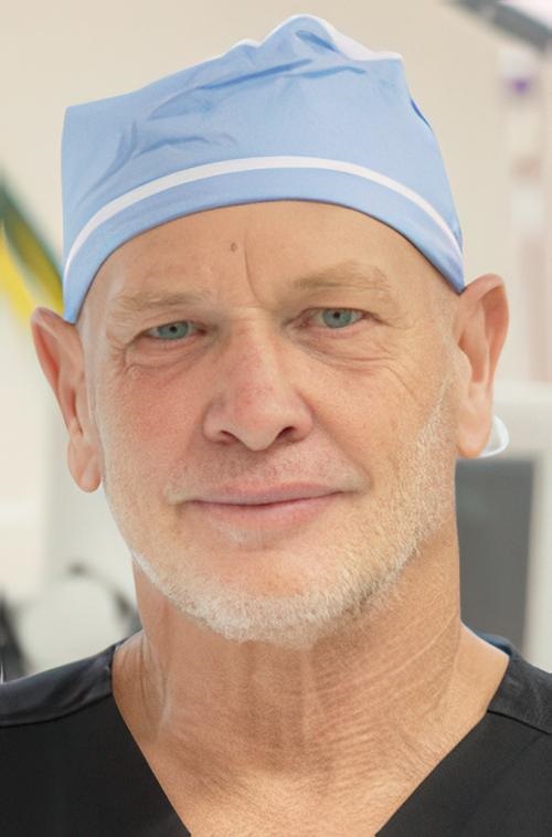
In what has been billed as a first, medical teams in the US and UK used Apple’s Extended Reality (XR) Vision Pro headset system to assist in surgical procedures. The surgeons themselves did not wear the $3,500 headset. Instead, surgical nurses used the device for touch-free access to a software application that assisted them in setting up, organizing, and performing the operations. For pathologists and clinical laboratories, in the histology laboratory, such an arrangement involving XR headsets could be used when a biopsy is at the grossing station as well.
The headset software the team used during surgery was developed by eXpanded eXistence, Inc. (eXeX), a Florida-based company whose primary product is an iOS (Apple mobile operating system) application that provides similar functions for mobile devices. eXeX adapted the iOS app to work on Apple’s Extended Reality headset.
Extended Reality is an umbrella term for augmented reality (AR) and virtual reality (VR). Apple refers to the technology as “spatial” computing.
Within the clinical laboratory, XR headsets could be used in the accessioning process as the accessioner works through the steps to confirm all required information accompanies the test requisition and that the patient’s specimen is processed/aliquoted appropriately.
“The eXeX platform, enhanced by artificial intelligence, is designed not as a medical device but as an organizational and logistics tool. It aims to streamline the management of tens of thousands of items, including equipment, tools, technologies, consumables, implants, and surgical products,” said neurosurgeon Robert Masson, MD, eXeX’s founder and CEO, in a February news release.
Masson first deployed the software in his own surgical practice. Then in March, eXeX announced that a surgical team at Cromwell Hospital in London used the system in two microsurgical spine procedures, according to a March new release.
That news garnered media coverage in the UK as well as in US-based publications that follow Apple.
“We are in a new era of surgery, and for the first time, our surgical teams have the brilliance of visual holographic guidance and maps, improving visuospatial and temporal orientation for each surgical team and for each surgery in all specialties,” said neurosurgeon Robert Masson, MD (above), eXeX’s founder and CEO, in a press release. Clinical laboratories may one day use XR headsets in the histology lab at the grossing station. (Photo copyright: Masson Spine Institute.)
Surgical Process Not Glamorous, But Important
Despite being on a cutting-edge XR platform, the eXeX software addresses “the least glamorous part” of the surgical process, Masson told Gizmodo.
“People assume that surgical healthcare has got to be sophisticated and modern,” he said. “The reality is the way we organize it is probably the most archaic of all the major industries on the planet. It’s all memorization and guesswork with scribbles on pieces of paper.”
The advantage of an XR headset is that it allows use of the eXeX software in a sterile environment, he added. “The ability to interact with digital screens and holograms and lists and maps and products unlocks all kinds of possibilities. Suddenly, you’ve got an interactive digital tool that you can use without violating the sanctity of sterility.”
Does he foresee a future when the surgeons themselves use XR headsets in the operating room? Not necessarily, Masson told Gizmodo.
“There’s always a tendency to say, ‘look at this amazing tech, let’s put a screw in with it,’” he said. “Well, we’re already putting screws in without the headset, so it doesn’t really solve a problem. People tend to think of floating spines, floating heights, you know, an overlay that tells you where to put a catheter in the liver. Honestly, it’s all unnecessary because we already do that pretty well. What we don’t do really well is stay organized.”
Other XR Apps for Healthcare
In a news release, Apple showcased other healthcare apps for its Vision Pro platform.
Epic Systems, an electronic health record (EHR) system developer, has an app called Epic Spatial Computing Concept that allows clinicians “to easily complete charting, review labs, communicate using secure chat, and complete in-basket workflows through intuitive gestures, like simply tapping their fingers to select, flicking their wrist to scroll, or using a virtual keyboard or dictation to type,” Apple stated in the news release.
Stryker, manufacturer of Mako surgical robotic arms for joint-replacement procedures, has an Apple iOS app called myMako that “allows surgeons to visualize and review patients’ Mako surgical plans at any time in a brilliant, immersive visual experience,” Apple said.
Cinematic Reality, from Siemens Healthineers, is an Apple iOS app that “allows surgeons, medical students, and patients to view immersive, interactive holograms of the human body captured through medical scans in their real-world environment,” Apple said.
New Era in Technology
For the past 20 years, manufacturing companies have installed systems at workstations with audio and video that show each step in a work process and with written checklists on the computer screen. This allows workers to check off each required step as proof that each required work element was performed.
This is similar to professional pilots who use checklists at every step in a flight process. One pilot will read the checklist items, the other will perform the step and confirm it was complete.
These procedures are generally completed on computer displays, but with the advent of XR headset technology, these types of procedures are evolving toward mobility.
To prepare for the emergence of XR-based healthcare apps, the US Food and Drug Administration (FDA) has organized a research team to devise best practices for testing these headset devices, CNBC reported.
It will be some time before XR headset technology finds its way into histology laboratories, clinical laboratories, and pathology practices, but since the rate of technology adoption accelerates exponentially, it might not take very long.
Following the loss of its histology accreditation, pressure on APS laboratory continues to mount
Government-run healthcare systems around the world often under-invest as demand grows and new healthcare technologies enter clinical practice. One such example is taking place in New Zealand, where public pathology and medical laboratory services are under extreme stress as physician test orders exceed the ability of the island nation’s clinical laboratories to keep up.
“The escalating pressure is complicating what was already a very difficult rescue job at one of the country’s busiest labs—Community Anatomic Pathology Services (APS),” RNZ reported. In 2023, APS lost its histology accreditation after it came to light that lab workers were not only exposed to toxic chemical levels at the facility, but that patients were waiting weeks for test results to return from the lab.
“The service is in crisis mode and, without urgent investment … there is a real risk that it will fail. The changes required are of such urgency that it is recommended that they be placed at the top of the agenda,” the report reads, RNZ reported.
“The size of New Zealand’s economy is restricting what our country spends on health. Health is already the second highest demand on the New Zealand tax dollar,” wrote Andrew Blair, CMInstD (above), then General Manager of Royston Hospital, Hastings, New Zealand, in an article he penned for Jpn Hosp, the journal of the Japan Hospital Association. “The tolerance of New Zealanders would be challenged if a government attempted to increase taxes further to meet the growing demands for expenditure on health, but at the same time the population’s expectations are increasing. This is the challenging situation we face today.” For New Zealand’s clinical laboratories, the demand for testing is increasing annually as the country’s population grows. (Photo copyright: Blair Consulting.)
Increased Demand on APS Leads to Problems
Established in 2015, APS tests thousands of anatomic and tissue samples yearly and is utilized by approximately a third of NZ’s population, according to RNZ.
The big story, however, is that from 2022 to 2023 utilization increased by a third. “The overall increasing demand is greater than the capacity of the service,” Te Whatu Ora (Health New Zealand), the country’s publicly-funded healthcare system, told RNZ.
As planned care increased, public hospitals started outsourcing operations to private surgical centers. A domino effect ensued when all of those samples then made their way to APS. There was an “increased volume of private surgery being carried out by 600 specialists in the region and 2,000 general practitioners, with up to 450 histology cases a day,” RNZ noted, adding, “The backlog has hit turnaround times for processing samples, which had been deteriorating.”
To make matters even more dire, working conditions at the country’s clinical labs is unfavorable and deteriorating, with short staffing, outdated workspaces and equipment, and exposure to dangerous chemicals.
“Conditions got so bad from 2019-2021 that workers were exposed to cancer-causing formaldehyde in cramped workspaces, and flammable chemicals were stored unsafely,” RNZ reported.
While pay increases and safety improvements have provided some relief, the memory of past incidences coupled with increasing delays continue to undermine confidence in New Zealand’s laboratory industry.
Patients Also at Risk Due to Long Delays in Test Results
“We recognize the concern and impact any delayed results can cause referrers and their patients,” Health New Zealand said in a statement, RNZ reported.
Nevertheless, a 2023 article in The Conversation noted that, “38,000 New Zealanders had been waiting longer than the four-month target for being seen by a specialist for an initial assessment.”
However, according to plastic surgeon and Melanoma Network of New Zealand (MelNet) Chair Gary Duncan, MBChB, FRACS, when patients return to their doctors for test results, those results often have not come back from the medical laboratory. Therefore, the physician cannot discuss any issues, which causes the patient to have to make another appointment or receive a melanoma diagnosis over the telephone, RNZ reported.
“Slow pathology services are unfair to patients. Such delays could result in the spreading of the melanoma to other parts of the body and require major surgery under anesthetic,” dermatologist Louise Reiche, MBChB, FRACS, told RNZ. “Not only will they suffer an extensive surgical procedure, but it could also shorten their life.”
Improvements at APS Underway
Changes are currently underway that may decrease the long delays in test results at New Zealand’s labs. “A business case was being done to set up an electronic ordering system to cut down on manual processing errors,” RNZ reported.
Additionally, “the situation is much improved due to dispersal of work around [the] city and country for now. The teamwork around the region has been a veritable lifesaver,” a source familiar with the work told RNZ.
Construction of a new lab for APS is also allegedly in the works. However, to date no announcement has been made, according to RNZ.
Time will tell if New Zealand’s government can repair its pathology system. News stories showcasing damage caused by lengthy delays in clinical laboratory test results—and the ensuing patient harm due to rationed care in general—continue to reveal the weakness in government-run healthcare systems.
MUSE microscope speeds up some anatomic pathology laboratory processes and removes exposure to toxic fixative chemicals
Because they handle tissue specimens, histotechnologists, anatomic pathologists, and hospital nurses are exposed to deadly chemicals such as formaldehyde, formalin, Xylene, and Toluene. The risks associated with these chemicals has been covered regularly by Dark Daily as recently as 2018 and as far back as 2011. (See, “Europe Implements New Anatomic Pathology Guidelines to Reduce Nurse Exposure to Formaldehyde and Other Toxic Histology Chemicals,” January 3, 2018; and, “Health of Pathology Laboratory Technicians at Risk from Common Solvents like Xylene and Toluene,” July 5, 2011.)
Now, scientists at the University of California at Davis (UC Davis) have developed a microscope that uses ultraviolet light (UV) to illuminate tissue samples. The UV microscope removes the need for traditional histology processes involved with preparation of tissue to produce conventional slides and makes it possible for anatomic pathologists to evaluate tissues without formalin fixation, according to a UC Davis news release.
“Here, we introduce a simple, non-destructive slide-free technique that, within minutes, provides high-resolution diagnostic histological images resembling those obtained from conventional hematoxylin and eosin histology,” the researchers wrote in their paper, published in Nature Biomedical Engineering.
High-resolution Biopsy Images in Minutes
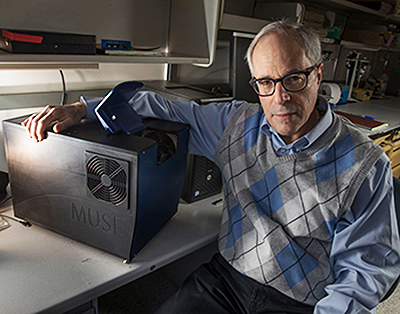
The UV microscope relies on technology that UC Davis researchers dubbed MUSE, which stands for Microscopy with Ultraviolet Surface Excitation. According to the researchers, MUSE produces high-resolution images of biopsies and other fresh tissue samples that are ready for a pathologist’s review within minutes.
“MUSE eliminates any need for conventional tissue processing with formalin fixation, paraffin embedding, or thin-sectioning. It doesn’t require lasers, confocal, multiphoton, or optical coherence tomography instrumentation. And the simple technology makes it well-suited for deployment wherever biopsies are obtained and evaluated,” stated Richard Levenson, MD, MUSE Microscopy CEO, Professor, and Vice Chair for Strategic Technologies in the Department of Pathology and Laboratory Medicine at UC Davis, in the news release.
Ultraviolet microscopy is distinguished by its ability to magnify samples and enable views with greater resolution. This is due to the shorter wavelength of ultraviolet light, which improves image resolution beyond the diffraction limit of optical microscopes using normal white light, according to News Medical.
The unique ultraviolet light microscope tool may soon enable clinical laboratories and anatomic pathology groups to accurately report on biopsies to physicians and patients faster, for less money, and without exposure to deadly chemicals. This would be timely considering the pressure on the pathology industry to switch to value-based reimbursement from fee-for-service billing, and to embrace personalized medicine.
“It has become increasingly important to submit relevant portion of often tiny tissue samples for DNA and other molecular functional tests,” notes Richard Levenson, MD, MUSE Microscopy CEO, Professor, and Vice Chair for Strategic Technologies in the Department of Pathology and Laboratory Medicine at UC Davis, shown above with MUSE. “Making sure that the submitted material actually contains tumor in sufficient quantity is not always easy and sometimes just preparing conventional microscope slices can consume most of or even all of small specimens. MUSE is important because it quickly provides images from fresh tissue without exhausting the sample.” (Photo and caption copyright: UC Davis.)
Traditional Microscopy is Time-Consuming, Hazardous, Expensive
Light microscopy, a time-honored technology, has been available to pathologists for more than 200 years. It is the cornerstone for cancer diagnostics and pathology, the UC Davis researchers acknowledged. But it requires time-consuming and expensive processes, which are especially glaring in a resource-challenged healthcare industry, they pointed out.
“Histological examination of tissues is central to the diagnosis and management of neoplasms and many other diseases. However, commonly used bright-field microscopy requires prior preparation of micrometer-thick tissue sections mounted on glass slides—a process that can require hours or days, contributes to cost, and delays access to critical information,” they wrote in their paper.
“MUSE promises to improve the speed and efficiency of patient care in both state-of-the art and low-resource settings, and to provide opportunities for rapid histology in research,” they continued.
No Histology Slide Preparation Needed
MUSE developers also called attention to the use of hazardous chemicals, such as formalin, in lab processes, which has been linked to cancers including myeloid leukemia, nasopharyngeal cancer, and sinonasal cancer, according to a National Academy of Sciences report. Still, more than 300 million slides are prepared in the US each year at a cost of several billion dollars to the healthcare industry, according to the MUSE Website.
MUSE, however, penetrates tissue samples by using ultraviolet light at short wavelengths—below the 300-nanometer range. The MUSE ultraviolet microscope can reach several microns-deep into tissues.
That’s enough, the researchers claim, to be comparable with the thickness of tissue slices anatomic pathologists use with traditional microscope slides. However, MUSE requires no conventional tissue processing associated with histology slides.
How Does it Work?
MUSE is comprised of an optical system with UV light-emitting diodes (LEDs), a UV compatible stage, and a conventional microscope. That’s according to Photonics Online, which described the process:
“UV light at 280 nanometer spectral range illuminates about one square millimeter of specimen;
“Surface is limited to a few nanometers deep to make high-contrast images possible;
“Excitation light, at sub-300 nanometer spectral region, elicits bright emission from tissue specimens;
“Specimens, which were stained with conventional florescent dyes, emit photons;
“Photons are captured using glass-based microscope optics;
“Images are comparable to the hematoxylin and eosin versions histologists and pathologists are accustomed to.”
The result, according the MUSE website, “is stunning detailed images conveying a degree of resolution, structure, and depth unachievable until now by any single technology.”
Other Alternative Histology Processes Under the Microscope
MUSE is not the only approach being studied that could create cellular images without sectioning tissue samples. Anatomic and histopathology laboratory leaders looking to differentiate their labs should keep watch on the development of MUSE and other alternatives to current histology methods, especially once these new devices become green-lighted by the Food and Drug Administration (FDA) for use in patient care.
University of Turin study in Italy shows under-vacuum sealing systems reduce exposure to formaldehyde by 75% among nurses handling tissue biopsy specimens during surgery
Histology technicians and anatomic pathology (AP) laboratories regularly handle dangerous chemicals such as formaldehyde. They understand the risks exposure brings and take precautions to minimize those risks. However, in operating suites worldwide, nurses assisting surgeons also are being exposed to this nasty chemical.
Nurses must place biopsies and other tissues into buckets of formaldehyde to preserve the tissue between the operating room (OR) and histology laboratory. Formaldehyde, along with toluene, and xylene, is used to process and preserve biopsy tissue, displace water, and to create glass slides. It is an important substance that has long been used to maintain the viability of tissue specimens. Thus, exposure to formaldehyde among nurses is well-documented.
According to a National Academy of Sciences report, formalin, a tissue preservative that is a form of formaldehyde, has been linked to:
Now, motivated by increasing formaldehyde regulations in Europe, as well as the need to increase awareness of exposure risks, the University of Turin (Unito), and other hospitals in Italy’s Piedmont region, conducted a cross-sectional study of 94 female nurses who were being potentially exposed to formaldehyde.
Researchers Aim for “Formalin-Free” Hospitals
The Unito study showed that nurses using an under-vacuum sealing (UVS) system in ORs are exposed to levels of formaldehyde 75% lower than those who did not use the system. This study differs from other similar tests because the level of exposure is not just potential, due to environmental contamination, but confirmed with analytic data from specific urine analyses.
The researchers divided the nurses into two groups:
· One group immersed samples in containers of formaldehyde following standard procedures;
· The other group worked in operating rooms equipped with a UVS system.
The researchers described a UVS system that called for the tissue removed during surgery to be sealed in a medical grade vacuum bag and refrigerated at four degrees centigrade before being transferred to the lab for fixation.
One example of a UVS system is TissueSAFE plus, developed by Milestone Medical, located in Bergamo, Italy, and Kalamazoo, Mich. According to the company’s website, the system, “Eliminates formalin in the operating theatre and allows a controlled formalin-free transfer of biospecimens to the laboratory.”
The image above is from a research paper by Richard J. Zarbo, MD, Pathology and Laboratory Medicine, Henry Ford Health System. It describes “five validation trials of new vacuum sealing technologies that change the approach to the preanalytic ‘front end’ of specimen transport, handling, and processing, and illustrate their adaptation and integration into existing Lean laboratory operations with reduction in formalin use and personnel exposure to this toxic and potentially carcinogenic fixative.” (Image copyright: Henry Ford Health System/Springer International Publishing.)
Increased Scrutiny Leads to New Pathology Guidelines
In a paper published in Toxicology Research, a journal of The Royal Society of Chemistry, the researchers noted a marked difference related to the adoption of the under-vacuum sealing procedure, as an alternative to formaldehyde for preserving tissues. “Nurses, operating in surgical theatres, are traditionally exposed to formaldehyde because of the common and traditional practice of immersing surgical samples, of a size ranging between two and 30 centimeters, in this preservative liquid (three to five liters at a time) to be later transferred to a [histopathology] lab,” the authors wrote. “We evaluated the conditions favoring the risk of exposure to this toxic reagent and the effect of measures to prevent it.”
Throughout Europe, increased scrutiny has forced medical pathology associations to write new guidelines that accept alternative methods to formaldehyde-based tissue preservation methods.
“In Europe, and in Italy in particular, the level of attention to formaldehyde exposure in the public health hospital system has become very high, forcing pathology associations to rewrite guidelines,” Marco Bellini, General Manager of the Medical Division at Milestone Medical, told Dark Daily. “What makes this study unique from many other similar tests is that the level of exposure has been confirmed with data from specific urine analyses,” he added.
The main topic of these guidelines is the preanalytical aspects of specimen collection, transportation, and preservation, where the vacuum method has been indicated as a valid alternative to improve the standardization of these crucial steps in pathology. By moving the starting point for specimen fixation from the OR to the histology labs, parameters can be controlled and documented, with the main advantage of reducing formaldehyde exposure by operators at the collection point.
These guidelines will be presented at the European Society of Pathology (ESP) with the intent to extending them throughout Europe.
Toluene’s and Xylene’s Effects Studied
Formaldehyde is not the only potentially harmful substance in the clinical laboratory. As previously noted, common solvents toluene and xylene also are potentially hazardous.
Medical laboratory leaders are reminded to initiate processes that ensure safe specimen handling, transport, and processing, as well as workflow changes that eliminate chemical odors in the lab. Studies, such as those cited above, may provide information necessary to affect change.
Once thought to be separate components, the new model of a contiguous mesentery could lead to new medical laboratory tools for diagnosing and treating digestive diseases such as Crohn’s and colorectal cancer
For more than a century, pathology professionals have treated the network of tissue folds surrounding the human digestive system, known as the mesentery, as separate entities. However, new research indicates the mesentery is in fact a single, continuous organ and therefore reverses that thinking. This could impact the way pathologists and medical laboratories currently perform diagnostics and testing of digestive diseases.