VCU scientists used the technique to measure mutations associated with acute myeloid leukemia, potentially offering an attractive alternative to DNA sequencing
More accurate but less-costly cancer diagnostics are the Holy Grail of cancer research. Now, research scientists at Virginia Commonwealth University (VCU) say they have developed a clinical laboratory diagnostic technique that could be far cheaper and more capable than standard DNA sequencing in diagnosing some diseases. Their method combines digital polymerase chain reaction (dPCR) technology with high-speed atomic force microscopy (HS-AFM) to generate nanoscale-resolution images of DNA.
The technique allows the researchers to measure polymorphisms—variations in gene lengths—that are associated with many cancers and neurological diseases. The VCU scientists say the new technique costs less than $1 to scan each dPCR reaction.
“We chose to focus on FLT3 mutations because they are difficult to [diagnose], and the standard assay is limited in capability,” said physicist Jason Reed, PhD, Assistant Professor in the Virginia Commonwealth University Department of Physics, in a VCU press release.
Reed is an expert in nanotechnology as it relates to biology and medicine. He led a team that included other researchers in VCU’s physics department as well as physicians from VCU Massey Cancer Center and the Department of Internal Medicine at VCU School of Medicine.
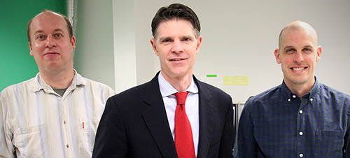
“The technology needed to detect DNA sequence rearrangements is expensive and limited in availability, yet medicine increasingly relies on the information it provides to accurately diagnose and treat cancers and many other diseases,” said Jason Reed, PhD (above center, with Andrey Mikheikin, PhD, on left and Sean Koebley, PhD, on right), in a press release from Virginia Commonwealth University (VCU). “We’ve developed a system that combines a routine laboratory process with an inexpensive yet powerful atomic microscope that provides many benefits over standard DNA sequencing for this application, at a fraction of the cost.” (Photo copyright: Virginia Commonwealth University.)
Validating the Clinical Laboratory Test
The physicists worked with two VCU physicians—hematologist/oncologist Amir Toor, MD, and hematopathologist Alden Chesney, MD—to compare the imaging technique to the LeukoStrat CDx FLT3 Mutation Assay, which they described as the “current gold standard test” for diagnosing FLT3 gene mutations.
The researchers said their technique matched the results of the LeukoStrat test in diagnosing the mutations. But unlike that test, the new technique also can measure variant allele frequency (VAL). This “can show whether the mutation is inherited and allows the detection of mutations that could potentially be missed by the current test,” states the VCU press release.
“We plan to continue developing and testing this technology in other diseases involving DNA structural mutations,” Reed said. “We hope it can be a powerful and cost-effective tool for doctors around the world treating cancer and other devastating diseases driven by DNA mutations.”
“In our approach we first used digital PCR, in which a mixed sample is diluted to less than one target molecule per aliquot and the aliquots are amplified to yield homogeneous populations of amplicons,” he said. “Then, we deposited each population onto an atomically-flat partitioned surface.”
The VCU researchers “scanned each partition with high-speed atomic force microscopy, in which an extremely sharp tip is rastered across the surface, returning a 3D map of the surface with nanoscale resolution,” he said. “We wrote code that traced the length of each imaged DNA molecule, and the distribution of lengths was used to determine whether the aliquot was a wild type [unmutated] or variant.”
In Diagnostics World, Reed said the method “doesn’t really have any more complexity than a PCR assay itself. It can easily be done by most lab technicians.”
Earlier Research
A VCU press release from 2017 noted that Reed’s research team had developed technology that uses optical lasers (similar to those in a DVD player) to accelerate the scanning. The researchers previously published a study about the technique in Nature Communications, and a patent is currently pending.
“DNA sequencing is a powerful tool, but it is still quite expensive and has several technological and functional limitations that make it difficult to map large areas of the genome efficiently and accurately,” Reed said in the 2017 VCU press release. “Our approach bridges the gap between DNA sequencing and other physical mapping techniques that lack resolution. It can be used as a stand-alone method or it can complement DNA sequencing by reducing complexity and error when piecing together the small bits of genome analyzed during the sequencing process.”
Using CRISPR technology, the team also developed what they described as a “chemical barcoding solution,” placing markers on DNA molecules to identify genetic mutations.
New DNA Clinical Laboratory Testing?
Cancer diagnostics are constantly evolving and improving. It is not clear how long it will be before VCU’s new technique will reach clinical laboratories that perform DNA testing, if at all. But VCU’s new technique is intriguing, and should it prove viable for clinical diagnostic use it could revolutionize cancer diagnosis. It is a development worth watching.
‘Prime editing’ is what researchers are calling the proof-of-concept research that promises improved diagnostics and more effective treatments for patients with genetic defects
Known as Prime Editing, the scientists developed this technique as a more accurate way to edit Deoxyribonucleic acid (DNA). In a paper published in Nature, the authors claim prime editing has the potential to correct up to 89% of disease-causing genetic variations. They also claim prime editing is more powerful, precise, and flexible than CRISPR.
The research paper describes prime editing as a “versatile and precise genome editing method that directly writes new genetic information into a specified DNA site using a catalytically impaired Cas9endonuclease fused to an engineered reverse transcriptase, programmed with a prime editing guide RNA (pegRNA) that both specifies the target site and encodes the desired edit.”
And a Harvard Gazette article states, “Prime editing differs from previous genome-editing systems in that it uses RNA to direct the insertion of new DNA sequences in human cells.”
Assuming further research and clinical studies confirm the
viability of this technology, clinical laboratories would have a new diagnostic
service line that could become a significant proportion of a lab’s specimen
volume and test mix.
In that e-briefing we wrote that Liu “has led a team of scientists in the development of a gene-editing protein delivery system that uses cationic lipids and works on animal and human cells. The new delivery method is as effective as protein delivery via DNA and has significantly higher specificity. If developed, this technology could open the door to routine use of genome analysis, worked up by the clinical laboratory, as one element in therapeutic decision-making.”
Now, Liu has taken that development even further.
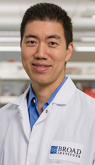
“A major aspiration in the molecular life sciences is the ability to precisely make any change to the genome in any location. We think prime editing brings us closer to that goal,” David Liu, PhD (above), Director of the Merkin Institute of Transformative Technologies in Healthcare at the Broad Institute, told The Harvard Gazette. “We’re not aware of another editing technology in mammalian cells that offers this level of versatility and precision with so few byproducts.” (Photo copyright: Broad Institute.)
Cell Division Not Necessary
CRISPR stands for Clustered Regularly Interspaced Short Palindromic Repeats. It is considered the most advanced gene editing technology available. However, it has one drawback not found in Prime Editing—CRISPR relies on a cell’s ability to divide to generate desired alterations in DNA—prime editing does not.
This means prime editing could be used to repair genetic mutations in cells that do not always divide, such as cells in the human nervous system. Another advantage of prime editing is that it does not cut both strands of the DNA double helix. This lowers the risk of making unintended, potentially dangerous changes to a patient’s DNA.
The researchers claim prime editing can eradicate long lengths of disease-causing DNA and insert curative DNA to repair dangerous mutations. These feats, they say, can be accomplished without triggering genome responses introduced by other forms of CRISPR that may be potentially harmful.
“Prime editors are more like word processors capable of
searching for targeted DNA sequences and precisely replacing them with edited
DNA strands,” Liu told NPR.
The scientists involved in the study have used prime editing to perform over 175 edits in human cells. In the test lab, they have succeeded in repairing genetic mutations that cause both Sickle Cell Anemia (SCA) and Tay-Sachs disease, NPR reported.
“Prime editing is really a step—and potentially a significant step—towards this long-term aspiration of the field in which we are trying to be able to make just about any kind of DNA change that anyone wants at just about any site in the human genome,” Liu told News Medical.
Additional Research Required, but Results are Promising
Prime editing is very new and warrants further
investigation. The researchers plan to continue their work on the technology by
performing additional testing and exploring delivery mechanisms that could lead
to human therapeutic applications.
“Prime editing should be tested and optimized in as many cell types as researchers are interested in editing. Our initial study showed prime editing in four human cancer cell lines, as well as in post-mitotic primary mouse cortical neurons,” Liu told STAT. “The efficiency of prime editing varied quite a bit across these cell types, so illuminating the cell-type and cell-state determinants of prime editing outcomes is one focus of our current efforts.”
Although further research and clinical studies are needed to
confirm the viability of prime editing, clinical laboratories could benefit
from this technology. It’s worth watching.
With $191 million in startup capital, the genomics startup will draw on existing genetic databases to create personalized medicine therapies for chronic diseases
Why do some people get sick while others do not? That’s what genetic researchers at Maze Therapeutics want to find out. They have developed a new approach to using tools such as CRISPR gene editing to identify and manipulate proteins in genetic code that may be the key to providing personalized protection against specific diseases.
If viable, the results of Maze’s research could mean the development of specific drugs designed to mimic genetic code in a way that is uniquely therapeutic to specific patients. This also would create the need for clinical laboratories to sequence and analyze patients’ DNA to determine whether a patient would be a candidate for any new therapies that come from this line of research.
Based in San Francisco, Maze Therapeutics (Maze) is studying modifier genes—genes that affect the phenotype or physical properties of other genes—and attempting to create drugs that replicate them, reported MIT Technology Review. Maze believes that genetic modifiers could afford a “natural form of protection” against disease.
“If you have a disease-causing gene, and I have the disease-causing gene, why is it that you may be healthy and I may be sick? Are there other genes that come into play that provide a protective effect? Is there a drugging strategy to recover normal phenotype and recover from the illness?” Maze Chief Executive Officer Jason Coloma, PhD, asked in an interview with FierceBiotech.
In 2019, Maze received $191 million in financing from Third Rock Ventures, ARCH Venture Partners, and others, to find ways to translate their findings into personalized medicines, according to a news release. And with the availability of international public genetic databases and CRISPR gene editing, now may be good timing.
“This was the perfect time to get into this space with the tools that were being developed and the amount of data that has been accumulated on the human genetic side,” Charles Homcy, MD, Third Rock Ventures Partner and Maze Scientific Founder, told Forbes, which noted that Maze is tapping existing population-wide genetic databases and large-scale studies, including the United Kingdom’s Biobank and Finland’s Finngen.
To help find genetic modifier drug targets, Maze is accessing CRISPR gene editing capabilities. Jonathan Weissman, PhD, Maze Scientific Founder and Professor of Cellular Molecular Pharmacology at University of California, San Francisco (UCSF), told MIT Technology Review: “You take a cell with a disease-causing gene and then see if you can turn it back to normal. We can do 100,000 experiments at once because each cell is its own experiment.”
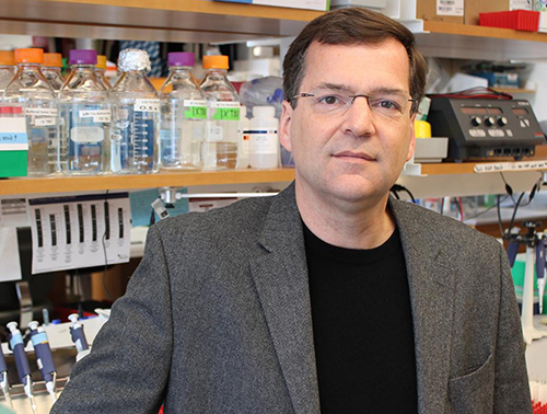
“At Maze, we are focused on expanding our understanding of the natural disease protection provided by genetic modifiers through an integrated approach that combines studying natural human genetic variation across the globe and conducting large-scale experiments of gene perturbations,” Charles Homcy, MD (above), Founder and interim CEO of Maze and a partner at Third Rock Ventures, said in a news release. “Through our integrated approach, we believe we will create novel medicines based around those modifiers to treat a number of diseases.” (Photo copyright: Forbes.)
Using CRISPR to Identify the Cause of Disease
One drug research program reportedly progressing at Maze involves developing gene therapy for the neurogenerative disease amyotrophic lateral sclerosis (ALS). The program borrows from previous research conducted by Aaron Gitler, PhD, Professor of Genetics at Stanford University and Maze co-founder, which used CRISPR to find genetic modifiers of ALS. The scientists found that when they removed the protein coding gene TMX2 (Thioredoxin Related Transmembrane Protein 2), the toxicity of proteins building the disease was reduced, reported Chemical and Engineering News.
“We used the CRISPR-Cas9 system to perform genome-wide gene-knockout screens for suppressors and enhancers of C9ORF72 DPR toxicity in human cells,” Gitler and colleagues wrote in Nature Genetics. “Together, our results demonstrate the promise of using CRISPR-Cas9 screens in defining the mechanisms of neurodegenerative diseases.”
“We have the flexibility to think differently. We like to
think of ourselves as part of this new breed of biotech companies,” Coloma told
FierceBiotech.
It’s an exciting time. Clinical laboratories can look
forward to new precision medicine diagnostic tests to detect disease and
monitor the effects of patient therapies. And the research initiatives by Maze
and other genetic companies represent a new approach in the use of genetic code
to create specific drug therapies targeted at specific diseases that work best
for specific patients.
The companion diagnostics that may come from this research would
be a boon to anatomic pathology.
CRISPR-Cas9 connection to cancer prompts research to investigate different approaches to gene editing
Dark Daily has covered CRISPR-Cas9 many times in previous e-briefings. Since its discovery, CRISPR, or Clustered Regularly Interspaced Short Palindromic Repeats, has been at the root of astonishing breakthroughs in genetic research. It appears to fulfill precision medicine goals for patients with conditions caused by genetic mutations and has anatomic pathologists, along with the entire scientific world, abuzz with the possibilities such a tool could bring to diagnostic medicine.
All of this research has contributed to a deeper understanding of how cells function. However, as is often the case with new technologies, unforeseen and problematic questions also have arisen.
“Here we report significant on-target mutagenesis, such as large deletions and more complex genomic rearrangements at the targeted sites in mouse embryonic stem cells, mouse hematopoietic progenitors, and a human differentiated cell line,” wrote the authors in their introduction.
Another study, this one conducted by biomedical researches at Cambridge, Mass., and published in Nature, describes possible toxicity caused by Cas9.
“Our results indicate that Cas9 toxicity creates an obstacle to the high-throughput use of CRISPR-Cas9 for genome engineering and screening in hPSCs [human pluripotent stem cells]. Moreover, as hPSCs can acquire P53 mutations, cell replacement therapies using CRISPR-Cas9-enginereed hPSCs should proceed with caution, and such engineered hPSCs should be monitored for P53 function.”
Essentially what both groups of researchers found is that CRISPR-Cas9 cuts through the double helix of DNA, which the cell responds to as it would any injury. A gene called p53 then directs a cellular “first-aid kit” to the “injury” site that either initiates self-destruction of the cell or repairs the DNA.
Therefore, in some instances, CRISPR-Cas9 is inefficient because the repaired cells continue to function. And, the repair process involves the p53 gene. P53 mutations have been implicated in ovarian, colorectal, lung, pancreatic, stomach, liver, and breast cancers.
Though important, some experts are downplaying the significance of the findings.
Erik Sontheimer, PhD (above), Professor, RNA Therapeutics Institute, at the University of Massachusetts Medical School, told Scientific American that the two studies are important, but not show-stoppers. “This is something that bears paying attention to, but I don’t think it’s a deal-breaker,” he said. (Photo copyright: University of Massachusetts.)
“It’s something we need to pay attention to, especially as CRISPR expands to more diseases. We need to do the work and make sure edited cells returned to patients don’t become cancerous,” Sam Kulkarni, PhD, CEO of CRISPR Therapeutics, told Scientific American.
Both studies are preliminary. The implications, however, is in how genes that have become corrupted are used.
A team from the Salk Institute may have found a solution. They are investigating a different enzyme—Cas13d—which, in conjunction with CRISPR would target RNA rather than DNA. “DNA is constant, but what’s always changing are the RNA messages that are copied from the DNA. Being able to modulate those messages by directly controlling the RNA has important implications for influencing a cell’s fate,” Silvana Konermann, PhD, a Howard Hughes Medical Institute (HHMI) Hanna Gray Fellow and member of the research team at Salk, said in a news release.
The Salk team published their findings in the journal Cell. The paper describes how “scientists from the Salk Institute are reporting for the first time the detailed molecular structure of CRISPR-Cas13d, a promising enzyme for emerging RNA-editing technology. They were able to visualize the enzyme thanks to cryo-electron microscopy (cryo-EM), a cutting-edge technology that enables researchers to capture the structure of complex molecules in unprecedented detail.”
The researchers think that CRISPR-Cas13d may be a way to make the process of gene editing more effective and allow for new strategies to emerge. Much like how CRISPR-Cas9 led to research into recording a cell’s history and to tools like SHERLOCK (Specific High-sensitivity Enzymatic Reporter unLOCKing), a new diagnostic tool that works with CRISPR and changed clinical laboratory diagnostics in a foundational way.
Each discovery will lead to more branches of inquiry and, hopefully, someday it will be possible to cure conditions like sickle cell anemia, dementia, and cystic fibrosis. Given the high expectations that CRISPR and related technologies can eventually be used to treat patients, pathologists and medical laboratory professionals will want to stay informed about future developments.
Three innovative technologies utilizing CRISPR-Cas13, Cas12a, and Cas9 demonstrate how CRISPR might be used for more than gene editing, while highlighting potential to develop new diagnostics for both the medical laboratory and point-of-care (POC) testing markets
CRISPR (Clustered Regularly Interspaced Short Palindromic Repeats) is in the news again! The remarkable genetic-editing technology is at the core of several important developments in clinical laboratory and anatomic pathology diagnostics, which Dark Daily has covered in detail for years.
Now, scientists at three universities are investigating ways to expand CRISPR’s use. They are using CRISPR to develop new diagnostic tests, or to enhance the sensitivity of existing DNA tests.
One such advancement improves the sensitivity of SHERLOCK (Specific High Sensitivity Reporter unLOCKing), a CRISPR-based diagnostic tool developed by a team at MIT. The new development harnesses the DNA slicing traits of CRISPR to adapt it as a multifunctional tool capable of acting as a biosensor. This has resulted in a paper-strip test, much like a pregnancy test, that can that can “display test results for a single genetic signature,” according to MIT News.
Such a medical laboratory test would be highly useful during pandemics and in rural environments that lack critical resources, such as electricity and clean water.
One Hundred Times More Sensitive Medical Laboratory Tests!
MIT News highlighted the high specificity and ease-of-use of their system in detecting Zika and Dengue viruses simultaneously. However, researchers stated that the system can target any genetic sequence. “With the original SHERLOCK, we were detecting a single molecule in a microliter, but now we can achieve 100-fold greater sensitivity … That’s especially important for applications like detecting cell-free tumor DNA in blood samples, where the concentration of your target might be extremely low,” noted Abudayyeh.
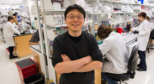
“The [CRISPR] technology demonstrates potential for many healthcare applications, including diagnosing infections in patients and detecting mutations that confer drug resistance or cause cancer,” stated senior authorFeng Zhang, PhD. Zhang, shown above in the MIT lab named after him, is a Core Institute Member of the Broad Institute, Associate Professor in the departments of Brain and Cognitive Sciences and Biological Engineering at MIT, and a pioneer in the development of CRISPR gene-editing tools. (Photo copyright: MIT.)
Creating a Cellular “Black Box” using CRISPR
Another unique use of CRISPR technology involved researchers David Liu, PhD, and Weixin Tang, PhD, of Harvard University and Howard Hughes Medical Institute (HHMI). Working in the Feng Zhang laboratory at the Broad Institute, they developed a sort of “data recorder” that records events as CRISPR-Cas9 is used to remove portions of a cell’s DNA.
They published the results of their development of CRISPR-mediated analog multi-event recording apparatus (CAMERA) systems, in Science. The story was also covered by STAT.
“The order of stimuli can be recorded through an overlapping guide RNA design and memories can be erased and re-recorded over multiple cycles,” the researchers noted. “CAMERA systems serve as ‘cell data recorders’ that write a history of endogenous or exogenous signaling events into permanent DNA sequence modifications in living cells.”
This creates a system much like the “black box” recorders in aircraft. However, using Cas9, data is recorded at the cellular level. “There are a lot of questions in cell biology where you’d like to know a cell’s history,” Liu told STAT.
While researchers acknowledge that any medical applications are in the far future, the technology holds the potential to capture and replay activity on the cellular level—a potentially powerful tool for oncologists, pathologists, and other medical specialists.
Using CRISPR to Detect Viruses and Infectious Diseases
Another recently developed technology—DNA Endonuclease Targeted CRISPR Trans Reporter (DETECTR)—shows even greater promise for utility to anatomic pathology groups and clinical laboratories.
Also recently debuted in Science, the DETECTR system is a product of Jennifer Doudna, PhD, and a team of researchers at the University of California Berkeley and HHMI. It uses CRISPR-Cas12a’s indiscriminate single-stranded DNA cleaving as a biosensor to detect different human papillomaviruses (HPVs). Once detected, it signals to indicate the presence of HPV in human cells.
Despite the current focus on HPVs, the researchers told Gizmodo they believe the same methods could identify other viral or bacterial infections, detect cancer biomarkers, and uncover chromosomal abnormalities.
Future Impact on Clinical Laboratories of CRISPR-based Diagnostics
Each of these new methods highlights the abilities of CRISPR both as a data generation tool and a biosensor. While still in the research phases, they offer yet another possibility of improving efficiency, targeting specific diseases and pathogens, and creating new assays and diagnostics to expand medical laboratory testing menus and power the precision medicine treatments of the future.
As CRISPR-based diagnostics mature, medical laboratory directors might find that new capabilities and assays featuring these technologies offer new avenues for remaining competitive and maintaining margins.
However, as SHERLOCK demonstrates, it also highlights the push for tests that produce results with high-specificity, but which do not require specialized medical laboratory training and expensive hardware to read. Similar approaches could power the next generation of POC tests, which certainly would affect the volume, and therefore the revenue, of independent clinical laboratories and hospital/health system core laboratories.