

New technology from researchers at the University of Texas Southwestern Medical Center enables the ability to study cancer cells in their native microenvironments
Imaging research is one step closer to giving clinicians a way to do high-resolution scans of malignant cells in order to diagnose cancer and help identify useful therapies. If this technology were to prove successful in clinical studies, it might change how anatomic pathologists and radiologists diagnose and treat cancer.
Researchers at the University of Texas Southwestern Medical Center developed a way to create near-isotropic, high-resolution scans of cells within their microenvironments. The process involves utilizing a combination of two-photon Bessel beams and specialized filtering.
New Imaging Approach Could be Useful to Both Pathologists and Radiologists
In a recent press release, senior author Reto Fiolka, PhD, said “there is clear evidence that the environment strongly affects cellular behavior—thus, the value of cell culture experiments on glass must at least be questioned. Our microscope is one tool that may bring us a deeper understanding of the molecular mechanisms that drive cancer cell behavior, since it enables high-resolution imaging in more realistic tumor.”
In a study in Developmental Cell, Erik S. Welf, PhD, et al, described the new microenvironmental selective plane illumination microscopy (meSPIM). When developing the technology, the team outlined three goals:
1. The microscope design must not prohibitively constrain microenvironmental properties.
2. Spatial and temporal resolution must match the cellular features of interest.
3. Spatial resolution must be isotropic to avoid spatial bias in quantitative measurements.
This new technology offers pathologists and medical laboratory scientists a new look at cancer cells and other diseases. The study notes that meSPIM eliminates the influence of stiff barriers, such as glass slide covers, while also allowing a level of control over both mechanical and chemical influences that was previously impossible.
Early meSPIM Research Reveals New Cell Behaviors
Early use of meSPIM in observing melanoma cells is already offering new insights into the relationship between the cell behavior of cellular- and subcellular-scale mechanisms and the microenvironment in which these cells exist. The study notes, “The ability to image fine cellular details in controllable microenvironments revealed morphodynamic features not commonly observed in the narrow range of mechanical environments usually studied in vitro.”
One such difference is the appearance of blebbing. Created by melanoma cells and lines, these small protrusions are thought to aid in cell mobility and survival. Using meSPIM, observers could follow the blebbing process in real-time. Formation of blebs on slides and within an extracellular matrix (ECM) showed significant differences in both formation and manipulation of the surrounding microenvironment.
The team is also using meSPIM to take a look at membrane-associated biosensor and cytosolic biosensor signals in 3D. They hope that investigation of proteins such as phosphatidylinositol 3-kinase (PI3K) and protein kinase C will help to further clarify the roles these signals play in reorientation of fibroblasts.
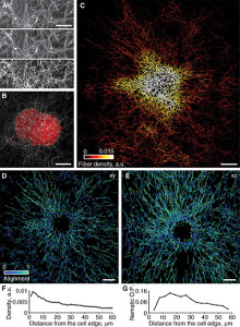
meSPIM combined with computer vision enables imaging, visualization, and quantification of how cells alter collagen fibers over large distances within an image volume measuring 100 mm on each side. (Photo Copyright: Welf and Driscoll et al.)
The research team believes this opens new possibilities for studying diseases at a subcellular level, saying, “Cell biology is necessarily restricted to studying what we can measure. Accordingly, while the last hundred years have yielded incredible insight into cellular processes, unfortunately most of these studies have involved cells plated onto flat, stiff surfaces that are drastically different from the in vivo microenvironment …
“Here, we introduce an imaging platform that enables detailed subcellular observations without compromising microenvironmental control and thus should open a window for addressing these fundamental questions of cell biology.”
Limitations of meSPIM
One significant issue associated with the use of meSPIM is the need to process the large quantity of data into useful information. Algorithms currently allow for automatic bleb detection. However, manual marking, while time consuming, still provides increased accuracy. Researchers believe the next step in improving the quality of meSPIM scans lie in computer platforms designed to extract and process the scan data.
Until this process is automated, user bias, sample mounting, and data handling will remain risks for introducing errors into the collected data. Yet, even in its early stages, meSPIM offers new options for assessing the state of cancer cells and may eventually provide pathologists and radiologists with additional information when creating treatment plans or assessments.
—Jon Stone
Related Information:
Seeing Cancer Cells in 3D (w/Video)
Quantitative Multiscale Cell Imaging in Controlled 3D Microenvironments
New 3-D Imaging Provides Unprecedented Views of Cancer in Its Natural Environment
New Imaging Technique, meSPIM, Illuminates Cells’ Microenvironments
