


Team of bioengineers succeeds in putting three different imaging technologies into a handheld probe that could be used by physicians to assess skin lesions in their offices
Dermatopathologists and pathology practice administrators will be keenly interested in a new, hand-held diagnostic device that is designed to reduce the need for skin biopsies. Because of high volume of skin biopsies referred to pathologists, any significant reduction in the number of such case referrals would have negative revenue impact on medical laboratories that process and diagnose these specimens.
This innovative work was done at the University of Texas at Austin’s Cockrell School of Engineering. The research team developed a probe that uses three different light modalities to detect melanoma and other skin cancer lesions in real-time, according to a news release.
New Device Is Moving Quickly into Clinical Trials
The device—dubbed a 3-in-1 spectroscopy probe—is apparently robust enough for clinical trials, which are reportedly under way. Thus, this device may be on a fast pace to gain clearance by regulators and then be ready for use by dermatologists in their offices. Developers are contacting funding agencies and start-up companies in order to bring this device to market.
If the 3-in-1 spectroscopy probe proves to be successful, there would be an downside and an upside for clinical laboratories. The downside, as noted earlier, is that wide adoption of a new skin cancer detection probe may eventually lead to a downturn in the number of biopsy referrals from dermatologists’ practices.
Conversely, on the upside, use of this technology may reduce the need for more dermatopathologists to read these specimens. Should a shortage of pathologists develop in coming years, reducing the number of biopsies that have to be read by a pathologist would be beneficial.
Issues Associated with Skin Biopsies and Patient Comfort Motivate Research
Developers of this device know that a skin biopsy is an invasive physical procedure. Another issue is that a biopsy takes an emotional toll on patients who often must wait days for results about cancer diagnosis, according to the researchers.
In addition to concerns about patient comfort, motivating the team’s research were the challenges faced by a physician when he or she decides which are the right lesions to biopsy in the first place. Biopsy, as the researchers observed, is an “imprecise art” with practical and financial consequences to skin cancer care.
“Currently, the only reliable method of distinguishing between dysplastic nevi and melanoma is via stained biopsies, which is invasive and expensive,” wrote the researchers in a paper published in the journal Review of Scientific Instruments.
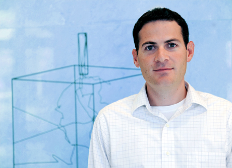
Motivated by the fact that many skin biopsies are collected needlessly and patients would benefit from a non-invasive method of evaluating skin lesions, a research team at the University of Texas at Austin’s Cockrell School of Engineering have developed a device that is about to undergo clinical trials. James Tunnell, Ph.D., (pictured above) associate professor in the Department of Biomedical Engineering at UT, led the research team that engineered a device that uses three different imaging technologies to assess skin lesions. (Photo copyright the University of Texas.)
In fact, for every case of skin cancer detected, about 25 unnecessary biopsies are done, according to the authors of the study. Biopsy’s imprecision generates costs to the U.S. healthcare system about $6 billion per year, reported the University of Texas in a press release.
Innovative Non-invasive Probe Uses Three Light Sources
Spectroscopic techniques rely on light’s interaction with biological tissues. The goal is to see the tissue’s “optical properties, which change with disease progression and can be used for diagnosis,” explained the journal article.
The 3-in-1 spectroscopy probe is reportedly different from other spectroscopy approaches to cancer detection. That’s because it combines—for the first time—(1) Raman spectroscopy, (2) diffuse reflectance spectroscopy, and (3) laser-induced florescence spectroscopy.
This play on light, so to speak, creates a “more complete picture of a skin lesion. And by revealing information invisible to the human eye, the probe could offer a faster, better screening tool for cancer and eliminate many biopsies,” according to the UT press release.

The 3-in-1 spectroscopy probe (pictured above on left) is about the size of a pen. Supporting it are spectroscopic and computer equipment that fit on a portable cart. Each reading takes about 4.5 seconds. The graphic on right shows the probe’s assembly with optical elements such as filters, fibers and front lens. (Photo copyright University of Texas.)
“As normal skin becomes cancerous, cell nuclei enlarge, the top layers of skin can thicken, and the skin cells can increase their consumption of oxygen and become disorganized. The changes alter the way the light interacts with the tissue,” observed James Tunnell, Ph.D., Associate Professor in the Department of Biomedical Engineering at the University of Texas and the lead researcher.
According to Tunnell and his co-authors, spectroscopic techniques detect skin changes like this: 1) Diffuse optical spectroscopy senses absorption of proteins such as hemoglobin; and 2) Raman spectroscopy detects vibrational modes of chemical bonds, such as those found in connective tissues.
Using Spectroscopy to Shed More Light on Cancer Diagnosis
Dark Daily readers recall that spectroscopy has long been in the news as a non-invasive approach to cancer answers. We reported on Duke University researchers’ “cancer flashlight” that uses spectroscopic techniques to detect pre-cancer cells in the colon. (See Dark Daily, “Duke University Researchers Demonstrate that Non-invasive Optical Biopsy Detects Cancer,” May 7, 2012.)
Similarly, in 2010, Dark Daily called attention to research under way at Massachusetts General Hospital regarding use of spectroscopic analysis to better identify prostate cancer without the need for a biopsy. (See Dark Daily, “Pathologists Pay Heed! New Spectroscopy Technique May Make Prostate Biopsy Obsolete,” February 19, 2010.)
Independent of these research initiatives, spectroscopy may have found its place in the realm of skin cancer diagnosis. That’s because, as real estate professionals say, “It’s all about location, location, location.” Simply said, skin is accessible to physicians. As the Cancer Journal put it in a story about progress in melanoma diagnosis, “Although pathologic examination remains the gold standard for diagnosis, this (malignant melanoma) cancer has potential to be diagnosed through noninvasive approaches because of its cutaneous location.”
Not only is skin the largest organ in the human body, but it can be studied with the specially-designed cancer detection probe under development at the University of Texas. Pathologists and laboratory professionals will have to wait and see how this new device performs in clinical trials. Until then, the pathologist’s view—albeit through a microscope and not through use of this new device—is deemed the “gold standard” for evaluating skin biopsies.
—By Donna Marie Pocius
Related content:
3-in-1 Optical Skin Cancer Probe
New Device Improves Skin Cancer Detection
Duke University Researchers Demonstrate that Non-invasive Optical Biopsy Detects Cancer
New Spectroscopy Technique May Make Prostate Biopsy Obsolete
The Evolution of Melanoma Diagnosis: 25 Years Beyond the ABCDs
